Ultrafiltration is simply a process by which toxic substances released during the body’s metabolism, along with some other essential components such as water and nutrients, are filtered in and out of the blood respectively, leading to the removal of those toxic substances from the body through the micturition process.
It occurs by the process of reverse osmosis through thin membranous pores of the glomerulus, which is a network of capillaries carrying the blood to filter and is situated in Bowman’s Capsule, which is a part of the nephrons present in the kidney. Hence it is also called “glomerular filtration”.
It is the primary process leading to urine formation in our body.
This phenomenon was first explained by a scientist named Richards in 1942.
Interesting Science Videos
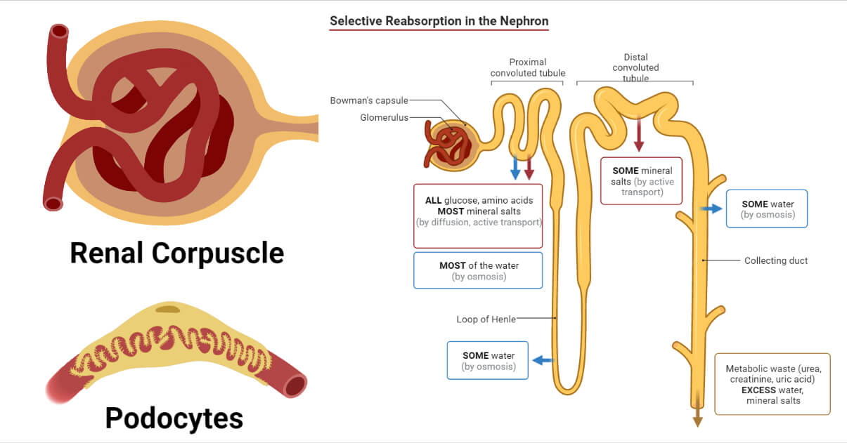
Bowman’s Capsule and its Structure
- The blood moves into the kidney through capillaries which form a bundle called the glomerulus in a cup-shaped vessel called Bowman’s Capsule.
- It is a double-layered structure with an outer parietal layer covered by squamous epithelium and an inner visceral layer lined by three layers:
- Inner layer – Endothelium
- Middle layer – Basement Membrane
- Outer layer – Epithelium
- The inner part, i.e., the surface of Bowman’s Capsule, comprises cells called Podocytes, where podo refers to the extensions. These extensions are termed pedicels which surround the surface of the glomerulus. There is the presence of a matrix made of glucose and protein in the gap between podocytes and glomerulus. The matrix is called the basement membrane, which leads to blood filtration.
- The capillaries in the glomerulus are porous, which leads to the exit of blood, and these pores are called filtration slits.
Glomerulus and its Structure
- It is formed by the capillaries originating from the afferent arteriole (renal arterioles are branches of renal arteries), which eventually leads to the efferent arteriole.
- These capillaries form a mesh-like structure inside Bowman’s Capsule.
- The lumen of the afferent arteriole is wider than that of the efferent arteriole.
- The capillaries contain tiny pores of size around 1000 Å.
Process Involved in Ultrafiltration
- The blood passes from the afferent arteriole through the network of the glomerulus and exits out through the efferent arteriole.
- During this transfer of blood, the filtration process occurs.
- The blood inside afferent arteriole has the presence of metabolic, toxic wastes such as urea, ammonia, uric acid, a large quantity of salts, creatinine, etc., and essential components such as vitamins, amino acids, glucose, and mineral salts as well. These substances have low molecular weights and can be easily filtered from the blood.
- As previously mentioned, the difference in the width of the lumen of the afferent and efferent arteriole has significance. The blood enters in large amounts and passes away in fewer amounts creating hydrostatic pressure of blood.
- Hydrostatic pressure is the main force that leads to the outer movement of fluid from the glomerulus.
- The fluid passes from the pores of capillaries to the basement membrane and eventually into the renal tubules through the filtrations slits of podocytes.
- As filtration occurs under the influence of pressure, hence this whole process is named ultrafiltration.
Hydrostatic Pressure
- The hydrostatic pressure values around +70 mm of Hg in the afferent arteriole (Ph).
- Some negative pressures oppose the hydrostatic pressure:
- Osmotic pressure exerted by plasma proteins (such as albumin proteins residing in glomerulus capillaries): It is around -30 mm of Hg. It is indicated by Pₒ.
- The pressure exerted by fluids in the renal tubules is around -10 mm of Hg. It is indicated by Pr.
- The pressure exerted by the intestinal fluid surrounding the renal tubule is around -10 mm of Hg. It is indicated by Pi.
- The net force to drive the ultrafiltration is provided by the sum of these pressures i.e., the pressure difference between the entry and exit of blood in the glomerulus and other opposing pressures. It is, as a whole, called Glomerular Filtration Pressure (GFP) or Effective filtration pressure (EFP).
GFP is given by the following formula:
GFP = Ph – (Pₒ+ Pr + Pi)
= 70 – (30+10+10) = 20 mm of Hg
Ultrafiltration Important Points
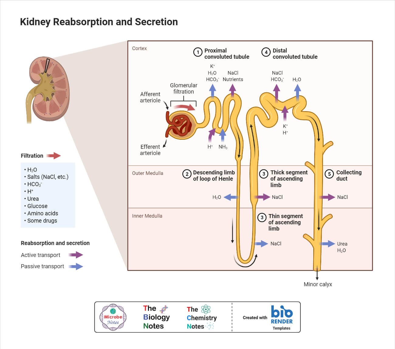
- GFP leads to the filtration of different substances such as water, smaller substances, and essential components such as vitamins, glucose, amino acids, sodium, etc., and also many toxic chemicals such as urea, uric acids, ammonia, and its salts, creatinine, pigments, potassium, etc. whereas some large sized molecules such as proteins, fats, and carbohydrates are not filtered out.
- The filtrate obtained after the glomerular filtration is called nephric or glomerular filtrate.
- The nephric filtrate thus obtained has a similar composition as blood without proteins and corpuscles.
Nephric filtrate = Blood – (Blood cells + Plasma Proteins) or (Plasma – Proteins)
- The process of ultrafiltration is totally physical.
- The amount of fluid that is filtered every day is around 180 liters, whereas the amount of urine produced from it is significantly less, i.e., around one to two liters.
Glomerular Filtration Rate (GFR)
It is the total amount of the nephric filtrate that is collected in a timeframe of one minute in all the present bowman’s capsules in both kidneys of a human body is called GFR. It is around 125 ml per minute and 180 liters per day.
References
- Keshari A.K. and Ghimire K.R. (2010) Excretory System. A Textbook of Higher Secondary Biology. 4th ed. Vidyarthi Pustak Bhandar. Kathmandu. Pg. 151-178.
- Accessed from: https://www.kidney.org/atoz/content/ultrafiltration. Accessed on: 19th Nov 2022.
- Koushanpor Esmail, 1986. Renal Physiology. Springer-Verlag. New York. Pg. 53-72.
- Ramirez J. 2016. Ultrafiltration: Methods, Application, and Insights. Nova Science Publishers.