Leukocytes, also known as white blood cells, are a cellular component of the blood that has a nucleus, absence of hemoglobin, and is capable of motility.
They are part of the immune system and help to defend the body against infection and diseases by destroying cancer cells and infection agents, ingesting foreign materials and cellular debris, and producing antibodies.
Leukocytes are produced by bone marrow and found in the blood and lymph tissue, spleen, liver, and kidney, which help in the regulation of leukocyte production levels.
The normal value of white blood cells ranges from 4000-11000 per microliter.
Leukocytes belong to at least five different categories and are distinguishable by their size, nuclear shape, and cytoplasmic inclusion.
Different types of leukocytes are involved in different functions. Some recognize intruders, some kill harmful bacteria, and some synthesize antibodies to protect the body against exposure to microorganisms (bacteria, viruses, and fungi).
Interesting Science Videos
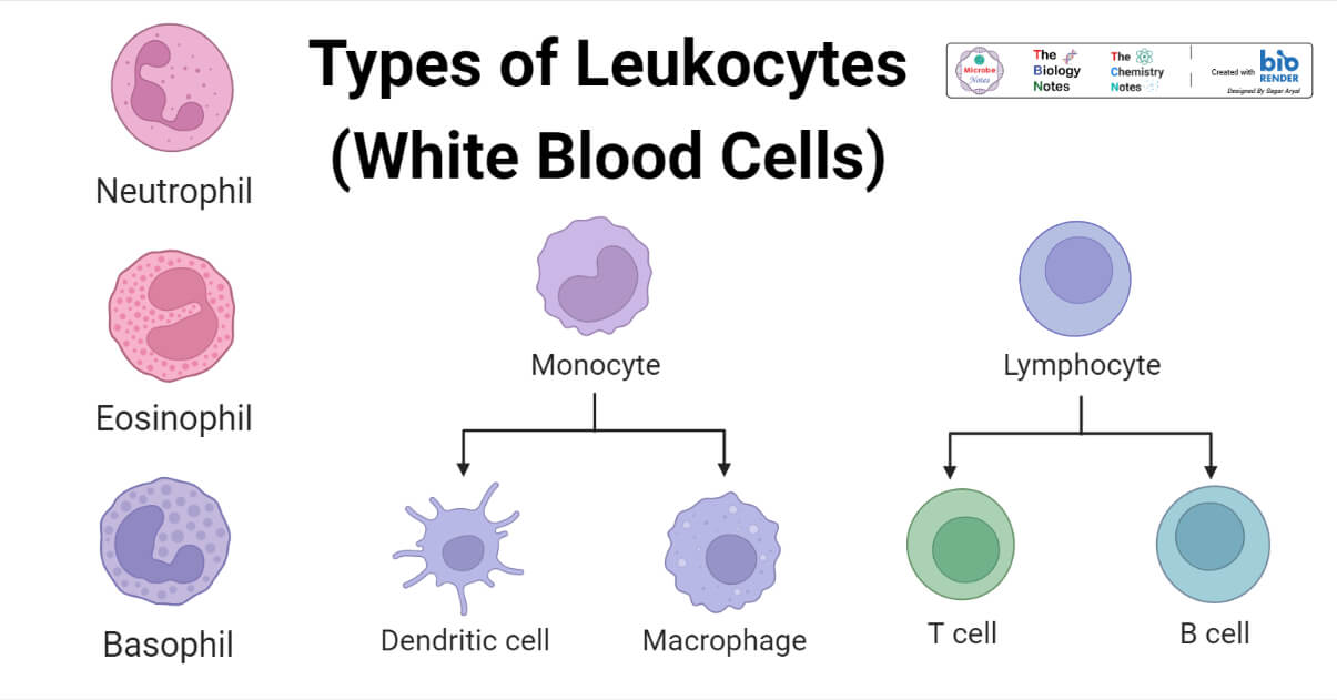
Types of Leukocytes
In practice, leukocytes are divided into two main groups:
1. Granulocytes, those with prominent stainable cytoplasmic granules, and
2. Agranulocytes are those without prominent stainable cytoplasmic granulocytes are mononuclear leukocytes.
Neutrophils, Eosinophils, and Basophils are types of granulocytes, whereas Monocytes and lymphocytes are agranulocytes leukocytes.
A. Granulocytes
Granulocytes are a type of leukocytes. it is an immune cell that contains granules with enzymes released during an allergic reaction, infection or disease, and asthma.
All granulocytes possess irregular or multilobed nuclei and belong to the myeloid sense of blood cells. Three types of granulocytes are described below:
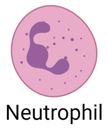
1. Neutrophil
Neutrophils are one type of leukocytes (white blood cells) that act as the first line of defense in the immune system.
Neutrophil Morphology/Anatomy
- Generally, they are spherical when at rest, but the shape can be changed to fight infection, and the size of neutrophils is 12-14µm dimeters.
- Neutrophils are also referred to as polymorphonuclear leukocytes because of their irregular segment (multilobed nuclei).
- In females, 3% of the nuclei of neutrophils show a conspicuous ‘drum-stick’ formation, which represents the sex chromatin of the inactive X chromosomes.
- Neutrophils are clear in color. When neutrophils react with dye, they change their color, so they are visible under a microscope. The cytoplasm of neutrophils is fine, neutrophilic granules are present, and in Leishman staining, the violet color of granules is seen.
Neutrophil Location
- Neutrophils are produced by hematopoiesis in the bone marrow, released into the peripheral blood, and circulate for 7-10 hours before migrating into the tissue, where they have a life span of only a few days.
Neutrophil Prevalence and normal range
- It is the most common type of white blood cell, and it consists of 40-75% of all white blood cells in an adult body.
- Neutrophils count is part of a complete blood count. The normal range of neutrophil counts ranges from 2500-7500 per µl of blood.
Neutrophil Defense mechanism
- During an inflammatory secretion, various substances are generated, which serve as chemotactic factors that promote the accumulation of neutrophils at an inflammatory site.
- They play an important role in defense of the body against microorganisms. They can phagocyte microbes and small particles in the circulation, and after extravasation, they carry out similar activities in other tissues.
- Actively phagocytic neutrophils can reduce oxygen enzymatically to form reactive oxygen species, including superoxide radicals and hydrogen peroxide, which enhance bacterial destruction probably by activation of some of the granule content.
- After the phagocytosis, they die, dead neutrophils, bacteria, tissue debris, and interstitial fluid from the characteristics green yellow pus of infected tissue.
A condition that affects neutrophils
- Neutropenia: It is the condition where neutrophils of body count are too lower than normal ranges, causing repeated infection and swelling. Cancer treatment, infection (hepatitis, tuberculosis, sepsis, Lyme disease), bone marrow disorder, vitamin deficiency, and autoimmune disease or infection are the main cause of neutropenia.
- Neutrophilia: It is the condition where neutrophils count is higher than normal ranges, also called neutrophilic leukocytosis. Bacterial infection, inflammation, injury, the reaction of certain drugs, and immature neutrophils leaving bone marrow too soon and reaching the bloodstream are the main cause of neutrophilia.
Neutrophil Function
- They migrate to the site of infection, where they kill or destroy the microorganisms by phagocytosis and releasing enzymes that kill them.
- Neutrophils boost the immune response of the body.
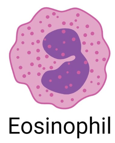
2. Eosinophil
Eosinophils are a type of granulocytes, leukocytes, or white blood cells that play an important role in the immune system, fight against disease, and boost inflammation in the infection site.
Eosinophil Morphology/ Anatomy
- Eosinophils are smaller in size (5-15µm) and motile but are present in small numbers in normal blood.
- which ranges from 100 to 400 per µl.
- The nucleus has two prominent lobes and appears U-shaped or spectacular-shaped in blood smear.
- The cytoplasm of eosinophils consists of coarse acidophilic granules.
Eosinophil Location
- Eosinophils are produced in the bone marrow and then travel to different tissue. Generally, found in the connective tissue of the stomach and intestine.
- The total lifespan of eosinophils is a few days, of which 10 hrs is spent in the circulation and the remainder in the extracellular tissues.
Eosinophil Prevalence and normal ranges
- Eosinophils consist of 1-4% of all white blood cells.
- Eosinophils count is part of a complete blood count, normal range of neutrophils counts ranges from 100 to 400 per µl of blood.
Eosinophil Defense mechanism
- The level of eosinophils rise in worm infection and also in certain allergic disorders, and it is thought that they evolved as a primary defense by destroying parasitic and cancer cell.
- They contain a surface receptor for Ig E that bind to Ig E- antigen complexes, triggering phagocytosis and release of granule contains.
- Different toxic molecules are released from their granules (e.g., eosinophil cationic protein and major base proteins) which are antiparasitic. They also release histaminase, which has a role in limiting the inflammatory consequences of mast cell degranulation.
A condition that affects eosinophilia
- Eosinopenia: It is the condition of eosinophils counts lower than the normal range in our body. Cushing’s syndrome and sepsis are common causes of Eosinopenia.
- Eosinophilia: It is the condition of eosinophils counts higher than the normal range.
- Alcohol intoxication, allergies, gastrointestinal disorder, leukemia, overproduction of cortisol, and parasitic infection are causes of eosinophil condition.
Eosinophil Functions
- Functions of eosinophils are killing cells, movement to inflamed areas, trapping substance, anti-bacterial and antiparasitic activity, involvement in an immediate allergic reaction, and modulating inflammatory reaction.
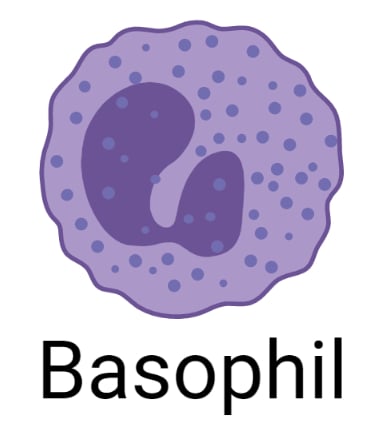
3. Basophils
Basophils are a type of granulocytes leukocytes. It is slightly smaller than other granulocytes but larger in cell size.
Basophils defend the body against pathogens (bacterial, fungal, and viral infection, allergens, and parasites, and Basophils release enzymes to prevent blood clots and improve blood flow.
Basophils Morphology/ Anatomy
- Basophils are spherical with 10-14µm in diameter.
- Distinguishing features of basophils is the presence of large, conspicuous basophils granules and kidney-shaped nucleus.
Basophils Location
- Basophils develop in the bone marrow, and after maturation, they flow through the bloodstream and migrate to damaged tissue to heal the area after injury.
Basophils Prevalence and normal range
- Form only 0.5 to 1% of total leukocytes or white blood cells, the normal range of count is 25-200 per µl.
Basophils Defense mechanism
- Basophilic granules contain heparin, histamine, and several other inflammatory agents. Basophils have high-affinity membrane receptors for Ig E and are coated with Ig E antibodies. If this binds to its antigen, it triggers degranulation of cells, producing vasodilation, increased vascular permeability, chemotactic stimuli for other granulocytes, and the symptoms of immediate hypersensitivity. Eg. In allergic rhinitis.
- Histamine improves blood flow by enlarging blood vessels and healing the affected area, and heparin prevents blood from clotting quickly.
A condition that affects basophils
- Basopenia: It is the condition in which the body does not synthesize enough basophil. Maybe Hyperthyroidism is a condition of basopenia.
- Basophilia: It is the condition when the body produces too many basophils cell. Leukemia, polycythemia vera, myelofibrosis, hypothyroidism, and autoimmune disease are a condition of basophilia.
Basophils Functions
- Basophils protect the body from external pathogens, parasites, and Allergens.
- Basophils granules contain an enzyme that is released and improves blood flow, and prevents blood clotting.
B. Agranulocytes leukocytes
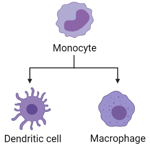
4. Monocytes
Monocytes are the largest type of white blood cell or leukocytes that reside in blood and tissue to find and destroy microorganisms and eliminate infected cells.
Monocytes Morphology/Anatomy
- The size of monocytes is near twice the size of red blood cells (15- 20 µm in diameter).
- but they form only in a small proportion (2-8% with a cell count of 100-700 per µl).
- Under a microscope, monocytes have a bilobed nuclei center that floats in a contained fluid called the cytoplasm; the nucleus changes shapes as the cell migrates throughout the body. the shape of the nucleus is oval, kidney, or horse-shaped, as observed under the microscope.
- Granules are absent in the cytoplasm.
Monocytes Location
- Monocytes develop in bone marrow and travel to the tissue where they protect the body from infection alongside other cells in the immune system.
Monocytes Prevalence and normal ranges
- It is the largest type of white blood cell and but they form only in a small proportion; it consists of 2-8% of all white blood cells in an adult body.
- Monocytes count is part of a complete blood count; the normal range of monocytes counts ranges from 100-700 per µl of blood.
Monocytes Defense mechanism
- Monocytes are actively phagocytic cells and contain numerous lysosomes; phagocytosis is triggered by the recognization of opsonized materials.
- Monocytes express class II MHC antigen.
- Most monocytes are thought to transit via the bloodstream from the bone marrow to the peripheral tissues, where they give rise to macrophage and dendritic cells; different monocytes’ subsets may target inflamed tissue.
- Dendritic cells recognize the antigen of invading germ; then, they release proteins called cytokines that inform other white blood cells to come to the site of infection and destroy antigen or infectious materials.
- Macrophages ingest microorganisms or infectious material and kill it with toxic enzymes within the cell, which also helps to remove dead cells from tissue.
A condition that affects monocytes
- Monocytopenia: When our monocyte count is too low, monocytopenia occurs. This condition occurs as a result of decreases in our leukocytes. Aplastic anemia, blood infection, burn injuries, HIV, and reaction to chemotherapy are common causes of monocytopenia.
- Monocytosis: It is the condition when the monocyte count is too high. Autoimmune disease (lupus disease, rheumatoid arthritis), blood disorders, cancer, cardiovascular disease, infection, and inflammatory disease are common causes of monocytosis.
Monocytes Functions
- Monocytes defend our body by either killing infectious agents or alerting other blood cells to help destroy them and boost immune response.
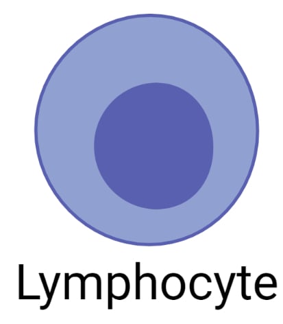
5. Lymphocytes
Lymphocytes are the second most numerous type of white blood cell in adulthood. In the immune system, it plays a mandatory role which helping the body to defend against disease and infection.
Two types of lymphocytes are present, which are T lymphocytes and B lymphocytes.
Lymphocytes Morphology/Anatomy
- Lymphocytes are larger than red blood cells. The size of lymphocytes varies ranges from 6-16µm. most circulating lymphocytes are 6-8 µm in diameter.
- Lymphocytes have a large nucleus, almost covered in the whole cytoplasm. Granules are absent in the cytoplasm.
Lymphocytes Location
- Lymphocytes form in the bone marrow. After maturation, lymphocytes reach the bloodstream and are found in blood and all part of the lymphatic system.
- Some lymphocytes travel to the thymus gland, which becomes a T cell and others travel to lymph nodes and organs, which become B cells.
Lymphocytes Prevalence and normal range
- The normal range of lymphocytes is between 1500 to 2700 per µl of blood; it consists of 20-30% of the total population of white blood cells.
- The lifespan of lymphocytes ranges from a few days to many years. long-lived lymphocytes are necessary for the maintenance of immunological memory.
Lymphocytes Defense mechanism
- Lymphocytes help our immune system fight against infection, disease, and cancer. Lymphocytes remember every antigen it comes in contact with. If the same antigen comes again, memory cells recognize it and quickly react. Due to this reason, we don’t get an infection like chickenpox or measles after one-time exposure, and getting vaccinated can protect or prevent certain diseases.
- Different types of T Cell help to kill the infected cell and regulate immune response to a foreign substance, such as cytotoxic T cell attached to an antigen on infected cell then kill the infected cell by making pore in their membrane, and enzymes are inserted into the cell, helper T cell help B cell for the production of antibodies against a foreign antigen.
- Antigen receptors surface present in B cell where Antigen attach. B cells recognized the antigen and produced specific antibodies against each one. B cells produce two types of immune response primary response and secondary response.
- when an antigen attaches to the receptor, B cells are stimulated. Among these, some are turned into memory cells, and some are plasma cells. Plasma cells produce antibodies specific to a particular antigen then a primary response is produced.
- When the B cell encounters the same antigen again, the memory B cell remembers it and multiplies. They turn into a plasma cell and immediately produce the correct antibody.
A condition that affects lymphocytes
- Lymphocytopenia: It is the condition of a low level of lymphocytes. Flu, mild infection, viral infection, tuberculosis, autoimmune disease, blood diseases, and radiation and chemotherapy treatment are the cause of low lymphocytes.
- Lymphocytosis: It is the condition of a high level of lymphocytes. Causes of higher lymphocytes are viral infection, syphilis, mononucleosis, tuberculosis, hypothyroidism, blood cancer, etc.
Lymphocytes Functions
- T cell attack and kills the infected cell and tumor cell and also regulate the body’s immune system.
- B cells synthesize antibodies against the virus, bacteria, and other foreign antigens.
Physiological variation of leukocytes
- Age: In infants, more than 20,000 WBC is present Per microliter. In children, the level ranges from 10,000 to 10,000.
- Sex: The male has a greater WBC count than the female.
- Emotional stress: emotional stress increases the leukocytes count,
- After exercise: Leucocyte count increase after exercise.
- Sleep: Leukocytes count decreases during sleep.
- Pregnancy: During pregnancy, leucocyte count increases.
References
- Guyton AC and Hall JE (2003) Medical Physiology, 10th 2. Standring S (2005). Gray’s Anatomy: The Anatomical Basis of Clinical Practice, 39 Edition, Indian Print: Saunders.
- Kindt TJ, Goldsby RA and Osborne BA (2006). Kuby Immunology, 6th 4. Edition, W. H. Freeman.
- https://www.cancer.gov/publications/dictionaries/cancer-terms/def/leukocyte.
- https://my.clevelandclinic.org/health/body/22313neutrophils
- https://www.osmosis.org/answers/eosinophils.
- https://my.clevelandclinic.org/health/body/23256-basophils.
- https://my.clevelandclinic.org/health/body/22110monocytes
- https://www.medicalnewstoday.com/articles/320987.
- Martin, H. E. (1932). Physiological leucocytosis: The variation in the leucocyte count during rest and exercise, and after the hypodermic injection of adrenaline. The Journal of Physiology, 75(2), 113.
really helpful notes. easy , quick and reliable !