- Within twenty-four hours after fertilization, the zygote initiates a rapid series of mitotic cell divisions called cleavage.
- These divisions are not accompanied by cell growth, so they subdivide the large zygote into many smaller daughter cells called blastomeres.
- The first cleavage division divides the zygote to produce two daughter cells.
- The second division, which is complete at about forty hours after fertilization, produces four equal blastomeres.
- By three days, the embryo consists of eight to sixteen cells, and by four days, it consists of sixteen to thirty-two cells.
- The embryo at this stage is called a morula.
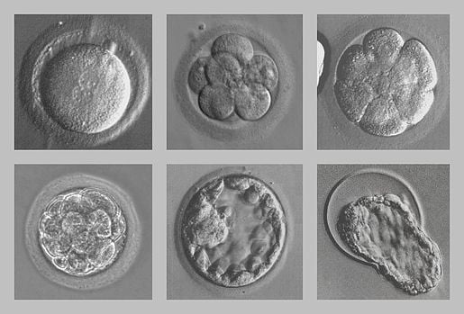
Image Source: nobelprize.org
Morula Stage
- An early stage in post-fertilization development when cells have rapidly mitotically divided to produce a solid mass of cells (16 or more) with a “mulberry” appearance is called the morula stage.
- The morula stage is the final stage prior to the formation of a fluid-filled cavity called the blastocoel cavity.
- It is distinct from a blastocyst in that a morula (3–4 days after fertilization) is a mass of totipotent cells in a spherical shape whereas a blastocyst (4–5 days after fertilization) has a cavity inside the zona pellucida along with an inner cell mass.
- A morula, if untouched and allowed to grow, will eventually develop into a blastocyst.
Development of Morula
- A key event prior to morula formation is “compaction”.
- During compaction, cells on the outer part of the morula become bound tightly together with the formation of desmosomes and gap junctions, becoming nearly indistinguishable.
- These changes in cell morphology and cell-cell adhesion initiates the formation of the solid ball of cells.
- With time, the cells on the outside and inside become differentially fated into trophoblast (outside) and inner cell mass (inside) progenitors.
Significance of Morula Stage
- The morula is the first embryonic stage where mammalian cells can be categorized as being either internal or external.
- The morula reaches the uterus between three and four days of development and greatly absorbs nutrients and fluid from the surrounding in preparation for the implantation process.
- In Assisted Reproductive Technology, the morula stage is when one of the earliest prenatal diagnostic tests can be carried out, by removing a single cell (blastomere) and carrying out genetic diagnosis on its DNA.
References
- Schoenwolf, G.C., Bleyl, S.B., Brauer, P.R., Francis-West, P.H. & Philippa H. (2015). Larsen’s human embryology (5th ed.). New York; Edinburgh: Churchill Livingstone.
- Sadler, T. W., & Langman, J. (2004). Langman’s medical embryology. Philadelphia, Pa: Lippincott Williams & Wilkins.
- Moore, K. L., Persaud, T. V. N., & Torchia, M. G. (2008). The developing human: Clinically oriented embryology. Philadelphia, PA: Saunders/Elsevier.
- https://embryology.med.unsw.edu.au/embryology/index.php/Morula_Development