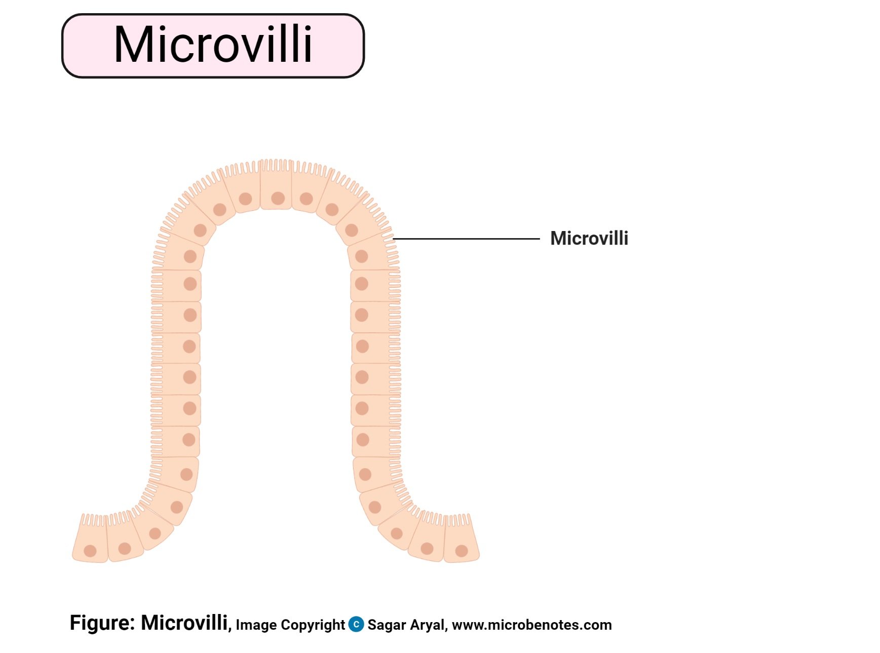
- Microvilli are small finger-like projections found on cells within the body that help the cells to get nutrition.
- Microvilli are membrane protuberances that arise from epithelial cells.
- They are usually about 0.1µm in diameter and up to 2 µm long.
- These microvilli are shorter and narrower than cilia but thicker than cilia.
- They are present in intestine mucosa, egg cells, white blood cells, and in the proximal convoluted tubules of the kidney.
- In light microscopy, they constitute the well-known striated or that microvilli are visible and look like a brush.
- So the microvillus border of intestinal epithelial cells is referred to as the brush border.
- A Brush border is a structure that forms when a large number of microvilli get together.
- A large amount of brush border is present on the epithelial cell of the small intestine which helps in absorption.
- They are not present only on absorptive surfaces. They can be found on secretory and other surfaces too.
- For example, in the liver, microvilli are found on both the secretory (bile canaliculus) pole of hepatocytes and absorptive (space of Disse).
- Particularly at the site of active absorption, they greatly increase the area of the cell surface (up to 40 times).
- In the intestine, they function in conjunction with villi.
- It is the projection of some of the mucous membranes, especially of the small intestine.
- They are the tiny fold that is projected out like the fingers.
- Since the area of the intestine is increased it aids in more absorption of the nutrients.
- There are little or no cellular organelles found in the microvilli, although they are cellular extensions.
- May be length and composition of microvilli within a certain group of homogenous cells, or different parts of the same organism are different. For example, the microvilli in the small intestine and large intestine in mice differ a bit slightly. They differ in length and amount of surface coat covering.
- In the immune system, microvilli are among the most common types of membrane protrusions found on lymphocytes.
Interesting Science Videos
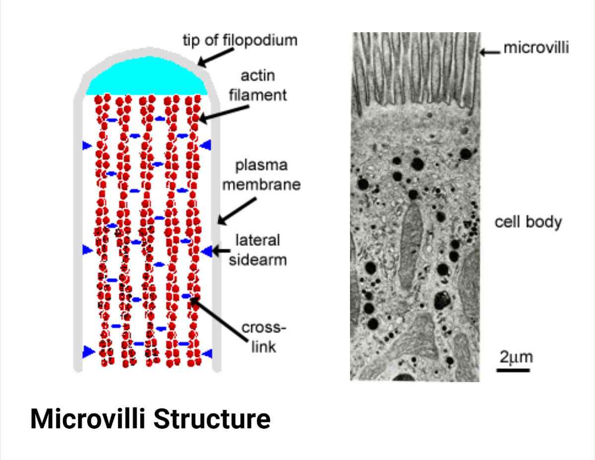
Microvilli Structure
- These are covered by a plasma membrane and supported internally by closely packed bundles of actin filaments, which serve as it is structural core.
- Twenty-Thirty tightly bundled actin – filaments are linked by cross-bridges of the actin-bundling proteins, fascin, and fimbrin.
- Another bridge composed of myosin I and calmodulin connect the filament bundles to the plasma membrane. Myosin I function through a binding site for filamentous actin on one side and lipid-binding domain on the other side.
- Actin filaments are most abundantly present in the cytosol near the cell surface.
- These filaments are assumed for the determination of the shape and movement of the plasma membrane.
- At the tip of each microvillus, a free end of microfilaments is inserted into a dense mass that includes the protein villin.
- These actin filaments of bundles of microvilli are embedded in the apical cytoplasm amongst a meshwork of transversely running actin filament stabilized by spectrin to form the terminal web, which is underlain by keratin intermediate filament.
- The web is anchored laterally to the tight junctions and zonula adherens of the apical epithelial junctional complex.
- Tropomyosin and myosin II are found in the terminal web, which may explain their contractile activity.
- The space which is found in between microvilli at the surface of a cell is known as the intermicrovillous space.
- This intermicrovillous space increases with the contractile activity of myosin II and tropomyosin.
- It then decreases when contraction ceases.
- In microvilli, basal granules are absent.
- They are cylindrical having the blunt ends.
- In the small intestine, Microvilli are usually covered with a very thick cell coat of glycocalyx.
- It reflects the presence of integral membrane glycoproteins, which include enzymes concerned with digestion and absorption.
- They have their own plasma membrane, cytoplasm. Some microfilaments are also present in it which gives them their structure.
- They have their own plasma membrane, inside of them, they have cytoplasm (cellular fluid) and some microfilaments, which give them their structure.
- Filopodia, irregular microvilli, are also found on the surface of many types of cells, particularly free macrophages and fibroblast, where they may be associated with phagocytosis and cell motility.
- Long and branching microvilli are called stereocilia, an early misnomer, as they are non-motile and lack microtubules. An appropriate name is stereovilli.
- Stereovilli is found on cochlear and vestibular receptor cells, where they act as sensory transducers and also in the absorptive epithelium of epididymis.
Microvilli Functions
- Microvilli present on the surface of epithelial cells like those found in the lining of the intestine increase the cell’s surface area, which facilitates the absorption of nutrients, ingested food, and a water molecule.
- In egg cells, microvilli help in anchoring the sperm to the egg, therefore allowing easier fertilization.
- They are an anchoring point in white blood cells.
- The membrane of microvilli consists of enzymes that play an important role in the conversion to simpler forms from the complex nutrients. Then they are easily absorbed.
- For Eg: On the surface of enterocyte microvilli, there is the presence of a higher concentration of glycosidases.
- Glycosidases are enzymes that help in the digestion of carbohydrates.
- There is an increment in the cell surface and digestive enzymes which can be found on the cell surface.
- Microvilli also help in detecting the sound in the ear. An electric signal is sent to the brain.
- From the clinical point of view, microvilli are very important. For example, congenital lack of microvilli in the intestinal tract causes a rare fatal condition found in newborn babies called microvillous atrophy.
- Microvilli present in the apical surface of hepatocytes participate in secretion.
References
- Drake, R. L., Vogl, W., Mitchell, A. W. M., & Gray, H. (2005). Gray’s anatomy for students. Philadelphia: Elsevier/Churchill Livingstone.
- Alberts, B. (2004). Essential cell biology. New York, NY: Garland Science Pub.
- Aramesh, M., Stoycheva, D., Sandu, I., Ihle, S. J., Zünd, T., Shiu, J. Y., Forró, C., Asghari, M., Bernero, M., Lickert, S., Oxenius, A., Vogel, V., & Klotzsch, E. (2021). Nanoconfinement of microvilli alters gene expression and boosts T cell activation. Proceedings of the National Academy of Sciences of the United States of America, 118(40). https://doi.org/10.1073/pnas.2107535118
- http://www.vivo.colostate.edu/hbooks/pathphys/digestion/smallgut/anatomy.html
- https://www.ncbi.nlm.nih.gov/pmc/articles/PMC2107317/
- https://study.com/academy/lesson/microvilli-definition-function.html
- https://www.sciencedirect.com/topics/biochemistry-genetics-and-molecular-biology/microvillus
- https://www.frontiersin.org/articles/10.3389/fimmu.2020.02187/full