Heart valves are the specialized flap or cusp-like structures inside the heart composed primarily of dense connective tissues that maintain the one-way flow of the blood inside the heart i.e., prevent the backward flow of the blood.
They are the essential anatomical structures inside the heart that control the direction and flow of the blood, ensuring efficient blood circulation through the heart chambers. The differential blood pressure inside the heart chambers causes the valves to open or close, controlling the direction of the movement of blood inside the heart.
Interesting Science Videos
Types of Heart Valves
The human heart has four valves classified – according to their anatomical location and function – into two categories, first the Atrioventricular valves (containing the bicuspid and tricuspid valves), and second the Semilunar valves (containing the pulmonary and aortic valves).
a. Atrioventricular Valves (AV Valves)
Those valves that are located in between the atrium and ventricles and prevent the backward flow of blood from the ventricle to the atrium are called the atrioventricular (AV) valves. Chordae tendineae hold them to the walls of the ventricles and attach them to papillary muscles preventing them from inverting. The fibrous ring known as the valve annulus serves as additional support for the AV valves. This annulus anchors the valve cusps and offers stability to them. There are two AV valves, the bicuspid valve, and the tricuspid valve. The closure of these valves produces the characteristic ‘lub’ sound of the heartbeat.
- Bicuspid Valve (Mitral valve)
It is also known as the mitral valve and is located between the left atrium and the left ventricle. It has 2 cusps (leaflets) (hence got the name bicuspid) resembling a bishop’s mitre (hence got the name mitral). This valve opens when the blood pressure inside the left atrium exceeds the pressure inside the left ventricle and allows the flow of blood to the left ventricle and closes at the end of the atrial contraction preventing the backflow of the blood in the atrium during the ventricular contraction.
- Tricuspid Valve
It is the valve located between the right atrium and the right ventricle. It has three cusps; hence got the name tricuspid. During the atrial systole, this valve opens and allows the blood to flow from the right atrium to the right ventricle and prevents the backward flow of the blood by being closed during the ventricular systole.
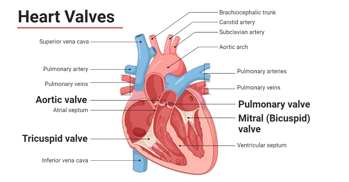
b. Semilunar Valves
Semilunar valves (SL valves) are the heart valves located in between the ventricles and the base of the two large arteries, viz. the aorta and the pulmonary artery. They prevent returning of blood back to the ventricles from the arteries after the ventricular systole. These valves have three half-moons (semilunar) shaped flaps (cusps); hence got the name ‘semilunar’. There are two semilunar valves, namely the pulmonary valve and the aortic valve. These valves lack chordae tendineae and are supported by the elastic arterial walls, sinuses of Valsalva (dilated area at the base of the aorta and pulmonary artery), and valve annulus. Closure of these two valves produces the characteristic ‘dub’ sound of a heartbeat.
- Pulmonary Valve
It is located at the base of the pulmonary artery. During the ventricular systole, this valve opens, due to pressure differences between the right ventricle and inside the pulmonary artery, allowing the deoxygenated blood to rush inside the pulmonary aorta. When the pressure inside the pulmonary artery exceeds the pressure inside the right ventricle, this valve closes preventing returning of the pumped blood to the ventricle.
- Aortic Valve
It is located at the base of the aorta. During ventricular systole, when higher pressure is buildup inside the left ventricle than in the aorta, this valve opens and allows the blood to be pumped inside the aorta. Soon at the end of ventricular systole, when the blood pressure becomes reversed, this valve closes and prevents the blood from returning to the left ventricle.
Structure of Heart Valves
- Heart valves are flap/cusp-like structures made of highly organized extracellular matrix (ECM) primarily containing collagen, elastin, and proteoglycans and valve interstitial cells (VIC) covered by a layer of endothelial cells.
- The mitral valve has two cusps, the anterior and the posterior cusp, and the tricuspid valve has three cusps, the anterior, the posterior, and the septal cusp. These cusps are attached to the papillary muscles by the chordae tendineae (fibrous chord of a tendon). The chordae tendineae provide anchorage and prevent the cusps from inverting during the ventricular systole.
- A fibrous ring called the valve annulus attaches all four valves to the myocardial wall providing support and stability.
- All the heart valves of humans are generally less than 1 mm thick. Structurally, the AV valves are slightly thicker than the SL valves, and the left-side valves are thicker than the right-sided valves. Despite their similar function, all valves are structurally and biochemically different than each other.
Functions of Heart Valves
- The mitral valve prevents the backflow of blood from the left ventricle to the left atrium.
- The tricuspid valve prevents the backflow of blood from the right ventricle to the right atrium.
- The pulmonary valve prevents the backflow of blood from the pulmonary artery to the right ventricle.
- The aortic valve prevents the backflow of blood from the aorta to the left ventricle.
Valve Disorders
Different cardiovascular diseases are associated with the disorder of valves. Such diseases of heart valves are called valvular heart diseases. Some common valvular heart diseases are tabulated below.
| Disease | Description |
| Stenosis (aortic stenosis, pulmonary stenosis, mitral stenosis, tricuspid stenosis) | A condition resulting from the narrowing of the heart valves, restricting the flow of blood. |
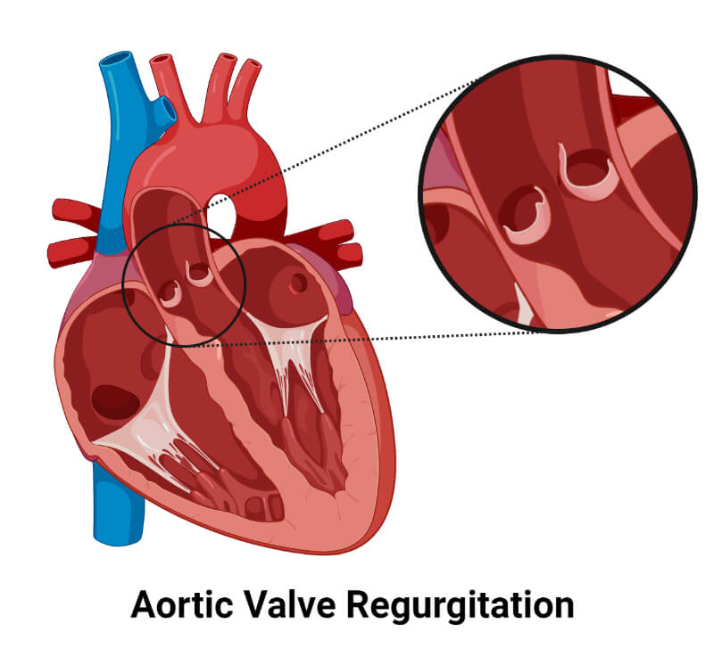
| Regurgitation | Incomplete closure of valves causing backflow of the blood. |
| Infective Endocarditis | Infection of heart valves. |
| Bicuspid Aortic Valve | A congenital disorder resulting in only two cusps in the aortic valve. |
| Pulmonary atresia | A congenital condition where the pulmonary valve is malformed and remains closed. |
References
- Ross & Wilson Anatomy & Physiology in Health and Illness. 13th ed. Churchill Livingstone Elsevier. ISBN 978-0-7020-7276-5
- Hinton, R. B., & Yutzey, K. E. (2011). Heart Valve Structure and Function in Development and Disease. Annual review of physiology, 73, 29. https://doi.org/10.1146/annurev-physiol-012110-142145
- Heart valve. (2023, May 24). In Wikipedia. https://en.wikipedia.org/wiki/Heart_valve
- 4 Heart Valves: What They Are and How They Work (clevelandclinic.org)
- Heart valves: Diagrams, types, function, diseases, and more (medicalnewstoday.com)
- Roles of Your Four Heart Valves | American Heart Association
- Heart valves anatomy: Tricuspid-aortic-mitral-pulmonary | Kenhub
- Heart valve disease – Symptoms and causes – Mayo Clinic
- Heart anatomy: Structure, valves, coronary vessels | Kenhub
- The Heart Valves – Tricuspid – Aortic – Mitral – Pulmonary – TeachMeAnatomy
- How the Heart Works – How Blood Flows through the Heart | NHLBI, NIH