Habit and Habitat of Earthworm
- The Earthworms are burrowers. It prefers to live in the burrow during the day and comes out at night and in damp cloudy weather for the search of food, reproduction, and exploring fresh habit. Thus, nocturnal in habit.
- The rainy season is the most favorable time for earthworms, after heavy rainfall, it leaves burrow and are seen in large numbers crawling on the ground.
- They breed during the rainy season.
- They are found in soil rich decaying organic matters usually in the garden, pastures, lawns, irrigated farmlands, near the banks of ponds, lakes, and rivers.
- They are cold-blooded or poikilothermal as their body temperature fluctuates with the surrounding temperature.
- They feed on dead organic matters, food and soil are ingested together and the latter, along with undigested food is finally egested in the form of worm casting.
- They are hermaphrodites, but they undergo copulation for exchange of their spermatozoa.
- Fertilization and development occur inside the cocoon.
- The natural life span of earthworm varies from 3 and a half years to 10 years.
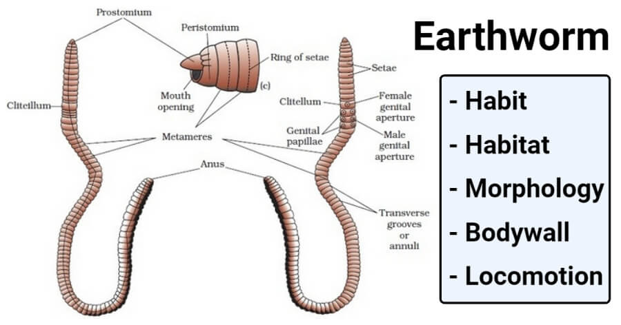
Image Source: Toppr.
External Morphology of Earthworm
The structures which are visible from outside are called external morphology of external features. The important external morphology of Earthworm ae as follows.
A. Shape and size
- Long, narrow, cylindrically elongated, pointed in front, blunt behind, and thickest a little behind the anterior end.
- Bilaterally symmetrical.
- The dorsal surface of the body is marked by the presence of a dark median line of a dorsal blood vessel that runs throughout the body just below the skin.
- The ventral surface is marked by the presence of genital openings and papillae in the anterior parts of the body.
- Size varies from species to species and individuals to individuals of the same species.
- A mature worm measures about 150mm in length and 3 to 5 mm in width.
B. Color
- Slimy to touch and glistening deep brown to clay color.
- The color is due to the presence of pigment porphyrin in its body wall which protects the body against bright and strong light.
- The dorsal surface is darker than the ventral surface.
C. Segmentation
- The body consists of about 100-120 small-ring-like segments or somites or metameres.
- Exhibit true segmentation.
- Segments are without parapodia.
- Segments are separated from each other by distinct ring-like grooves or furrows representing external segmentation.
- The interior of the body is divided by intersegmental septa or coelosepta into small chambers representing internal segmentation.
- All the segments of the body are similar except 1st and last segments.
D. Head
- Lacks distinct head and sense organs like yes, cirri, and tentacles.
- The buccal segment or peristomium is 1st segment at the anterior end of the body.
- Peristomium bears a terminal, crescentic mouth.
- They are prolonged into anteriorly into a fleshy lobe, the prostomium, which overhangs the mouth.
- The prostomium is called the boring part so it bores in soil.
E. Clitellum
- In mature earthworms, 14th, 15th, and 16th segments are enclosed by thick-collar or girdle-like glandular tissue called the clitellum.
- Cocoon formation takes place in clitellum.
- Absent in immature worm hence it indicates the maturity of the worm.
- The clitellum is glandular organs that secrete mucus, albumen, and a material for forming cocoons which assist in fertilization as the eggs are laid in them.
It divides the body into 3 regions such as:
1. Pre-clitellar region:
- Starts from the 1st to 13th segments. 1st segment is characteristically called peristomium which surrounds the mouth.
- At the anterior end of peristomium three is a small fleshly lobe called prostomium.
- The prostomium is not a segment. It is an extension of the peristomium at its dorsal side and ventral to it is the mouth.
2. Clitellar region: 14th, 15th, and 16th
3. Post-clitellar region: from 17th up to the posterior region.
- The clitellum is called Forest of Nephridia.
F. Setae
- All segments except first, last, and clitellum bears chitinous setae.
- They lie embedded in the middle of each segment which projects backwardly.
- About 80-120 setae are present on each segment.
- Each seta is minute, elongated, and S-shaped, and faint yellow in color.
- Each seta consists of 3 parts: The upperparts are the neck, the Middle swollen part is the modulus, and the inner part-is root or body, which is attached with setal sac or setigerous along with muscles.
- The movement of setae is controlled by special types of muscles.
- So, with the help of these muscles, the setae can be moved in any direction and extended or withdrawn at the will of the Earthworm.
- Called locomotory organs of earthworm.
- The clitellar segment possesses setae when the worms are immature, but setae are shed off before the clitellum is formed at maturity.
- Setae are formed of horny nitrogenous organic substances known as chitin.
- Setae are arranged in an annular row in the mid-ventral surface of each segment. This type of arrangement is known as perichaetine arrangement.
Video of Morphology of Earthworm (By Uniclass Content)

G. External Apertures of Earthworm
1. Mouth
- A crescentic anterior aperture.
- Situated just below the prostomium on the ventral side.
- Surrounded by 1st segment of the body- the peristomium or buccal segment.
2. Anus
- A vertical slit-like aperture at the posterior terminus.
- The exit of the alimentary canal.
- Undigested wastes are removed from it.
3. Genital openings
- The earthworm is hermaphrodites, so male and female generative apertures are found in the same individuals.
a. Male genital aperture
- Situated latero-ventrally in the 18th segment.
- A pair of crescentic openings of common prostatic and spermatic ducts.
b. Female genital aperture
- Median aperture situated at the ventral side in the 14th segment in the clitellar region.
- Female reproductive bodies are discharged from there.
4. Spermathecal pores
- 4 pairs of small ventrolateral spermathecal apertures
- Lies intersegmental between the grooves of 5/6, 6/7, 7/8, and 8/9 segments.
5. Nephridiopores
- A large number of very minute nephridiopores.
- They are scattered all over the body except for the first two segments.
- These pores are aperture of the integumentary nephridia, through which metabolic wastes of the body are removed.
6. Dorsal pores
- Minute apertures of coelomic chambers.
- Located mid-dorsally, one in each intersegmental groove, behind the 12th segment, except the last groove.
- Through these pores’ coelom communicates with the exterior.
7. Genital papillae
- These are pairs of conspicuous rounded elevation/circular and raised papillae in the same line, with the male pores, on the ventral side of each of the 17th and 19th segments.
- Each genital papilla bears a cup-like depression at the top but not any aperture.
- They function as suckers during copulation.
Read Also: Economic Importance of Earthworm
H. Body wall of Earthworm
The body wall of an earthworm is very thin, soft, shiny, elastic, and highly vascular. It consists of:
1. Cuticle
- It is a thin, elastic, and non-cellular and finely striated layer.
- It is a layer that is secreted by the columnar epithelial or supporting cells of the underlying epidermis.
- They remain perforated by a minute pore through which integumentary nephridial and epidermal glands open out.
- This layer is protective in function.
- It is made of 2 layers of collagenous proteins forming fibers and a polysaccharide with a small amount of gelatin.
2. Epidermis
- It is single-layered but multicellular.
- It lies just below the cuticle.
- It consists of tall and cylindrical epidermal cells which are of the following type, performing different functions:
a. Supporting cells- These are long columnar cells and are a major part of the epidermis. They have an oval nucleus nearly in the middle.
b. Gland cells- These are thicker consisting of numerous mucous cells and few albumen cells with secretory granules. These cells are situated between supporting cells and are of 2 types:
i. The mucous cells
- clubbed shaped and are large in number.
- secrete mucus which keeps the body moist.
- Helps in locomotion and prevents the animals from desiccation.
- Also known as goblet cells and they open at the surface of the cuticle by minute pores.
ii. Albumen cells
- Cylindrical, fewer in number.
- Have a uniformly distributed fine granule.
- and secretes albumen.
c. Basal cells
These are small conical cells, lying between supporting cells and gland cells. The cells are later on differentiated into supporting cells and gland cells hence it is also known as replacing cells.
d. Sensory receptor cells
These are narrow, columnar cells occurring in groups. These contain hair-like processes at their outer free ends and sensory in function. And these cells are also known as epidermal receptor cells.
3. Muscular Layer
- The muscle layer lies below the epidermis.
- It consists of an outer thin layer of circular muscle fibers running around the body
- And an inner longitudinal muscle fiber running along the length of the body.
- Longitudinal muscle fibers lie in parallel bundles, separated by connective tissue, and strengthened by collagen fibers.
- The contraction of circular muscles makes the body long and narrow.
- While the contraction of longitudinal muscles makes the body short and broad.
- The longitudinal muscle layer is further followed by a very thin strip of circular muscle fibers.
- The musculature of the body wall consists of smooth muscle fibers.
- Outer circular muscles consist of numerous scattered granules of porphyrin pigment.
- Two additional muscles are also found at the base of each setal sac. They are- A pair of protractor muscles and a single retractor muscle.
4. Coelomic epithelium
- It is the outer envelope of the coelomic cavity and hence called the coelomic epithelium or parietal peritoneum or parietal layer.
- It consists of flat cells. These flat cells are recognized by nuclei.
The function of the body wall of Earthworm
- It provides definite shape to the body (due to its elasticity).
- The mucus secreted by the mucous gland of the epidermis keeps the body smooth and moist helps in respiration and locomotion.
- Protects the internal delicate organ from injury.
- Secrete mucus which keeps the body surface slimy and kills harmful bacteria.
- Alternative contraction and relaxation of circular and inner longitudinal muscle help in movement.
- Albumen secreted by the clitellar gland helps in the nutrition of embryo developing inside the cocoon.
- Sensory epidermal cells are the only receptors to receive external stimuli.
- The coelomic epithelium or peritoneal layer secrets the coelomic fluid.
- The cuticle checks excess evaporation.
Locomotion of Earthworm
- No specialized locomotory organs are found in earthworms, even these are very active, and they crawl rapidly when out of the burrow.
- Movements of earthworm involve the musculature (a cumulative effect of contraction and relaxation of both the muscle layer) of the body wall and seta and the hydrostatic pressure created by the coelomic fluid.
- The increment in hydrostatic pressure of the anterior segments of the body (usually 9 segments) is responsible for forward locomotion.
- At the same time contraction of the circular muscle, the layer begins at the anterior end and passes backward.
- This results in the anterior region to extend forward and at the same time making it thinner in diameter.
- The extending and thinning passes backward as a wave of contraction at the rate of 2 to 3 cm per second; by this means worm is pushed forwards.
- The anterior end now grips the substratum and the setae act as hooks by their posteriorly directed points.
- This is followed by another wave of contraction affecting longitudinal muscle causing thickening and shortening of the body.
- This is again followed by the wave of thinning and the process is repeated alternately.
- Each wave of circular contraction causes the segments affected to move forward.
- At this stage, the segment in a state of longitudinal contraction does not move but remains as they are anchored to the ground by the protrude setae.
- During longitudinal contraction setae always protrude and during circular contraction it retracts.
- The worms move about 25cm distance in 1 minute it has been calculated by this method.
- The worm can move backward also when the direction of waves is reversed.
- They move backward usually during their withdrawal from the burrow and also during excavating it.
- During locomotion, the coelomic fluid serves as a kind of hydraulic skeleton because a decrease in its pressure results in the relaxation of muscles.
- The worm can move on a smooth and hard surface like glass by using mucus for adhesion as the setae cannot anchor the substratum.
References and Sources
- Kotpal RL. 2017. Modern Text Book of Zoology- Invertebrates. 11th Edition. Rastogi Publications.
- Jordan EL and Verma PS. 2018. Invertebrate Zoology. 14th Edition. S Chand Publishing.
- 13% – https://www.biologydiscussion.com/invertebrate-zoology/earthworms/pheretima-habit-and-habitat-and-external-features/29340
- 7% – https://microbiologynotes.com/external-morphology-earthworm/
- 1% – https://www.slideshare.net/tanvinaik165685/locomotion-in-annelids
- 1% – https://cdn1.byjus.com/wp-content/uploads/2019/07/Tamil-Nadu-class-8-Science-Book-Term-2-English.pdf
- 1% – https://biologyaipmt.com/2016/05/14/chapter-7-structural-organisation-in-animals/
- <1% – https://www.nationalgeographic.com/animals/invertebrates/c/common-earthworm/
- <1% – https://www.britannica.com/science/cuticle
- <1% – https://www.answers.com/Q/What_is_the_lifespan_of_an_earthworm
- <1% – https://orpons.wordpress.com/2015/05/16/5/
- <1% – https://link.springer.com/chapter/10.1007%2F978-1-4684-1224-6_17
- <1% – http://people.eku.edu/ritchisong/301notes3.htm
- <1% – http://education.med.nyu.edu/Histology/coursematerials/Presentations/Muscle-lec.html