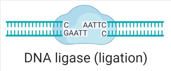
DNA ligase is an enzyme that catalyzes the ligation reaction, ligation is the process of joining various types of DNA fragments.
DNA ligase joins the DNA molecule covalently by catalyzing the formation of phosphodiester bonds between adjacent nucleotides.
The final phosphodiester linkage between the 5’-phosphate group on the DNA chain synthesized by DNA polymerase III and the 3’-hydroxyl group on the chain made by DNA polymerase I is catalyzed by DNA ligase.
The most organism requires energy for the cleavage of ATP and NAD+. Energy is required for the joining of nicks (nicks lack the phosphodiester linkage between the nucleotide sequence) of DNA. Whereas eukaryotic DNA ligases are ATP-dependent bacteria, archaea and virus DNA ligases are both NAD+ and ATP dependent.
DNA ligase forms a bond between the sugar-phosphate backbone to fully repair the DNA.
DNA ligase plays a vital role in DNA replication, DNA repair, and DNA recombination. Now day purified DNA ligase is isolated in the laboratory, which is used in gene cloning to join DNA molecules together to form recombinant DNA.
The enzyme DNA ligases were firstly isolated from E. coli in 1967, and several DNA ligases were discovered in 1967and 1968.
DNA ligase belongs to the nucleotide transferase superfamily, including RNA ligases and mRNA capping enzymes.
Interesting Science Videos
Structure of DNA Ligase
- The size of DNA ligases is different from organism to organism. Viral ATP-dependent DNA ligases are generally small in size.
- ATP-dependent and NAD+ dependent ligases have a catalytic core. Six conserved motifs (I, III, IIIa, IV, V, VI) are components of the catalytic core.
- For ATP or NAD+ co-factor binding, metal ion coordination, and ligation chemistry, these motifs are necessary.
- Many DNA ligases also contain a non-catalytic domain that is conserved, but the function of this domain is unknown.
ATP-dependent ligases (based on Bacteriophage T7)
- It is two domain ligase, the first one is the adenylation or nucleotide-binding domain. This domain serves site for ATP binding, which is located in a pocket beneath one of the β-sheets.
- Domain 1 is composed of 3 antiparallel β-sheets that are flanked by six α-helix.
- And the second domain is the oligonucleotide binding fold domain consists of a highly twisted antiparallel β-sheet with a single α helix running along one edge.
- These two domains are connected by a flexible linker.
NAD+ dependent ligases (based on E. coli LigA ligase)
- NAD+ dependent ligases have a central ligase catalytic core which is composed of oligonucleotide binding fold domain and nucleotidyltransferase domain.
- These two domains are located at the edge of the N-terminal domain (also known as domain la) and three C-terminal domains (which are a helix hairpin-helix (HhH) domain, zinc (Zn)-finger domain, and a BRTC domain).
- Domain la is required for the reaction of ligase with NAD+ to form the ligase-AMP intermediate.
Types of DNA Ligase
DNA ligases are classified into two categories based on source, the enzyme requires either ATP or NAD+ as a cofactor.
ATP dependent DNA ligases:
- Most eukaryotic DNA ligases use ATP as a cofactor, including bacteriophage, archaeal, and eubacteria DNA ligases.
- They vary in size in different organisms. The enzyme found in Haemophilus influenza consists of 268 amino acids, and larger cellular ligases such as human DNA ligase I consist of 912 amino acids, and IV consist of 844 amino acids.
1. Bacteriophage T7:
- It is the most widely used DNA ligase derived from the T7 bacteriophage. It is a monomeric form with a molecular weight 41KDa.
- It consists of two domains, namely 1 and 2. Domain 1 serves site for ATP binding it is composed of six alpha-helices that surround three antiparallel beta-sheets, and domain 2 is made up of antiparallel beta sheets and alpha helix
2. Bacteriophage T4:
- It can ligate either cohesive end or blunt ends of DNA, RNA -DNA hybrid, and also RNA.
- It is a monomeric form with a molecular weight 68KDa.
3. DNA ligases in Mammals:
- DNA ligase I: ligate Okazaki fragments during lagging strand DNA replication and some recombinant fragments
- DNA ligase II: purification or excision repair of alternatively spliced form of DNA ligase III found in non-dividing cells.
- DNA ligase III: It is the only mammalian ligase that is found in mitochondria. It ligates DNA during the process of nucleotide excision repair and recombinant fragment.
- DNA ligase IV: homologous recombination repair and important in immune cell development and other activities critical to a normal cell.
NAD+ dependent DNA ligases:
- Bacterial DNA ligases are NAD+ dependent ligases. They are also present in some mimivirus and entomovirus. While some bacteria, archaea, and viruses, DNA ligases are both NAD+ and ATP dependent.
- Mycobacterium tuberculosis codes for NAD+ dependent ligases and at least three different types of ATP-dependent ligases.
- They have a finely uniform size and consist of 656 – 837 amino acids.
1. E. coli DNA ligases:
- It is a monomeric enzyme of molecular weight 74KDa and requires NAD+ as a cofactor.
- It catalyzes the formation of the phosphodiester bond in double-stranded DNA containing cohesive ends.
- LigA and LigB (product of lig gene) are mostly used E. coli DNA ligases.
2. Taq DNA ligases:
- It is a thermostable ligase identified in several thermophilic bacteria, they require NAD+ as a cofactor.
- It is more stable and active at extreme temperatures than conventional DNA ligases. It is used in DNA amplification reactions to detect mutations in mammalian DNA.
Mechanism of DNA ligase
In the mechanism of DNA ligase, two covalent phosphodiester bonds between the 3’ hydroxyl end of acceptor nucleotide with the 5’ phosphate end of donor nucleotide are formed. In this process, two ATP molecules are consumed per one phosphodiester bond. The reaction can proceed in three steps or catalytic events.
1. Adenylation of DNA ligase/ Activation of DNA ligase
DNA ligase is activated using ATP. ATP breakdown into AMP releasing energy, and this Adenosine monophosphate nucleotide is attached to the amino group of lysine present on conserved active site of the enzyme by phosphoramidite bond leads to the formation of enzyme-AMP complex.
2. Activation of 5’ phosphate in nicks
The activated ligase-AMP complex is now transferred to the 5’ phosphate group at the breaking point, and AMP interacts with the phosphate group releasing the ligase enzyme, thus activating the 5’ end of single-strand DNA.
3. DNA ligation
The 5’ phosphate group is attacked by a 3’ hydroxyl group of another strand, forming a phosphodiester bond releasing free AMP and hydrogen ions. In this way, DNA and nicks are sealed.
Note: Bacterial ligase is adenylated by NADH2, which cleaved to form nicotinamide mononucleotide.
Functions of DNA Ligase
- DNA replication, DNA repair, and recombinant DNA experiment are incomplete without DNA ligases. DNA ligases join the nicks and form a phosphodiester bond between the nucleotide.
- Several laboratories purified DNA ligases from different organisms, which are used in gene cloning to join DNA molecules together to form recombinant DNA.
- They are used with restriction endonuclease enzyme to insert DNA fragments, often genes, into the plasmid.
- E. coli DNA ligases are used for high-efficiency cloning of full-length cDNA.
- Commercially available thermostable Taq ligases are used in amplification reactions because of their thermostable properties.
- Human DNA ligase IV is required for V(D) J recombination, the process that generates diversity in immunoglobulin and T-cell receptor loci during immune system development.
References
- Lewin B (2007), Genes IX, Oxford University Press, and Cell Press.
- Verma, P. S., & Agrawal, V. K. (2006). Cell Biology, Genetics, Molecular Biology, Evolution & Ecology (1 ed.). S . Chand and company Ltd.
- Ellenberger, T., & Tomkinson, A. E. (2008). Eukaryotic DNA ligases: structural and functional insights. Annu. Rev. Biochem., 77, 313-338.
- Doherty, A. J., Ashford, S. R., Subramanya, H. S., & Wigley, D. B. (1996). Bacteriophage T7 DNA Ligase: Overexpression, purification, crystallization, and characterization (∗). Journal of Biological Chemistry, 271(19), 11083-11089.
- Tomkinson, A. E., Vijayakumar, S., Pascal, J. M., & Ellenberger, T. (2006). DNA ligases: structure, reaction mechanism, and function. Chemical reviews, 106(2), 687-699.
- https://www.sciencedirect.com/science/article/pii/B0124437109001666
- https://www.sciencedirect.com/topics/neuroscience/dna-ligase
- https://pubs.acs.org/doi/full/10.1021/cr040498
- https://www.sigmaaldrich.com/NP/en/product/roche/dnaligro?gclid=Cj0KCQjwyMiTBhDKARIsAAJ-9Vu401zfGl26Qs4qB3qtJ7S_onnqCcG58mxtp72wtutLDAuMvzgmuYoaAntfEALw_wcB
- https://www.ncbi.nlm.nih.gov/pmc/articles/PMC97608/
- https://www.eurekaselect.com/article/9441
- https://www.cell.com/molecular-cell/fulltext/S1097-2765(07)00144-X