- The outer thin membrane or the layer of the living cell is known as the cell membrane.
- It is also known as the plasma membrane in animal cells.
- In the plant cells, it is known as plasmalemma.
- The term cell membrane was given by Nageli and Cramer (1885) for the membrane covering of the protoplast.
There are two types of cell membrane. They are:
Cytoplasmic membrane: which surrounds the whole protoplasm.
Internal membrane: which surrounds various cellular organelles and vacuoles.
- Inside constituents are proteins, carbohydrates and, nucleic acids. They are usually large in size. They are soluble in water and consists of charge.
- The outside environment of the cell is a bit different. The cell needs the nutrients too for its growth and development. Such nutrients along with the toxic substances which are harmful to the cell may be present in the outside environment.
- There can be the presence of ions, acids, and alkalis.
- Here the cell membrane acts as the barrier or like the checkpoints.
- The small molecules, solute, and lipid-soluble can go inside the cell membrane but the large molecules, water-soluble substances can’t go inside.
- It is impermeable to them. So it has got various mechanism by which such substances can be imported and exported.
- Different types of transport systems are available like active transport and passive transport.
- Similarly, there is the presence of facilitators and pumps.
- It allows only those things essential for cells to go inside whereas it limits the entry of such toxic substances.
- All the waste products of the cell are made exit to the outside.
- Cell membrane helps to maintain homeostasis.
- It provides protection to all the internal organelles of the cell.
Interesting Science Videos
Composition of Cell membrane
- Most of the cell membrane is composed of 40-50 % protein and 50-60 % lipids.
- Membrane lipids are of three types: a) Phospholipids b) Glycolipids c) Steroids
- In the different membrane, the proportion of the lipid varies:
The composition of the plasma membrane is:
Phospholipids- 55%
Glycolipids- 5%
Steroids- 20%
Other lipids- 20%
The composition of the bacterial membrane:
Cholesterol- 70%
Phospholipids- 30%
Structure of the cell membrane
The structure of the cell membrane is explained by the different models. They are as follows :
- Danielli-Davson Trilaminar sandwich model
- Fluid mosaic model of the Singer and Nicolson
- Unit membrane of Robertson
Danielli-Davson Trilaminar sandwich model
- It was proposed by James Danielli and Hugh Davsan in the year 1935.
- This model suggests the cell membrane as the solid and the stable structure.
- Four molecular layers are present in it i.e. two phospholipids and two protein layers.
- It consists of the phospholipid. It is based on the basis of physical and chemical properties of the plasma membrane.
- The plasma membrane is considered to be a protein-lipid-protein sandwich. The plasma membrane is of three-layer.
- The middle one is the phospholipid bilayer.
- Outer and inner denser protein is a monolayer.
- Protein and lipid are present in the P-L-L-P pattern.
- Protein molecules contain both polar and non-polar side chains.
- They are globular and aid in giving stability to the plasma membrane.
- Each phospholipid molecule is a polar molecule and consists of the hydrophilic and the hydrophobic end.
- Head (glycerols) of the phospholipid molecule: They are directed in the opposite directions.
- They are associated with the protein molecules by the Hydrogen bond, ionic bond, and electrostatic force of attraction.
- Tail end ( Fatty acids): They are held together by the weak Vander wal force. They lie facing each other.
- The plasma membrane is a porous membrane having microscopic pores of 7-10 A.
Drawbacks of Danielli-Davson Trilaminar sandwich model
- The cell membrane is the dynamic structure but not the stable structure.
- The cell membrane is not a solid structure. But rather it is the semisolid (quasifluid) structure.
- It doesn’t explain the functional specificity and variability in the biomembrane.
- It doesn’t explain the movements like active transport and movement of the water-soluble substances.
- Active and bulk transport of material through the membrane cannot be explained.
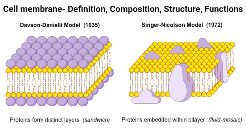
Image Source: BioNinja.
Fluid mosaic model of the Singer and Nicolson
- It was proposed by Singer and Nicholson in the year 1972 AD.
- This model suggests that the cell membrane is a quasifluid (semi-solid) and dynamic structure.
- This model is also known as the protein icebergs in a sea of phospholipids.
- In this model, the arrangement of the lipids and the integral proteins is in a mosaic style.
- Between the lipids and the proteins, there is interaction which results in the fluidity of the membrane.
- The interaction is hydrophobic. Two types of globular proteins are present which are embedded in the phospholipid bilayers.
Membrane proteins are of two types:
- Intrinsic protein
- Extrinsic protein
Intrinsic protein
- Within the phospholipid bilayer, those intrinsic proteins are found to be embedded inside.
- Intrinsic or integral proteins or tunnel proteins are soluble in nature. Integral proteins form a channel for the passage of water, ions, and other water-soluble small-sized solutes.
- It has got two parts:
Hydrophilic head: It is polar in nature. It protrudes out from the cell membrane.
Hydrophobic tail: It is non-polar in nature. They are present inside of the membrane facing towards the center.
Extrinsic protein
- They are present on two surfaces of the membrane in the floating form.
- Extrinsic proteins are attached to the phosphoryl surface.
- They are electrically charged too. Ionic bonds or calcium bridges help in the attachment.
- They are loosely attached.
- They are also called peripheral proteins.
- They are located outside of the lipid bilayer.
- They are soluble in nature.
Membrane protein are of five types:
- Structural protein: It helps in maintaining stability.
- Channel protein: It is involved in the transport of water and some dissolved substances.
- Carrier proteins: For active transport
- Enzymes: For different metabolic activities
- Receptor protein: For transport of hormones and conduction of nerve impulses.
Advantages of the fluid mosaic model
- This model explains the dynamic and quasifluid structure.
- Variability of the cell membrane is explained.
- It explains the transport of solute and solvent through the cell membrane.
- It explains the active and bulk transport of material through the cell membrane.
- Lipid: protein ratio supports this model.
This fluid mosaic model is the scientific and widely accepted model of the cell membrane.
Functions of the cell membrane
Cell recognition and communication
- In the cell membrane, glycolipids and glycoproteins are present. It helps in recognizing the cell.
- It is essential during the defense against microbes and tissue formation.
- The binding of the ligands to the specific receptors induces signal transduction.
Blood grouping
- Based on the antigens present in the cell membrane, blood grouping can be done and separated as A, B, AB, O.
Digestion
- In the gastrointestinal tract, microvilli are present.
- They are the modified cell membrane that helps in the digestion of food materials.
Locomotion
- In the amoeba, pseudopodia are present for locomotion which is the modified structure of the cell membrane.
Active transport
- The carrier protein present in the cell membrane helps in the active transport of materials.
Bulk transport
- By the process of endocytosis and pinocytosis, bulk materials are transported.
Exocytosis
- It helps in the removal of the waste materials and the secretory materials.
Osmosis
- It shows semi-permeable activity for the transport of water by osmosis.
Diffusion
- The exchange of gases with the external environment occurs by diffusion.
- Diffusion is of two types: simple diffusion and facilitated diffusion.
Metabolism
- Cell membrane performs metabolic functions.
- Several enzymes are present on the cell surface which is involved in the breakdown of extracellular nutrients.
- Some are involved in the biosynthesis of the cell wall.
- Incase of prokaryotes, respiratory enzymes are found in the plasma membrane.
References and Sources
- Shakya M, Mehata KR, Gautam MK, Pokhrel KR and Khanal K (2020 ) “ Principles of Biology”, Asmita Books Publisher and Distributors Ltd, Bhotahity, Nepal
- 1% – https://www.researchgate.net/publication/20685751_Lipid_regulation_of_membrane_structure_and_function
- 1% – https://www.mechanobio.info/what-is-the-plasma-membrane/what-types-of-proteins-are-found-in-the-plasma-membrane/
- 1% – https://www.khanacademy.org/science/high-school-biology/hs-cells/hs-the-cell-membrane/a/hs-the-cell-membrane-review
- 1% – https://www.britannica.com/science/cell-membrane
- 1% – https://quizlet.com/325193549/ap-bio-unit-3-ch-3-7-11-flash-cards/
- 1% – https://organismalbio.biosci.gatech.edu/biodiversity/prokaryotes-bacteria-archaea-2/
- 1% – https://biologyeducare.com/plasma-membrane/
- <1% – https://www.microscopemaster.com/plasma-membrane.html
- <1% – https://www.golifescience.com/the-plasma-membrane/
- <1% – https://www.frontiersin.org/articles/10.3389/fendo.2019.00470/full
- <1% – https://www.differencebetween.com/difference-between-hydrophilic-and-vs-hydrophobic/
- <1% – https://www.differencebetween.com/difference-between-carrier-and-vs-channel-proteins/
- <1% – https://www.biology-questions-and-answers.com/cell-structure.html
- <1% – https://www.biologydiscussion.com/questions/questions-on-cell-the-unit-of-life-for-class-11-biology/86100
- <1% – https://www.biologydiscussion.com/cell/plasma-membrane/plasma-membrane-meaning-structure-and-functions/35731
- <1% – https://science.sciencemag.org/content/175/4023/720
- <1% – https://quizlet.com/70511129/bio-111-chapter-5-post-hw-1-flash-cards/
- <1% – https://gut.bmj.com/content/gutjnl/13/9/735.full.pdf
- <1% – https://en.wikipedia.org/wiki/Cell_membranes
- <1% – https://courses.lumenlearning.com/suny-ap1/chapter/the-cell-membrane/