Aurelia is commonly referred to as jellyfish because it is made of a jelly-like substance. A jellyfish is not true fish which is a vertebrate animal with a backbone. It belongs to the class Scyphozoa of phylum Coelenterata. The most common scyphozoan jellyfish is Aurelia aurita (popularly known as moon jelly). It represents the dominant medusoid stage.
Interesting Science Videos
Systematic Position
Phylum: Coelenterata
Class: Scyphozoa
Order: Semaeostomae
Family: Ulmaridae
Genus: Aurelia
Species: aurita
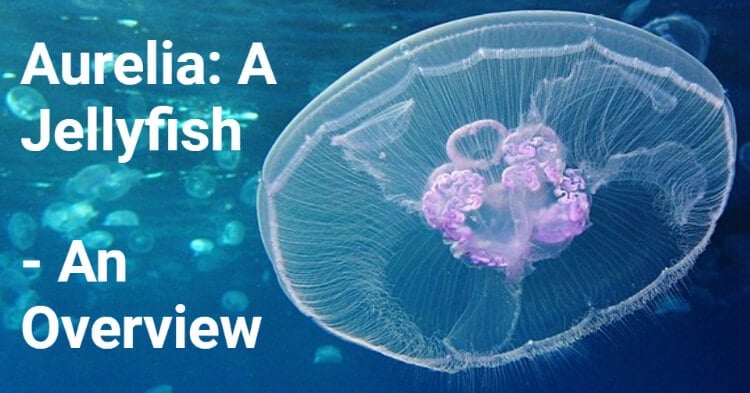
Image Source: Alexander Vasenin.
Habit and Habitat of Aurelia (Jellyfish)
- It is cosmopolitan jellyfish occurring in warm and temperate seas ranging in temperature from 6-19ºc all over the world.
- It lives in coastal waters singly or in large shoals.
- It can live in waters with a salt content as low as 0.6%. Salt content and water temperature affect the shape, size, and reproductive strategies.
- They thrive best in waters with temperatures are above 17.5°C and salinity greater than 38.0%, largest.
- It ranges in depth between the epipelagic zone and the mesopelagic zone (200 to 1,000 m).
- It is found either floating with water currents or waves or swimming feebly by the contraction movements of its bell.
- It is carnivorous, feeds on small organisms with the help of its long oral arms.
- It responds to various stimuli and is most active in diffuse light.
External morphology of Aurelia (Jellyfish)
1. Shape and size
- It is a large version of the medusa of Obelia.
- It looks like a soft bell or umbrella-shaped body with 4 red or purple horseshoe-shaped gonads on its upper surface and 4 long and narrow oral lobes hanging downwards from the lower surface.
- Its circular body measures about 90mm in diameter and presents a convex aboral or exumbrellar surface and a concave oral or subumbrellar surface.
2. Colouration
- Its body is perfectly transparent and bluish-white in color.
- The reddish or pinkish horseshoe-shaped gonads are clearly visible from the body surface.
3. Manubrium, mouth, and oral arms
- A very short and inconspicuous manubrium is present in the center of the subumbrellar surface.
- At its free distal end is a square mouth, from each corner of which hangs down a long, tapering much-frilled and delicate process, the oral arm.
- Each of the four oral arms has a ventral ciliated groove leading into the mouth and its edges are armed with nematocysts.
- The radii, along which angles of the mouth and oral arms lie, are referred to as perradii.
- Midway between two adjacent perradii is an interradius, and between each perradius and its adjacent interradius, on either side, is adradius.
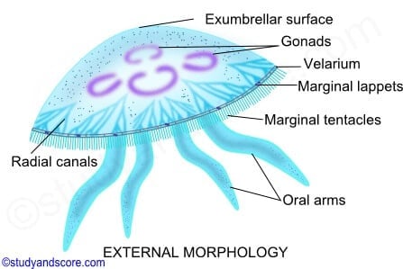
Image Source: Study and Score.
4. Subgenital pits
- On each interradius, a little distance from the mouth, the sub-umbrellar surface bears a circular aperture. It leads into a small shallow cavity, the subgenital pit, lying immediately between a gonad and of uncertain function.
5. Gonads
- A horseshoe-shaped and frilled gonad are present just above each subgenital pit within the umbrella.
- It is red or purple in color.
- Free arms of all the four gonads are directed towards the center of the umbrella. There is no connection between gonads and subgenital pits.
6. Lappets and tentaculocysts
- The circular margin of the umbrella or bell is broken into 8 lobes by 8 indentations or notches, 4 of which are perradial and the other 4 interradial.
- In each notch, there are two delicate leaf-like processes, called the marginal lappets.
- Between lappets lies small sensory organs, the tentaculocyst or rhopalium.
7. Marginal tentacles
- Between notches or rhopalia, the free edge of the umbrella is beset closely with a row of numerous small, delicate and hollow threads or marginal tentacles.
- The tentacles bear a number of stinging cells or nematocysts.
8. Velarium
- The margin of the subumbrellar surface, bearing lappets and tentacles, forms a thin and flexible flap, called velarium or pseudovelum.
- It differs from the true velum of Obelia in having gastrodermal canals running into it. Such medusa with a pseudovelum(e.g., Aurelia) is called acraspedote medusa, while a medusa with atrue velum (e.g.,Obelia) is called craspedote medusa.
Histology of Aurelia (Jellyfish)
The basic histological plan of Aurelia medusa is more or less the same as that of the Obelia medusa. It is diploblastic and derived from two embryonic layers, ectoderm and endoderm.
a. Epidermis
- All the exposed parts of an umbrella, that is, exumbrellar surface, velarium, tentacles, subumbrellar surface including subgenital pits, oral arms, and manubrium, are covered by epidermis.
- The gullet is formed by the evagination of the epidermis.
- It consists of epithelial cells (on the exumbrellar surface), epithelio-muscle cells (confined to subumbrellar surface) beside sensory cells, nerve cells, gland cells, and cnidoblasts. Sensory cells form a sensory epithelium between epithelial cells.
b. Gastrodermis
- Gastrodermis has columnar ciliated epithelial cells, they have no muscle processes; all the parts of the gastrovascular canal system, except the gullet, are lined by the gastrodermis.
- The gastric filaments are formed by the thin core of the mesogloea and a double layer of gastrodermis.
- Gonads are also gastrodermal structures.
- The cavities of tentaculocysts and marginal tentacles, being an extension of the gastrovascular canal system, are also lined by the gastrodermis.
- Gastrodermis mainly consists of flagellated columnar endothelial cells.
- The gland cells are present but nerve cells and muscle processes are wanting. Cindoblasts are confined to gastric filaments.
- In the interspace between gastrovascular canals, the gastrodermis of ex-and sub-umbrellar surface fuse to form a thin sheet, of the gastrovascular lamella.
c. Mesogloea
- It constitutes the main bulk of the body, forming a thick gelatinous layer between the epidermis and gastrodermis.
- The mesoglea of Aurelia is not structureless, contains numerous branching elastic fibers and wandering amoeboid cells derived from the epidermis.
- This type of mesogloea is more or less like connective tissue snd is known as collenchyma.
- It is ectodermal in an origin and endo-mesodermal as in higher metazoans.
- The mesogloeal fluid resembles seawater except that it has more potassium and less sulfate.
Nematocysts
They occur on oral arms, ex- and subumbrellar surface, marginal tentacles, as well as gastric filaments. They are of 3 types:
1. Atrichous isorhizas: Capsule is elongated. The thread tube is open at the tip and is without a butt and spines.
2. Holotrichous isorhizas: Capsule is oval and butt is narrow. The thread tube is long, open at the tip, and armed with a spiral row of small spines.
3. Heterotrichous microbasic euryteles: Capsule is small. The thread tube is open at the tip and covered by minute spines. The butt is short nad its distal dilated portion bears unequal spines.
Musculature and locomotion of Aurelia (Jellyfish)
a. Musculature
- Aurelia has a well-developed musculature mainly confined to the subumbrellar surface.
- The musculature is formed by muscle processes of epithelio-muscle cells of the epidermis.
- A broad, circular, and peripheral muscle band, known as coronal muscle, extends along the periphery of the sub-umbrella.
- The conspicuous longitudinal muscle are present in the tentacles, manubrium, and oral arms.
- The radial muscle extends from manubrium to coronal muscle along the main radii of the umbrella.
- Coronal and longitudinal muscles of tentacles are striated, while others are unstriated
b. Locomotion
- The highly developed musculature brings about swimming movements of Aurelia, during which the ex-umbrellar surface is kept upwards.
- Rhythmic contractions of circular muscles force water out from the sub-umbrellar cavity, like a jet. As a result, the body is propelled forward or upward. This type of jet-propulsion is known as hydropropulsion.
- When the contractions stop, the body gradually sinks to the bottom.
- Horizontal movements depend on wave action and help in buoyance because of their low density.
- If the body is tilted, equilibrium is maintained with the help of 8 tentaculocysts.
Digestive system of Aurelia (Jellyfish)
1.Gastrovascular canal system
- The rectangular mouth leads into a short gullet, within manubrium, which opens into a spacious four-lobed stomach occupying the center of the umbrella.
- Extending laterally from the stomach are four wide interradial gastric pouches. Within each gastric pouches is a C-shaped gonad and arrow of small gastric filaments or phacellae, bearing nematocysts.
- The gullet communicates with each gastric pouch through a groove-like gastro-genital canal that runs between the two free ends of the gonad.
- Around each gonad runs an exhalent channel that also communicates with the gullet through the gastro-oral canal.
- Radial canals branch out from pouches to join the circular or ring canal in the bell margin.
- From the periphery of the stomach as well as from the gastric pouches, sixteen radial canals originate, four are per-radials, four inter-radials, and eight adradials.
- Both per radials and inter-radials are branched, while the adradials are un-branched.
- All the radial canals are lined by ciliated gastrodermis, which ultimately open into a ring or circular canal situated at the margin of the body.
2. Nutrition
a. Food
- Aurelia is exclusively carnivorous. It is mainly a suspension or ciliary feeder. It feeds upon the planktonic organisms and small marine invertebrates, such as crustaceans, worms, their eggs, and larvae.
b. Ingestion
- The small planktonic organisms are carried directly into the stomach with the entering water current.
- Some small organisms that may get entangled in the mucus of the sub-umbrellar surface are collected by oral arms.
- Sometimes, as medusa sinks slowly or swims gently downwards, the prey is captured in contact with tentacles and oral arms.
- Nematocysts of oral arms paralyze and entangle these organisms, which are then swept up along the lateral tracts of oral arms and passed into the mouth.
- Undesirable particles are rejected and dropped on the way.
- If the prey, still in living condition, reaches the gastric pouches, it is paralyzed and killed by the nematocysts of gastric filaments.
c. Digestion
- The digestion takes place in the stomach and gastric pouches, where the gland cells of these regions secrete enzymes for the extracellular digestion of protein, fats, carbohydrates, and even chitin.
- Partly digested food particles, circulate through the canal system and are ingested by gastrodermal cells for intracellular digestion in food vacuoles.
- The undigested material passes out of the body with the outgoing current of water.
d. Distribution
- The water circulating through the canal system transports digested food to the gastrodermis of all parts. Further, wandering amoeboid cells in mesogloea transports food from the gastrodermis to the epidermis.
e. Reserve food
- The food is reserved in the gastrodermal cells of gastric filament in the form of glycogen and fat droplets.
Circulation of water in Aurelia (Jellyfish)
- The circulation of water is due to the beating of cilia of the gastrodermal cell lining of the gastrovascular canal. It circulates through the gastrovascular canal system along a fixed route.
- The inhaled water current enters the mouth and passes through the narrow gastrogenital canal into gastric pouches, and finally through the unbranched adraial canal enters the circular canal.
- From the circular canal, exhalent water current returns through perradail and interradial canals.
- The Preradial canal conveys it directly into the stomach from where it passes into the gullet and finally exists along the basal grooves of oral arms.
- Interradial canals convey it to the basal grooves of the oral arms via exhalent and gastro-oral canals. This arrangement greatly prevents the mixing of water currents entering and leaving the gastrovascular canal system.
- The contraction of the bell during locomotion and movements of oral arms helps in circulation.
- One complete circulation takes about 20 minutes.
- The circulation of water helps in nutrition, respiration, excretion, and reproduction.
Respiration and excretion of Aurelia (Jellyfish)
- Aurelia has no special organs for respiration and excretion. Oxygen dissolved into water diffuses directly into the epidermis as well s the gastrodermis, both of which are constantly bathed by water.
- Carbondioxide and nitrogenous wastes diffuse out into surrounding water.
- Some workers are of the opinion that the subgenital pits facilitate gaseous exchange. This is based on the observation that, at the time of swimming, foul water constantly leaves the pits and freshwater with dissolved oxygen) enters into them.
- 99% weight of jellyfish is formed by water.
- Nothing is known about the nature of excretory products.
Nervous system of Aurelia (Jellyfish)
It consists of the a. main nerve net, b. a diffuse nerve net, and c. 8 rhopalial ganglia
a. Main nerve net
- Each nerve net or plexus consists of nerve cells and fibers.
- The main nerve net is more developed and lies in the subumbrellar surface and extends into tentacles, rhopalia, manubrium, and oral arms.
- The presence of the main nerve net on the subumbrellar side is correlated with the presence of well-developed musculature on that side; the former co-ordinates muscular movements during locomotion.
- Its nerve elements form a sort of nerve ring along the margin of the umbrella near-circular canal.
- The main nerve net is thickened along per- and interradii due to the concentration of its nerve element along these radii.
- Each radial thickening, near the margin of the umbrella, is connected with the rhopalial ganglion, situated near the rhopalium on that radius.
b. Diffuse nerve net
- It lies in the epidermis of the subumbrella as well as the exumbrella.
- Its nerve elements consist of smaller cell bodies.
- It is also connected with rhopalial ganglia.’It controls the local responses, like feeding, and can inhibit contractions of the umbrella because the 2 nerve nets are joined through tentaculocysts.
c. Rhopalial ganglia
- These are formed by the aggregation of nerve cells.
- There are 8 such rhopalial ganglia, one near each sense organ or rhopalium.
- Nerve impulses received by the sense organs are conducted through nerve nets to the muscle fibers which react accordingly.
Sense organs of Aurelia (Jellyfish)
The sense organs of Aurelia are 8 rhopalia which are situated one in each of the per-and interradial marginal notches. each rhopalium comprises tentaculocyst or statocyst, 2 ocelli, and 2 olfactory pits.
1. Tentaculocyst or statocyst
- It is a hollow club-shaped structure, situated in the marginal notch between two marginal lappets.
- It is covered on the outer side by the process of bell margin, termed hood, which also connects the bases of 2 marginal lappets.
- Just below the club is a pad of tall ciliated sensory epithelial cells which are connected with the subumbrellar nerve net lying below the epidermis.
- Tentaculocyst is hollow tentacles. Projecting into tentaculocyst is an extension of a circular canal lined by gastrodermis. Lying in the distal part of the tentaculocyst is a mass of polygonal statolith cells of gastrodermal origin.
- Each statolith cell contains a self-secreted particle, the statolith (Gr., statos, standing + lithos, stone), composed of calcium sulfate and calcium phosphate.
- Statoliths act as a weight, causing the club of tentaculocyst to bend up and down at its base, whenever the animal tilts to one side or other, during swimming.
- Tentaculocysts control the equilibrium of the umbrella during swimming. If the umbrella is tilted, the clubs of tentaculocysts press against their sensory pad beneath, the sensory cells which become stimulated. Higher the tilt greater the stimulation. The impulse is conducted through the subumbrellar nerve net to muscle fibers which react accordingly.
- In response, the upper half of the umbrella drives less water than the lower half at each beat, so that the umbrella automatically rights itself.
2. Ocelli
- There are two ocelli, one of ectodermal and the other of endodermal origin.
- The former, known as pigment spot ocellus, consists of a patch of pigmented and sensory epidermal cells on the outer side of the club of tentaculocyst.
- The latter, known as pigment cup ocellus, consists of a cup-shaped cavity lined by pigmented and sensory gastrodermal cells and is situated on the inner side of tentaculocyst, in association with statoliths.
- The sensory cells of both the ocelli are connected with their respective underlying nerve nets.
- Ocelli are photoreceptors.
3. Olfactory pits
- These are in the form of depression of thickened epidermis containing sensory cells.
- One such depression lies at the base of the hood. It is termed the outer or aboral olfactory pit.
- The other, known as the inner or adoral olfactory pit, is situated on the inner side of the tentaculocyst at the base of the pad of ciliated sensory epithelial cells
- These olfactory pits are probably chemoreceptors.
Reproduction and life history of Aurelia (Jellyfish)
Aurelia is dioecious i.e., the male and female sexes are separate but there is no sexual dimorphism.
1. Sex organs
- Testes and ovaries are similar in appearance. A medusa has 4 horseshoe-shaped gonads radially lying on the floor of the stomach periphery, that is, one in each gastric pouch.
- The gonads are endodermal in origin.
- They are reddish violet in color.
- They are visible through the semitransparent jelly of the umbrella as frilled organs with their concavities facing inwards.
- On maturity, ova nad sperms break into the gastrovascular cavity and pass out of the mouth with the outgoing water current. The ova and eggs are lodged in the frill of oral arms.
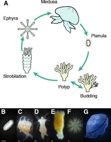
Figure: Life-cycle stages of Aurelia. (A) Life-cycle scheme depicting sexual reproduction of mature medusa and asexual proliferation of the polyp. (B − G) Photographs of the six analyzed stages: planula (B), polyp (C), early strobila (D), advanced strobila (E), ephyra (F), and mature medusa (G). The dashed line in G represents the excised part of the medusa that was used for RNA-seq. Bar in B, 50 μm, and in C − F, 500 μm. Image Source: Vera Brekhman et al. 2015.
2. Fertilization
- Spermatozoa, swimming about in the water, reach the ova and fertilize them either in the stomach of females or in the frills of oral arms. Thus, fertilization is either internal or external.
3. Formation of planula larva
- The frills of oral arms serve as temporary brooding members where fertilized egg or zygote undergoes early development into a ciliated larval stage called planula.
- The zygote undergoes holoblastic and equal segmentation to produce a solid ball-like morula.
- Morula becomes a single-layered blastula soon by the accumulation of fluid in its interior.
- The two-layered gastrula develops by invagination, having outer ectoderm and inner endoderm lining an enteron cavity, with its blastopore or gastral mouth not completely closed. Thus, it differs from the gastrula of Hydrozoa (e.g., Obelia) which develops by the process of delimitation and multipolar regression of cells into blastocoel having no blastopore.
- The embryo, now elongates, its outer cell becomes ciliated, blastopore closes and the typical planula larva is formed. At this stage, masses of planulae are visible as minute opaque patches on the oral arms of female individuals.
4. Formation of scyphistoma
- The ciliated planula eventually escapes and after a short free-swimming existence attaches itself to a stone or seaweed by its aboral end.
- The cilia are lost and a mouth opens at its free distal end where the blastopore had closed.
- The larva now becomes elongated and metamorphosed into a small trumpet-shaped or Hydra-like polyp or hydratuba, about 5mm high.
- The larva proximal part is narrowed into a stalk-like organ, attached to the substratum by an adhesive basal disc.
- Its tentacles bud out around the mouth where 4 tentacles are perradial, subsequent 4 interradial, and then 8 adradial in position. Thus, 16 long and slender tentacles are formed.
- Its mouth becomes square in outline and its edges become elongated to form a short manubrium.
- The larva now looks like a trumpet-shaped polyp or Hydra and is called hydratuba or young scyphistoma (Gr., skyphos, cup+ stoma, mouth).
- The endoderm of its cavity is raised into 4 interradial longitudinal gastric ridges or taenioles, characteristic of Scyphozoa, which divides the enteric cavity into four perradial diverticula or pouches.
- Simultaneously, the ectoderm between mouth and tentacles also becomes invaginated as four interradial funnel-like depressions, known as septal funnels or infundibula, which sink into four gastric ridges.
- Scyphistoma feed and grows up to 12mm in height and may survive in this stage for several months. Sometimes, it multiplies either by lateral budding or by growing horizontal creeping stolons, which bud off fresh hydratubae. These buds eventually separate from the parent, as in Hydra.
5. Formation of ephyrae( strobilation)
- Scyphistoma undergoes a remarkable process of budding or transverse fission of oral end in autumn and winter, called strobilation.
- Distally, the body develops a series of ring-like transverse constrictions or furrows which gradually deepens so that the organism resembles a pile of minute saucers or disc, placed one above the other. At this stage, scyphistoma with a segmented body is called strobila and each of the segments is called ephyra larva.
- The ephyrae or ephyrulae are connected together by muscular strands. As they grow older, they constricted off one by one and swim away as little medusae or jellyfish.
- About a dozen ephyrae are formed in single strobilation.
- When the food is plentiful and the temperature is low, several ephyrae are produced at one time (polydisc strobilation). When food is scarce and the temperature is high, a single ephyra is produced at a time (monodisc strobilation).
- When all the ephyrae get detached, the basal unsegmented part of the scyphistoma grows new tentacles and continues to live as a polyp or hydratuba. It may live for several years, feeding, growing, and multiplying by budding in summers, but producing ephyrae by strobilations in winter.
- The scyphistoma may be metamorphosed into a single adult Aurelia, without forming ephyrae under exceptional circumstances.
6. Ephyra larva
- A newly released ephyra is a microscopic gelatinous creature, about 1mm in diameter and with a well-developed tetramerous symmetry.
- The edge of its umbrella is greatly fluted, being produced into 8 bifid lobes or arms( 4 per and 4 interradial), separated by 8 deep adradial indentations or clefts.
- Each lobe has 2 exaggerated marginal lappets with a groove or notch between them is a sensory recess or niche bearing short tentacles, which becomes tentaculocyst or rhopalium.
- Ephyra contains a small segment of the stomach of scyphistoma with gastric ridges.
- It remains opens on the subumbrellar side and grows out to form a short manubrium baring a four-sided mouth at its apex.
- Spacious enteric cavity projects into 8 arms as branched perradial and interradial canals. Adradial canals appear later on.
- The gastric ridges or mesenteries are replaced by gastric filaments. 4 interradial septal invaginations probably persist as 4 subgenital pits.
- The first ephyra differs from the rest in bearing the original tentacles of scyphistoma.
- Ephyrae swim actively in sea-water feeding on minute organisms, such as protozoans, which are caught by lappets and transferred to the mouth.
7. Metamorphosis
- As the ephyra grows in size, the space between the notched lobe fills up, mesogloea increases enormously so that two layers of endoderm(gastrodermis)f fuse to form a solid gastrodermal lamella, except in regions of gastrovascular canals.
- The adradial region grows more rapidly, gradually filling up their wide clefts, so that the umbrella of 8 -8-rayed ephyra becomes circular and saucer-shaped ], as in an adult medusa.
- With the appearance of 4 oral arms and numerous tentacles, ephyra finally transferred into adult Aurelia.
- An ephyra formed in winter becomes a sexually reproducing adult medusa by spring or summer.
Alternation of generation (metagenesis) in Aurelia (Jellyfish)
- There are 2 contrasting views regarding the life history of Aurelia.
- According to the older view, the development of Aurelia illustrates in a sense, the phenomenon of alternation of generations.
- The free-swimming adult Aurelia represents the sexual generations, producing ova or spermatozoa, like the medusoid phase of Obelia. Here, the fertilized egg gives rise to a small fixed polyploid form, or scyphistoma, after passing through a free-swimming ciliated planula larval stage.
- Scyphistoma represents the asexual generation, equivalent to the polypoid Obelia colony. It reproduces asexually by strobilation, budding of ephyrae, each of which metamorphoses into an adult Aurelia.
- According to the modern view, the development of Aurelia does not show true alternation of generation or metagenesis.
- However, the alternation of generation shown by Aurelia does not truly correspond with that of Obelia because, the medusoid stage in Obelia develops as a bud on a branched polyploid colony, whereas the Aurelia medusa is formed by the metamorphosis of an ephyra larva, produced as one of several transverse segments of the polyploid scyphistoma.
- Thus, the life cycle of Aurelia may be considered as a simple continuous process involving a prolonged metamorphosis further complicated by the multiplication of scyphistoma larva into several ephyrae.
References and Sources
- Brekhman, V., Malik, A., Haas, B. et al. Transcriptome profiling of the dynamic life cycle of the scypohozoan jellyfish Aurelia aurita. BMC Genomics 16, 74 (2015). https://doi.org/10.1186/s12864-015-1320-z.
- https://sbancollege.org/study-material/99549259418.%20Zoology_Ravi%20Ranjan_28.04.2020.2020- 7%
- https://www.iaszoology.com/aurelia/- 6%
- https://www.studyandscore.com/studymaterial-detail/aurelia-general-characters-and-life-cycle- 6%
- https://gurujistudy.com/bsc-lower-non-chordates-aurelia-question-answers/- 5%
- https://www.biologydiscussion.com/invertebrate-zoology/phylum-coelenterata/aurelia-aurita-habitat-nutrition-and-life-history-with-diagram/28702- 4%
- https://www.notesonzoology.com/phylum-cnidaria/aurelia-jelly-fish-structure-histology-and-nutrition/1471- 3%
- https://sbancollege.org/study-material/90006989515.%20Zoology_Ravi%20Ranjan_29.04.2020.2020- 1%
- https://courses.lumenlearning.com/boundless-biology/chapter/pollination-and-fertilization/- <1%
- https://brainly.in/question/16242941- <1%