- The teleost fish Danio rerio is commonly known as the zebrafish.
- It is a common small‐bodied tropical, freshwater species originally from South Asia.
- The fish is a part of the Cyprinidae family, which includes carps, true minnows, barbs and related genera.
- The Danio genus (formerly Brachydanio) contains more than 40 related species along with the zebrafish (Danio rerio).
Interesting Science Videos
Zebrafish as a Vertebrate Development Model Organism
In recent years, zebrafish has become a favorite organism of those who wish to study vertebrate development for the following reasons:
- Zebrafish have large broods.
- They breed all year round.
- They can be easily maintained.
- Zebrafish have transparent embryos that develop outside the mother which is an important feature for microscopy.
- They can be raised so that mutants can be readily screened and propagated.
- In addition, they develop rapidly, so that at 24 hours after fertilization, the embryo has formed most of its tissues and organ primordia and displays the characteristic tadpole-like form.
- Zebrafish have a similar genetic structure to humans and share about 70% of the genes.
Zebrafish Fertilization and Development
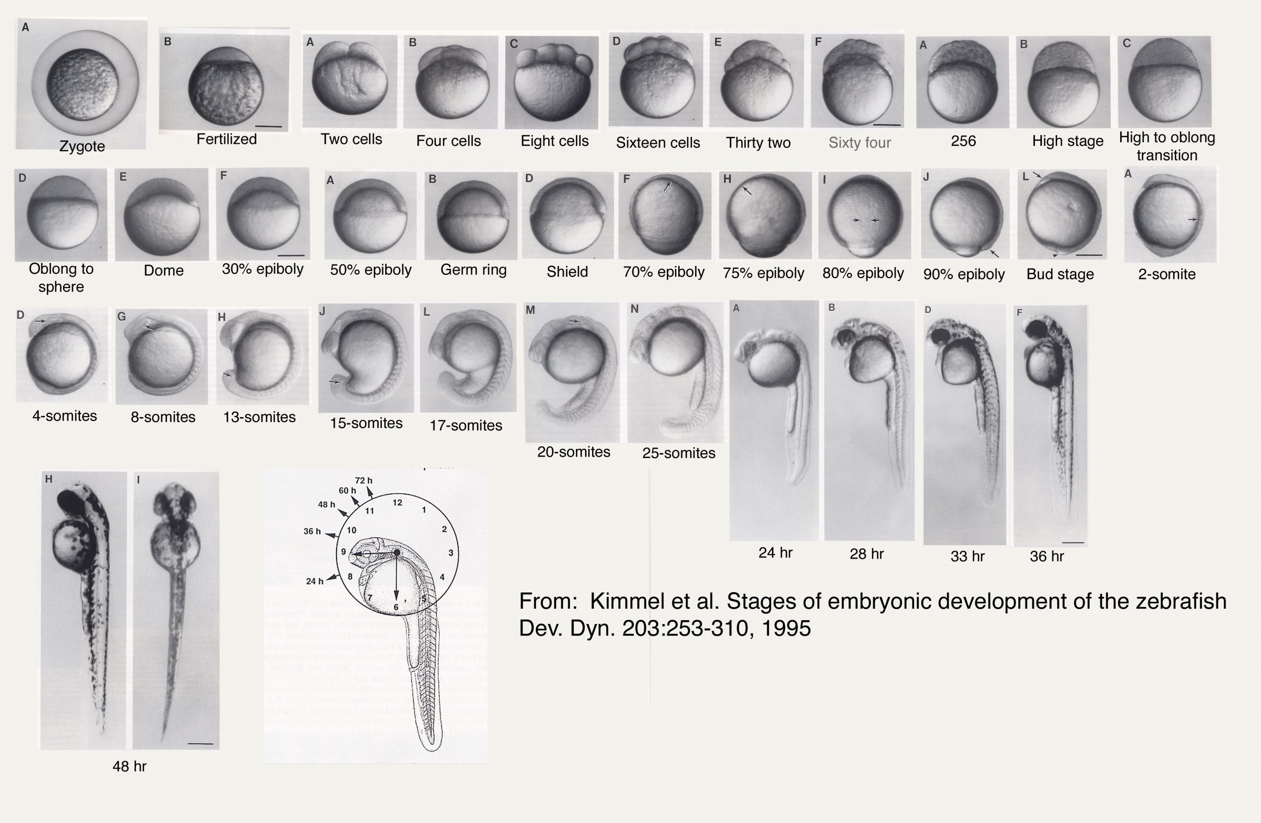
Image Source: Duke University
Fertilization
- Male and female zebrafish show distinct courtship displays that lead to coordinated egg‐laying and sperm release resulting in fertilization.
- A gravid female can release hundreds to over a thousand oocytes, making zebrafish prolific breeders.
- The calcium waves initiated at fertilization stimulate the contraction of the actin cytoskeleton to squeeze the non-yolky cytoplasm into the animal pole of the egg. This converts the spherical egg into a more pear-shaped structure, with an apical blastodisc.
- After fertilization, the chorion lifts away from the egg and clear cytoplasm moves to the animal pole of the embryo, separating the blastodisc from the larger underlying yolk mass.
- The zebrafish embryo develops rapidly, with precursors to all major organs appearing within 36 hours of fertilization.
Cleavage
- Beginning around 45 min after fertilization, the blastodisc undergoes cleavage.
- In fish eggs, cleavage occurs only in the blastodisc, a thin region of yolk-free cytoplasm at the animal cap of the egg. Most of the egg cell is full of yolk.
- The cell divisions do not completely divide the egg, so this type of cleavage is called meroblastic (Greek, meros, “part”). Since only the cytoplasm of the blastodisc becomes the embryo, this type of meroblastic cleavage is called discoidal.
- Each cell cycle takes around 15 min and the divisions occur synchronously.
- The first 12 divisions occur synchronously, forming a mound of cells that sits at the animal pole of a large yolk cell. These cells constitute the blastoderm.
- Initially, all the cells maintain some open connection with one another and with the underlying yolk cell so that moderately sized (17-kDa) molecule can pass freely from one blastomere to the next.
Formation of yolk syncytial layer and enveloping layer
- Also around this time, the marginal cells that retain connections with the yolk collapse, pushing their nuclei and cytoplasmic contents into the yolk forming a multinucleate yolk syncytial layer (YSL).
- The YSL is formed at the ninth or tenth cell cycle when the cells at the vegetal edge of the blastoderm fuse with the underlying yolk cell.
- This fusion produces a ring of nuclei within the part of the yolk cell cytoplasm that sits just beneath the blastoderm.
- Later, as the blastoderm expands vegetally to surround the yolk cell, some of the yolk syncytial nuclei will move under the blastoderm to form the internal YSL, and some of the nuclei will move vegetally, staying ahead of the blastoderm margin, to form external YSL.
- The YSL will be important for directing some of the cell movements of gastrulation.
- The second cell population distinguished at the midblastula transition is the enveloping layer.
- It is made up of the most superficial cells of the blastoderm, which form an epithelial sheet a single cell layer thick.
- The EVL eventually becomes the periderm, an extraembryonic protective covering that is sloughed off during later development.
- Between the EVL and the YSL are the deep cells. These are the cells that give rise to the embryo proper.
Epiboly and Gastrulation
- The fate of the blastoderm cells appears to be fixed shortly before gastrulation begins.
- At this time, cells in specific regions of the embryo give rise to certain tissues in a highly predictable manner, allowing a fate map to be made.
- The first cell movement of fish gastrulation is the epiboly of the blastoderm cells over the yolk.
- It is coordinated by myosin motors in the yolk syncytial layer, polarized cell divisions within the epithelial enveloping layer, and rearrangements among the deep cells of the blastoderm.
- In the initial phase, the deep blastoderm cells move outwardly to intercalate with the more superficial cells.
- Later, these cells move over the surface of the yolk to envelop it completely.
- During migration, one side of the blastoderm becomes noticeably thicker than the other. Cell-labeling experiments indicate that the thicker side marks the site of the future dorsal surface of the embryo.
- After the blastoderm cells have covered about half the zebrafish yolk cell, a thickening occurs throughout the margin of the epibolizing blastoderm.
- This thickening is called the germ ring, and it is composed of a superficial layer, the epiblast, and an inner layer, the hypoblast.
- Once formed, the cells of both the epiblast and hypoblast intercalate on the future dorsal side of the embryo to form a localized thickening, the embryonic shield.
- As the cells undergo epiboly around the yolk, they are also involuting at the margins and converging anteriorly and dorsally toward the embryonic shield.
- The hypoblast cells of the embryonic shield converge and extend anteriorly, eventually narrowing along the dorsal midline of the hypoblast. This movement forms the chordamesoderm, the precursor of the notochord.
- The cells adjacent to the chordamesoderm, the paraxial mesoderm cells, are the precursors of the mesodermal somites.
- The concomitant convergence and extension in the epiblast bring the presumptive neural cells from all over the epiblast into the dorsal midline, where they form the neural keel. The rest of the epiblast becomes the skin of the fish.
- The fish hatch from their chorion typically between 48 to 72 hpf and 72 hpf marks the conventional end of the embryonic period.
- The swim bladder inflates, allowing upright swimming throughout the water column between days 3 and 4 and the fish begin feeding behaviors shortly thereafter.
Post Embryonic Growth
- After the period of embryonic growth, the zebrafish spend the next ∼4 weeks in a larval state.
- Growth during the larval state varies widely based on temperature, density, and individual differences.
- Once the fish reach a size of ∼11 mm (typically ∼4 weeks), they are classified as juveniles.
- They typically become sexually mature 10 to 12 weeks post‐fertilization (usually >17 mm in size).
References
- Gilbert, S. F. (2000). Developmental biology. Sunderland, Mass: Sinauer Associates.
- https://www.yourgenome.org/facts/why-use-the-zebrafish-in-research
- https://currentprotocols.onlinelibrary.wiley.com/doi/full/10.1002/cpet.19
- https://www.ncbi.nlm.nih.gov/pubmed/8589427
- https://www.jove.com/science-education/5151/zebrafish-reproduction-and-development
- https://www.tankarium.com/zebra-danio/