Wuchereria bancrofti is a filarial nematode that causes Wuchereriasis or filariasis(commonly called elephantiasis) in human beings. The name of this worm is given Wuchereria bancrofti in honor of the two scientists Wucherer and Bancroft who made a considerable contribution in studying the disease caused by these worms.
Image Source: Study and Score and Wikipedia.
Interesting Science Videos
Systematic Position of Wuchereria bancrofti
Phylum: Nematoda
Class: Phasmidia
Order: Filaroidea
Family: Filariidae
Genus: Wuchereria
Species: bancrofti
Habits and Habitat of Wuchereria bancrofti
- Wuchereria bancrofti (Filarial worm) is a dreaded endoparasite of humans.
- It is a digenetic parasite completing its life cycle in 2 hosts. The final host is man harboring the adult worms, while the intermediate host is blood-sucking insects, the female mosquitoes of genus Culex, Aedes, or Anopheles.
- Adult worms live coiled up in the lymph glands and lymph passage of man, where they often obstruct the flow of lymph.
- The larvae i.e. microfilariae are found in the peripheral blood, occasionally they are also found in chylous urine or in hydrocele fluid.
Morphology of Wuchereria bancrofti
1. Adult worms
- These are long, hair-like, transparent, translucent, thread-like worms with smooth cuticle and tapering ends.
- These are filiform and cylindrical in shape with both ends tapering.
- Sexes are separate with distinct sexual dimorphism.
- The female is larger (70–100 × 0.25 mm) than the male (25–40 × 0.1 mm).
- The posterior end of the female worm is narrow and pointed that bears an anus, while that of the male is curved vertically and contains numbers of genital papillae two copulatory spicules of unequal length.
- Males and females remain coiled together usually in the abdominal and inguinal lymphatics and in the testicular tissues.
- The head end is slightly enlarged.
- Mouth aperture is simple, without lips.
- The pharynx is divisible into an anterior muscular portion and a posterior glandular portion. An oesophageal bulb is lacking.
- The intestine is simple.
- The vulva of the female is located ventrally in the pharyngeal region and provided with a pyriform ejector mechanism.
- The female worm is viviparous and directly liberates sheathed microfilariae into the lymph.
- The adult worms live for many years, probably 10–15 years or more.
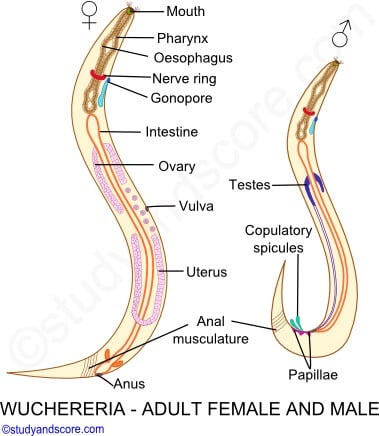
Image Source: Study and Score.
2. Microfilaria
- The first stage larva is called microfilaria.
- They are colorless, transparent bodies with blunt anterior ends and pointed tails.
- They are very active and can move forward and backward within the sheath which is much longer than the embryo.
- They are microscopic and measure about250–300 μm in length and 6–10 μm in thickness.
- Its body is covered with a hyaline sheath followed by a cuticula being lined by flattened subcuticular cells or epidermis and an inner column of cytoplasm containing nuclei. Its cuticle has well-marked striations.
- Somatic cella or Nuclei appear as granules in the central axis of the body extended from head to tail except for the 5% terminal end of the tip.
- Their head-end has clear space devoid of granules known as cephalic space.
- Important structures from the anterior end downwards are future mouth or oral stylet, nerve ring: oblique area devoid of granules, nephridiopore, renette cell, dark-colored inner mass, and 4 cells of the future anus.
- They do not undergo further development in the human body unless they are taken up by their suitable host (mosquitoes).
- Their life span in the human body is probably 70 days.
Image Source: Study and Score.
Periodicity of Microfilariae
- The microfilariae circulate in the bloodstream.
- In India, China, and many other Asian countries, they show a nocturnal periodicity in peripheral circulation; being seen in large numbers in peripheral blood only at night (between 10 pm and 4 am), but they disappear inside during the rest of the day. It is believed that during the daytime they retire inside the deeper blood vessel. This correlates with the night biting habit of the vector
Culex mosquito. - In the Pacific islands and some parts of the Malaysian archipelago, the microfilariae are nonperiodic or diurnal subperiodic, such that they occur in peripheral circulation at all times, with a slight peak during the late afternoon or evening. This is related to the day-biting habits of the local vector mosquitoes.
3. Third stage of the larva (infective form)
- The third stage larva is the infective form of the parasite is found only in mosquitoes.
- They are elongated, filariform, measures1.5mm in length, and 18-23 µm in diameter.
Life cycle of Wuchereria bancrofti
- Wuchereria bancrofti is digenetic i.e. its life history is completed in two hosts
- Definitive host: Man. No animal host or reservoir is known for W. bancrofti
- Intermediate host: Female mosquitoes, belonging to genus Culex, Aedes, and Anopheles.
- Infective form: Actively motile third-stage filariform larva is infective to man.
- Mode of transmission: Humans get the infection by the bite of a mosquito carrying a filariform larva.
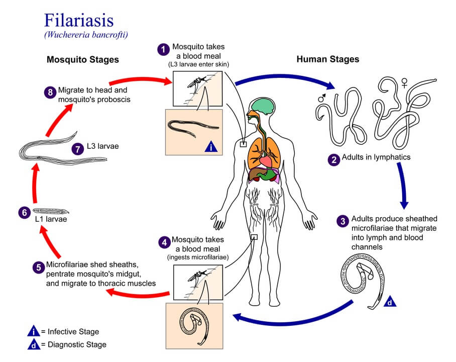
Figure: The life cycle of Wuchereria bancrofti. Image Source: Wikipedia.
a. Copulation
- Copulation takes place when individuals of both sexes are present in the same lymph gland.
b. Larval development in man
- The female worm is viviparous or ovoviviparous, it releases numerous larvae called microfilariae into the bloodstream.
- The microfilariae are born in a very immature stage. However, they find their way into the lymph vessels, soon enter into blood vessels and circulate with blood showing active movements.
- They do not undergo further development until sucked by the intermediate host. i.e. female mosquitoes.
- Microfilariae show nocturnal and diurnal periodicity in the blood of man.
- During the daytime, the microfilariae tend to stay in the deep blood vessels of man. But, at night time they travel near the surface in peripheral or superficial blood vessels. This behavior enables them to get ingested by the night biting mosquito(Culex and Aedes) which serves as an intermediate host.
- These microfilariae circulate in the blood for 6 months to 2 years and then die if not taken by a mosquito.
c. Development in mosquito
- Microfilariae are sucked from the peripheral blood of man.
- Microfilariae lose their sheath within 2 to 6 hours in the stomach of the mosquito and then they penetrate the stomach wall and within 4 to 17 hours migrates to thoracic muscles or wing musculature where they undergo metamorphosis and grow.
- In the next 2 days, they become short and thick like sausages having short spiky tails and measures 124 to 250µm in length and 10 to 17µm in diameter, also possess rudimentary digestive tract. These are first-stage larvae (L1).
- within the next 3 to 7 days they grow rapidly and moult once or twice to become the second stage larvae (L2) which measure 225 to 33oµm in length and 15 to 3o µm in diameter.
- Metamorphosis finally completes by 10-11 days into third-stage filariform larvae (L3) which measure about 1500 to 2000 µm in length and 18-23 µm in diameter.
- The third stage larvae are actively motile and infective.
- These larvae migrate through the hemocoel to the mosquito’s labium(proboscis).
d. Infection of the new human host
- When this infected mosquito pierces its proboscis in the warm and moist skin of man, the larvae creep out of labium to human skin,
- Then, it penetrates the skin and finally come to settle down into lymphatics.
- In the lymph vessels, they grow and become fully adult and sexually mature within a period of 5 to 18 months.
- The sexually mature worms start reproduction to repeat life history.
References and Sources
- Kotpal RL. 2017. Modern Text Book of Zoology- Invertebrates. 11th Edition. Rastogi Publications.
- Jordan EL and Verma PS. 2018. Invertebrate Zoology. 14th Edition. S Chand Publishing.
- 2% – https://www.cdc.gov/parasites/lymphaticfilariasis/biology_b_malayi.html
- 2% – https://www.biologydiscussion.com/parasitology/parasitic-worm/filarial-worm-histology-habitat-and-structure-with-diagram/62317
- 2% – https://www.biologydiscussion.com/invertebrate-zoology/phylum-aschelminthes/wuchereria-bancrofti-habitat-structure-and-life-history/29013
- 1% – https://www.slideshare.net/ssuser4037c1/filarial-tissuenematodes
- 1% – https://www.msdsonline.com/resources/sds-resources/free-safety-data-sheet-index/wuchereria-bancrofti/
- 1% – https://www.mcdinternational.org/trainings/malaria/english/DPDx5/HTML/Frames/A-F/Filariasis/body_Filariasis_mic2
- 1% – https://www.biology-today.com/general-zoology/invertebrate-zoology/life-cycle-of-wuchereria-bancrofti/
- 1% – https://www.biologydiscussion.com/parasitology/parasitic-worm/life-history-of-filarial-worm-with-diagram/62303
- <1% – https://www.cdc.gov/westnile/transmission/index.html
- <1% – https://web.stanford.edu/group/parasites/ParaSites2004/Filariasis/transmission.htm
- <1% – http://lanwebs.lander.edu/faculty/rsfox/invertebrates/ascaris.html
Nice 👍🏻😊