Vorticella (L.,vortex=whirl pool) is a microscopic, unicellular eukaryotic ciliate. It occurs as a sessile attached by a stalk to the substratum. Its body is almost bell-shaped or campanulate with a free anterior end surrounded by cilia. The commonest species is Vorticella campanula. Some of the species included in this genus are V. microstoma, V. picta, V. nebulifera, V. monliata.
Interesting Science Videos
Systematic position of Vorticella (Bell Animalcule)
Phylum: Protozoa
Subphylum: Ciliophora
Subclass: Peritricha
Order: Peritrichida
Suborder: Sessilina
Family: Vorticellidae
Genus: Vorticella
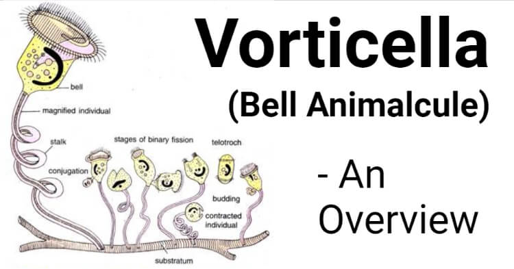
Image Source: Biology Discussion.
Habit and Habitat of Vorticella (Bell Animalcule)
- Vorticella is an extremely common, solitary, and stalked ciliate found in freshwater ponds, lakes, rivers, streams, etc.,
- They often occur socially in large groups anchored by their large contractile stalks to aquatic plants, animals, stones, twigs, etc. The contractile stalk permits the body to expand and contract frequently.
- Sometimes their body may be detached from the stalk and swims freely.
- They can also be found in saline environments (salty waters) as well as aquatic vegetation.
- They are solitary, occurs in large groups but never colonial.
- They are found abundantly in stagnant water containing organic matter, feeding largely on bacteria. But some species, such as V. campanula and V. nebulifera, can live in uncontaminated waters where the bacterial growth does not become too great.
- Some species are epizoic and few are parasitic.
Structure of Vorticella (Bell Animalcule)
1. Shape, size, and coloration
- It has a campanulate or bell-shaped symmetrical body. To its base is attached a long contractile stalk. Both the body and stalk are capable of great individual variation in size and form.
- The bell of the largest species ( V. campanula) varies in size up to 160µ long and 100µ broad, while the length of stalk varies from 55 to 4150µ.
- The body of smaller species, V. microstoma, measures about 35µ by 55µ.
- V. nebulifera is greenish and V. campanula is blue in color.
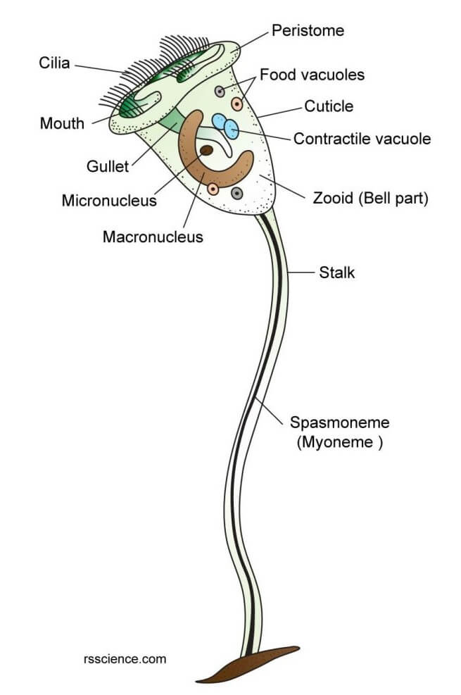
Figure: Structure of Vorticella (Bell Animalcule). Image Source: Rs’ Science.
2. Body or bell
The body of each individual is shaped like a solid inverted bell. The detailed structure of the bell is as follows
a. Peristome
- The broader free end of the bell, with a slightly convex area, known as a peristomal disc.
- The outer margin of the broader free end of the bell is thickened to form a prominent rim called a peristomial collar, border, or lip.
- It is separated from the peristomial disc by a shallow, circular, and marginal depression known as oral groove or peristome, where cilia are inserted.
- The collar can close over the disc when the animal is retracted.
b. Food passage
- On one side of the disc, the peristomial groove is deeply invaginated forming a funnel-like vestibule or buccal cavity or infundibulum.
- At, its inner end, the buccal cavity opens through a cell mouth or cytostome into the cell gullet or cytopharynx, which ends in a food vacuole in the endoplasm.
c. Oral ciliature
- There are no somatic or body cilia in Vorticella. Oral or aboral cilia is only found in the peristomial grooves which are arranged in a whirlpool manner.
- The peristomial groove form three concentric rows of ribbon-like membranes (aboral cilia), which are arranged in a circlet.
- The inner circlet includes two rows of cilia. which remain straight and always moving.
- The outer circlet is single and has short cilia. The cilia of this row hang horizontally towards the outer side of the lip. They direct the food particles towards the vestibule.
- The bases of all the 3 cilia are fused together but their distal ends are free.
- The three cilia lie in a counter-clockwise direction around the margin of the disc and then continue down into the buccal cavity.
- Inside the buccal cavity, the ciliary membrane separates. Two inner membranes run along the inner wall of the buccal cavity, the outer membrane follows its outer wall.
- The cilia of the outer membrane become long and fused together forming an undulating membrane.
- Cytopharynx has no cilia. Due to the movement of cilia, the food particles are sent to the cytopharynx by the vestibule.
- In the cytopharynx, a small food vacuole is formed around the food particles.
d. Cytoplasm
- The body of cytoplasm is differentiated into an outer layer of clear ectoplasm an inner granular fluid of endoplasm.
Ectoplasm
It has no trichocysts. It consists of an elastic outer pellicle and a thicker inner layer of contractile fibers or myonemes.
a. Pellicle
- It forms an outer envelope of the bell and also lines the buccal cavity.
- It is especially thick at the base of the bell.
- It is devoid of cilia but its basal granules are present in circles showing that its cilia are lost. Basal granules can be demonstrated by the Klein silver-line method.
- In the stalk, the pellicle is covered by an external cuticle.
b. Myonemes
- Myonemes form a system of variously running fibers that function differently on contraction.
- The longitudinal myonemes shorten the body, oblique myonemes pull the disc inwards, and circular myonemes contract and close the peristomial border over the cilia and disc.
- Myonemes running parallel down the sides of the bell are more visible at the base where they converge before entering the stalk.
Endoplasm
The granular endoplasm contains nuclei, contractile and food vacuoles, etc.
a. Nuclei
- Endoplasm contains a large, elongated,rod-like, and horseshoe-shaped macro-nucleus.
- The macronucleus is highly polyploid with a large amount of chromatin material scattered in the nucleoplasm.
- A tiny rounded micronucleus occurs in close association with the macronucleus. It is rarely seen in living animals. It can be seen only stain with acetocarmine or any other permanent stains.
b. Contractile vacuoles
- It is located near the buccal cavity, a clear and permanent pulsating space.
- It is about 7µ in diameter when fully distended.
- It shows diastole 9expansion) and systole(contraction) in a rhythmic manner and pours its contents into an efferent canal, which leads into the buccal cavity.
- In V. picta and V. monilata there are 2 contractile vacuoles.
c. Food vacuoles
- Many large and small food vacuoles are present in the endoplasm containing food particles.
- They are formed at the inner end of the cytopharynx and constrict off as rounded spheres into the endoplasm.
d. Cytopyge or cytoproct
- It is a temporary or permanent opening. The undigested food particles are passed into the buccal cavity through it.
Stalk
- The bell-shaped body of Vorticella remains attached to the substratum by a long, uniformly, thin, unbranched, and highly contractile stalk.
- It has no cilia.
- The unbranched stalk distinguishes Vorticella from the other ciliates.
- The older view is that the stalk is formed only by ectoplasm. But, electron microscopy has revealed that the stalk consists of 2 parts, the central canal, and outer sheath.
- The central canal consists of endoplasm, a contractile cord which is a specialized myoneme, called spasmoneme, and some granules, possibly mitochondria.
- Spasmoneme is a bundle of spiral fibrils continuous with the myonemes of the bell. It is believed that the contractility of the stalk is due to the contractile ability of spasmoneme.
- When the stalk is contracted the spiral spasmoneme coils up tightly and looks like spring when viewed under the microscope.
Locomotion of Vorticella (Bell Animalcule)
- Normally, the cilia of Vorticella remain very active and the movement of the stalk are frequent and rapid.
- When the animal is feeding, its stalk remains fully extended and the bell sways to and fro like a flower in the breeze.
- Individuals do not move in unison; each individual sways to its own rhythm.
- But, Vorticella is extremely sensitive to any mechanical stimulus. When irritated, all activities cease instantly. The stalk is retracted into a close and delicate spiral, cilia are stilled, the disc is pulled in and closed over by peristomial lip and the rounded body is brought close to the bottom, where it rests motionless until the danger has passed.
Nutrition in Vorticella (Bell Animalcule)
- The mode of nutrition is holozoic as in Paramecium.
- A water current produced by the adoral cilia of the peristome brings bacteria and other small organic particles into the peristomial groove, from where they are further carried into the buccal cavity.
- From the buccal cavity, food are carried by undulating membranes to the cytopharynx, movement of food is aided by undulations of the inner rows of cilia.
- The terminal end of the cytopharynx with food particles is pinched off along with some water into the endoplasm forming food vacuoles one after another.
- The formation of food vacuoles may be compared with the formation of soap bubbles at the end of a pipe.
- The food vacuole in the endoplasm moves in an irregular cyclosis (unlike Paramecium).
- Digestion is similar to that of Paramecium and the medium, the medium of food vacuoles is the first alkaline, then acidic, and then it becomes alkaline till absorption.
- The excess digested food forms refractile glycogen granules stored in the endoplasm.
- The undigested remains are expelled in the vestibule through the cytoproct.
- Both food and fecal matters pass through the same passage in Vorticella.
Respiration, Excretion, and Osmoregulation in Vorticella (Bell Animalcule)
- Respiration and excretion take place by diffusion through the general body surface as in Paramecium and Amoeba.
- Osmoregulation is performed by the contractile vacuole. A single, large, and pulsating contractile vacuole is present in the endoplasm between the disc and the buccal cavity. It opens into the buccal cavity through a permanent opening and pulsates rhythmically showing diastole and systole phases. At the diastole phase, the excess water from the endoplasm is secreted into it and at the systole phase, the water is expelled into the buccal cavity.
Contractility and irritability in Vorticella (Bell Animalcule)
- Vorticella exhibits a high degree of contractility and irritability.
- It very readily responds to external stimuli. Even a flash of change in its surrounding water or touch of a minute particle will bring about change in the relative position of its parts.
- When irritated, the first response is always the stalk, which becomes coiled into a close spiral reduces its size, then the disc is withdrawn and the peristome closes over it. As a result, the body form becomes somewhat globular.
Reproduction in Vorticella (Bell Animalcule)
- Reproduction in Vorticella takes place asexually by longitudinal binary fission and at intervals by conjugation which is the sexual mode of reproduction.
- Encystment also occurs under unfavorable conditions.
1. Longitudinal binary fission
- The organism splits into two by longitudinal binary fission.
- During fission, Vorticella closes its peristome over the disc and the body becomes depressed and transversely elongated.
- The elongated, curved macronucleus shortens to become straight and comes to lie transversely in the middle of the body.
- Macronucleus now divides into two daughter nuclei amitotically, whereas the tiny micronucleus divides by mitosis.
- A vertical constriction develops at the free distal end and then passes down the length of the bell to one side of the stalk.
- This constriction divides the animals into two unequal parts or daughter individuals.
- The smaller daughter individual is without a stalk, while the larger daughter individual retains the parental stalk.
- The smaller individuals acquire a ring of aboral cilia and develop a new contractile vacuole. It becomes cylindrical, gets detached basally, and is now called telotroch.
- The telotroch swims about rapidly by its posterior circlet of cilia keeping its posterior or aboral end which has a short adhesive disc or scopula.
- The scopula is concavity bordered by a projecting rim and stiff cilia-like projection.
- The scopula secretes a stalk by which telotroch gets fixed. Now, the scopula is lost, the bell expands and a new peristomial disc is formed so that it metamorphoses soon into an adult.
- The whole process of binary fission is completed in about 20 to 30 minutes.
- The larger individual, retaining old disc, may be called the parent, while the smaller individual, or telotroch, the offspring.
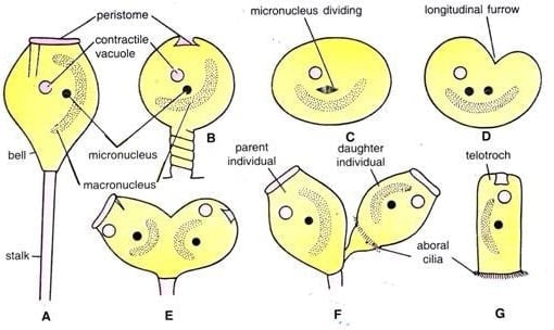
Figure: Longitudinal binary fission in Vorticella (Bell Animalcule). Image Source: Biology Discussion.
2. Conjugation
- Sexual reproduction takes place by conjugation. After a number of generations produced by binary fission, the individuals appear weakened and unhealthy and conjugation takes place at this stage.
- It is also known as anisogamontogony because two conjugating individuals (anisogametes) are dissimilar in size.
- Macroconjugant is the larger, stalked, and sessile individual(the female)looking like an ordinary vegetative individual.
- Microconjugant is the smaller motile which arises by special division.
a. Formation of micro and macroconjugants
- Vorticella first divides asexually by longitudinal binary fission into two very unequal parts without being separated. The larger part is the ordinary somatic individuals, while the smaller part is called the microconjugant.
- More than one microconjugant are produced in some species( V. nebulifera) by repeated division.
- Each microconjugant acquires a girdle of cilia at the posterior end, becomes detached from the stalk, and swims about actively in water.
- Swimming is an adaptation ensuring conjugation in sessile species.
- Microconjugant differs from telotroch in being smaller in size, in never metamorphosing into an adult, and in never forming stalk.
- Microconjugant never feeds or encyst and only survive for 24 hours after which they die.
- Macroconjugants are produced by normal stalked individuals, in which certain nuclear modifications have taken place.
- Macroconjugants are morphologically similar to the normal trophic individuals, but are specialized physiologically and can attract microconjugants for about 2 hours.
- Macroconjugants are stationary and passive, while microconjugants are motile and active.
b. Fusion of conjugants
- A microconjugant swims and fuses by its aboral end with a macroconjugant in the lower third of its body.
- After attachment, the microconjugant loses cilia and throws off its pellicle.
- The two conjugants differ in their further development. Macronuclei of both conjugants disintegrate and finally disappear.
- In microconjugant, 3 nuclear divisions take place forming 8 nuclei, out of which 7 disintegrate and the remaining one becomes the male pronucleus.
- In macroconjugant, micronucleus undergoes two divisions forming 4 nuclei, 3 of which disintegrate and the remaining one becomes female pronucleus.
- In both cases, the first division is reduction division and hence the male, as well as female pronuclei, are haploid.
- Later, the male pronucleus of the microconjugant migrates into the cytoplasm of the macroconjugant and fuses with its female pronucleus, resulting in a diploid zygote nucleus or synkaryon.
- After this, the microconjugant shrivels and perishes. Fertilized macroconjugant is now called a zygote.
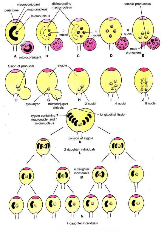
Figure: Conjugation in Vorticella (Bell Animalcule). Image Source: Biology Discussion.
c. Post-zygotic development
- Synkaryon of zygote undergoes 3 mitotic divisions to form 8 nuclei. Out of which, 7 become macronuclei and the remaining 1, the micronucleus.
- Micronucleus now divides mitotically producing 2 daughter nuclei followed by cytokinesis. Thus, 2 daughter cells are formed, one with 4 macronuclei and one micronucleus.
- Each daughter cell and its micronucleus divide twice. The daughter cell with 4 macronuclei forms 4 daughter individuals, and the daughter cell having 3 macronuclei gives rise to only 3 individuals, each having one micronucleus and one macronucleus.
- The 7 daughter cells thus produced begin to grow, acquire stalks, and finally become adults.
Dispersal and Encystation of Vorticella (Bell Animalcule)
- Under favorable conditions, a normal individual of Vorticella may also grow a posterior ring of cilia to become a telotroch.
- The telotroch then breaks loose from its stalk, swims away to a more favorable spot, and there becomes fixed again by growing a stalk.
- Vorticella is also known to tide over adverse conditions by undergoing encystation.
- The whole bell-body encysts on the stalk from which it finally breaks off. The stalk contracts to its maximum and the bell-body loses water, rounds off, and secretes around it a gelatinous ectocyst.
- It soon becomes a wrinkled membrane and underneath it develops a double-layered endocyst.
- Myonemes become the indistinct and pulsating rate of contractile vacuole goes very low, till it disappears.
- After some time, the peristome is absorbed. The completed cyst of V. microstoma measures about 38µ in diameter and its poles are marked by two scars.
- One scar represents the point of attachment with stalk, while the other scar indicates the last point of escape for excreted water.
- On the return of favorable conditions, encystation takes place.
- Contractile vacuole becomes enlarged and the organism grows an aboral circlet of cilia to become a telotroch.
- After some time of free swimming, telotroch gets fixed to some time substratum and grows into an adult Vorticella.
References and Sources
- Kotpal RL. 2017. Modern Text Book of Zoology- Invertebrates. 11th Edition. Rastogi Publications.
- Jordan EL and Verma PS. 2018. Invertebrate Zoology. 14th Edition. S Chand Publishing.
- https://www.notesonzoology.com/term-paper/vorticella/term-paper-on-vorticella-protozoa-microorganisms-zoology/9122- 19%
- https://www.biologydiscussion.com/invertebrate-zoology/protozoa/vorticella-campanula-habitat-structure-and-locomotion/28340- 9%
- https://www.sakshieducation.com/Vidhya/30-01-2011-IstYearZoology.pdf- 1%
- https://www.notesonzoology.com/protozoa/vorticella-structure-and-reproduction-with-diagram-protozoa/5671- 1%
- https://rsscience.com/vorticella/- 1%
- https://www.pirx.com/droplet/gallery/vorticella.html- <1%