A normal human usually has 23 pairs of chromosomes, with two sex chromosomes that determine sex and 44 autosomal chromosomes, which are responsible for growth and function.
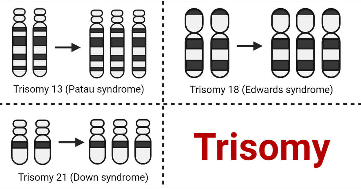
Trisomy is a genetic condition caused by an alteration in the number of chromosomes where the affected person has three copies of one of the chromosomes instead of two. The affected person possesses 47 chromosomes instead of 46.
Trisomy occurs in at least 4% of all clinically recognized pregnancies and frequently results in various birth defects in children, including intellectual impairments and delayed development.
Despite the possibility of a full-term birth, trisomy most frequently results in a miscarriage in the first three months of pregnancy.
Interesting Science Videos
Risk factors for Trisomy
- The most important risk factor for trisomy conditions is maternal age.
- Women in their late 30s and 40s have a higher chance of trisomy conditions occurring.
- Paternal age does not seem to be associated with the incidence of trisomy.
Cause of Trisomy
- A normal egg cell and normal sperm cell start with 46 chromosomes when a baby is conceived.
- The egg and sperm cells divide in half, each possessing 23 chromosomes.
- After fertilization, the baby will have a complete set of 46 chromosomes.
- But sometimes, an error can occur when dividing 46 chromosomes in half. Either egg or sperm cell may keep both copies of a chromosome instead of just one copy.
- The baby will have three copies of chromosomes if this egg or sperm is fertilized. The cause is unknown, and prevention is not possible.
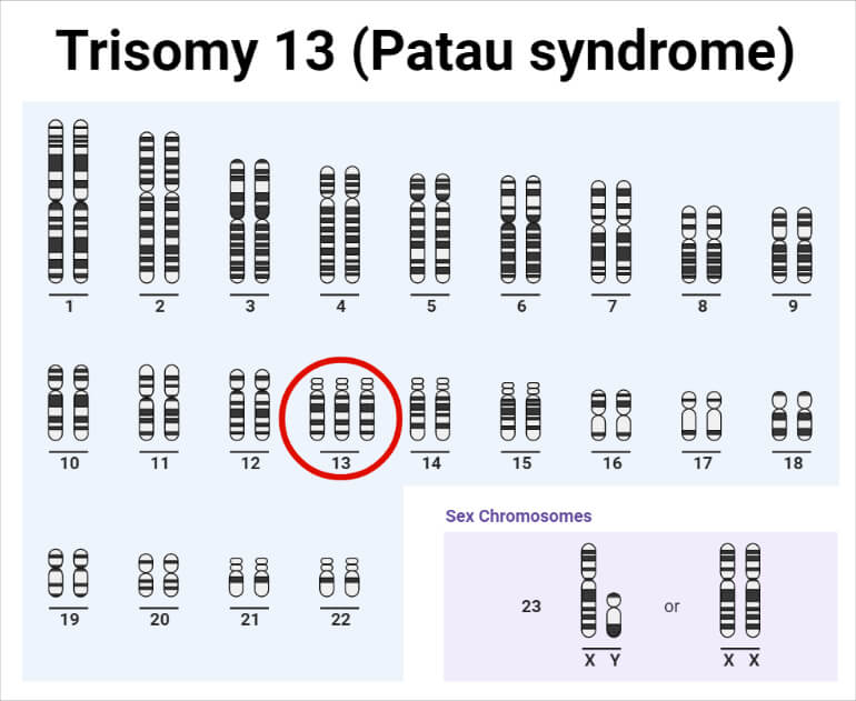
- Trisomy 13 occurs when the baby has three copies of chromosome number 13.
- Trisomy 18 occurs when the baby has three copies of chromosome number 18.
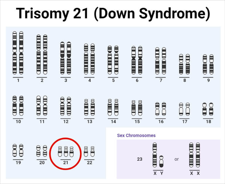
- Trisomy 21 occurs when the baby has three copies of chromosome number 21.
- This extra copy of a chromosome is then present in every cell in the body.
- Sometimes the extra chromosome, or part of it, is attached to another chromosome in the egg or sperm. This is called translocation. This is the only form of trisomy that may be inherited from a parent. Some parents may have balanced translocation.
- This means the chromosome is attached to another chromosome. However, it has no impact on their health.
Types of Trisomy in Humans
The most common trisomy conditions include:
- Trisomy 13 (Patau syndrome)
- Trisomy 18 (Edward syndrome)
- Trisomy 21 (Down syndrome)
1. Trisomy 13 (Patau syndrome)
- Trisomy 13 is also known as Patau syndrome.
- It was described in 1960 by Klaus Patau and coworkers.
- It occurs in about 1 in 8000 to 12000 newborns.
Some of the characteristics of Trisomy 13 include:
- Low birth weight
- Small skull (microcephaly)
- An abnormal opening in the skull
- Structural problems of the brain, such as the front of the brain not divided normally (holoprosencephaly)
- Eyes that are close together
- Low-set ears that have unusual shape
- Scalp abnormalities that resemble ulcers
- Cleft lip or Cleft palate
- Additional toes or fingers (polydactyly)
- Congenital heart disorders, such as ventricular septal defect
- Neural tube defect where the spinal cord, meninges and blood vessels protrude through a gap in the vertebrae (myelomeningocele)
- In boy babies, testes do not descend into the scrotum
- In girl babies, uterus that forms in 2 branches (bicornuate uterus) is formed
It is rare for babies to survive past the neonatal stage with Patau syndrome.
Most babies born with trisomy 13 die by age 1. There are a few reports of babies with trisomy 13 surviving to their teens.
2. Trisomy 18 (Edward syndrome)
- Trisomy 18 is also known as Edward syndrome.
- It occurs in about one out of every 6000 to 8000 newborns.

Some of the characteristics of Trisomy 18 include:
- Small skull (microcephaly)
- Physical irregularity of the kidneys, ureters, heart, lungs and diaphragm
- Cleft lip or Cleft palate
- Malformations of the hands and feet such as missing thumbs, club feet and webbing between the fingers and toes (syndactyly)
- Neural tube defect where the spinal cord, meninges and blood vessels protrude through a gap in the vertebrae (myelomeningocele)
- In boy babies, testes do not descend into the scrotum
It is rare for babies to survive past the neonatal stage with Edward syndrome.
3. Trisomy 21 (Down syndrome)
- Trisomy 21 is also known as Down syndrome.
- Down syndrome is named after the physician J. Langdon Down who first described this genetic defect in 1866. It was formally called mongolism or mongolian idiocy.
- It affects about 1 in 800 babies.
- The average lifespan of an adult with Down syndrome is 60 years, though this can vary.
- Trisomy 21 is found to be affected either by senescence of oocytes, virus infection, radiation damage, etc.
- Pregnant women who previously had infectious hepatitis may have a threefold increased risk of giving birth to children with Down syndrome.
There are three types of Down syndrome. They are:
- Standard Trisomy 21
About 95% of people with Down syndrome have Trisomy 21.
Instead of the normal 2 copies, each cell in the body possesses 3 copies of chromosome 21.
- Mosaic Down syndrome
This type affects about 2% of the people with Down syndrome.
It occurs during an error in cell division where the extra chromosome spontaneously appears as the embryo develops.
Both normal cells and some cells with an extra copy of chromosome 21 are present in individuals with this condition.
- Translocation Down syndrome
This type accounts for a small percentage of people with Down syndrome (about 3%) and is inheritable.
This occurs when an extra part or a whole extra chromosome 21 is present, but it is attached or “trans-located” to a different chromosome rather than being a separate chromosome 21.
Some of the characteristics of Trisomy 21 include:
- Eyes that slant upward
- Small white patches on the edge of the iris of the eye (Brushfield spots)
- Small ears that may fold over slightly at the top
- Small mouth that makes the tongue appear large
- Small nose with a flattened bridge
- Short neck
- Small hands with short fingers
- 2 palm creases instead of 3, including one across the palm and one around the base of the thumb
- Short height
- Loose joints
Adults with Down syndrome are commonly smaller than adults who do not have Down syndrome.
Types of Sex Cell Trisomy in Humans
It includes:
- Trisomy X (XXX)
- Klinefelter syndrome (XXY)
- Jacob’s syndrome (XYY)
1. Trisomy X
- It is a genetic condition where a female is born with an extra X chromosome.
- This condition occurs only in females.
- It occurs in about 1 in 1,000 female newborns.
- It is not usually inherited from an affected parent.
- The majority of cases are entirely random occurrences that are caused by errors in chromosome replication and division during the formation of egg or sperm cells.
Some of the characteristics of Trisomy X include:
- Taller than girls of the same age and/or taller than predicted by their parents’ heights
- Wide-spaced eyes known as hypertelorism
- Vertical skin folds that cover the inner corners of the eyes (known as epicanthal folds)
- Curved or bent little fingers (known as clinodactyly)
- Poor muscle tone (known as hypotonia)
- Premature ovarian aging or failure
- Genito-urinary deformities
- Attention deficit disorder
2. Klinefelter Syndrome
- It is a genetic condition where a male is born with an extra X chromosome.
- It affects about 1 in 650 newborn boys.
- Klinefelter syndrome is not inherited; the addition of an extra X chromosome occurs during the formation of reproductive cells (eggs or sperm) in one of an affected person’s parents.
Some of the characteristics of Klinefelter syndrome include:
- Hypertension (high blood pressure)
- Type 2 diabetes
- High cholesterol and fats in the blood
- Gynecomastia (breast growth in a male)
- Breast cancer
- Tremors
- Osteoporosis (weakened bones)
- Autoimmune diseases (Thyroid disease, Lupus and Rheumatoid arthritis)
3. Jacob’s syndrome
- It is a genetic condition characterized by an extra copy of the Y chromosome in each of an individual’s cells.
- It affects about 1 in 1,000 newborns.
- It is not inherited.
- During the formation of sperm cells, a random chromosomal change may occur. An error in cell division called nondisjunction can result in sperm cells with an extra copy of the Y chromosome.
Some of the characteristics of Jacob’s syndrome include:
- Hypotonia (weak muscle tone)
- Delayed motor skill development, such as with walking or crawling
- Delayed or difficult speech
- Autism diagnosis
- Attention difficulties
- Hand trembling or involuntary muscle movements
- Learning disabilities
- Taller-than-average height
Signs of Trisomy During Pregnancy
- Too much amniotic fluid surrounding the baby (polyhydramnios)
- Only one umbilical cord artery
- Smaller than expected placenta
- The baby is small for its gestational date.
- Baby is less active than expected
- Congenital defects, including cleft palate or heart irregularities, are picked up during ultrasound scans.
Diagnosis of Trisomy conditions
Prenatal tests that can help detect trisomy disorders include:
- Ultrasound scans: Sound waves are used to create a picture.
- Maternal Serum Screening: A specialized blood test. Two serum screening tests are available to all women.
- Combined First Trimester Screening (CFTS)
- Second Trimester Maternal Serum Screening (2TMSS)
- Amniocentesis: Between 15 and 20 weeks of pregnancy, a sample of the amniotic fluid is taken and examined.
- Chorionic villus sampling (CVS): Between 10 and 13 weeks of pregnancy, a sample of cells from the chorion, the tissue that will ultimately become the placenta, is taken and examined.
- Non-invasive prenatal testing (NIPT): After ten weeks of pregnancy, a screening test that measures fetal DNA circulating in the mother’s blood.
- Percutaneous umbilical blood sampling (PUBS): A small blood sample from a baby’s umbilical cord to test for health conditions.
Treatment of Trisomy
- Medical Care
- Drug therapy is not a component of the standard of care for trisomy.
- Nasogastric and gastrostomy supplementation for feeding problems.
- Orthopedic management of scoliosis may be needed.
- Cardiac management is primarily medical. Most of the children require a diuretic and digoxin for congestive heart failure.
- Neonatal intensive care (NICU) management
- Periodic screenings
Periodic screenings throughout childhood and into adulthood may be advised by the healthcare professional.
Treatment can be given immediately for any health issues, learning difficulties, or developmental delays.
- Early intervention services
These services may start in the first few months of life or as soon as requirements are recognized and may include speech, occupational, physical, or developmental therapy.
- Educational assistance
If the child has a learning disability, educational help to learn techniques and strategies to be successful in school and daily life can be provided.
- Psychosocial management
About issues of diagnosis and survival.
Parents should be informed properly about the syndrome, including its cause, implications, and possible outcomes.
- Assistance and support in daily functioning
If a child’s difficulties interfere with everyday functioning, this support and assistance may include help with social interactions, work, and daily living activities.
Trisomy in Non-humans
Trisomy-22 has been reported in chimpanzees which shows Down syndrome-like phenotypic features. Trisomy-21 has been reported in the gorilla.
References
- Better Health Channel. (2021). Trisomy Disorder. Accessed from:
https://www.betterhealth.vic.gov.au/health/conditionsandtreatments/trisomy-disorders - Boston’s Children Hospital. (2022). Trisomy 18 and 13. Accessed from:
https://www.childrenshospital.org/conditions/trisomy-18-and-13 - CDC. (2021). Facts about Down Syndrome. Accessed from;
https://www.cdc.gov/ncbddd/birthdefects/downsyndrome.html - Cleveland Clinic. (2022). Trisomy. Accessed from:
https://my.clevelandclinic.org/health/diseases/22912-trisomy - Cleveland Clinic. (2022). Trisomy X Syndrome. Accessed from:
https://my.clevelandclinic.org/health/diseases/17892-triple-x-syndrome - Hassold, T., Hunt, P. A., & Sherman, S. (1993). Trisomy in humans: incidence, origin and etiology. Current opinion in genetics & development, 3(3), 398–403.
https://doi.org/10.1016/0959-437x(93)90111-2 - Healthline. (2022). XYY Syndrome. Accessed from:
https://www.healthline.com/health/xyy-syndrome - Mayoclinic. (2022). Diagnosis and Treatment of Triple X Syndrome. Accessed from:
https://www.mayoclinic.org/diseases-conditions/triple-x-syndrome/diagnosis-treatment/drc-20350981 - MedlinePlus. (2022). 47, XYY Syndrome. Accessed from:
https://medlineplus.gov/genetics/condition/47xyy-syndrome/#frequency - MedlinePlus. (2019). Klinefelter Syndrome. Accessed from:
https://medlineplus.gov/genetics/condition/klinefelter-syndrome/#inheritance - MedlinePlus. (2021). Trisomy 13. Accessed from:
https://medlineplus.gov/genetics/condition/trisomy-13/#references - Medscape. (2022). Trisomy 18 Treatment & Management. Accessed from:
https://emedicine.medscape.com/article/943463-medication - Murdoch Children’s Research Institute. (2022). Maternal Serum Screening. Accessed from:
https://www.vcgs.org.au/tests/maternal-serum-screening - Stanford Medicine. (2022). Trisomy 13 and Trisomy 18 in Children. Accessed from:
https://www.stanfordchildrens.org/en/topic/default?id=trisomy-18-and-13-90-P02419 - Verma P.S. and Agarwal V.K. (2005). Trisomy. In Cell Biology, Genetics, Molecular Biology, Evolution and Ecology. Multi-color Edition. S. Chand & Company Ltd. Ram nagar, New Delhi, pg 196-197