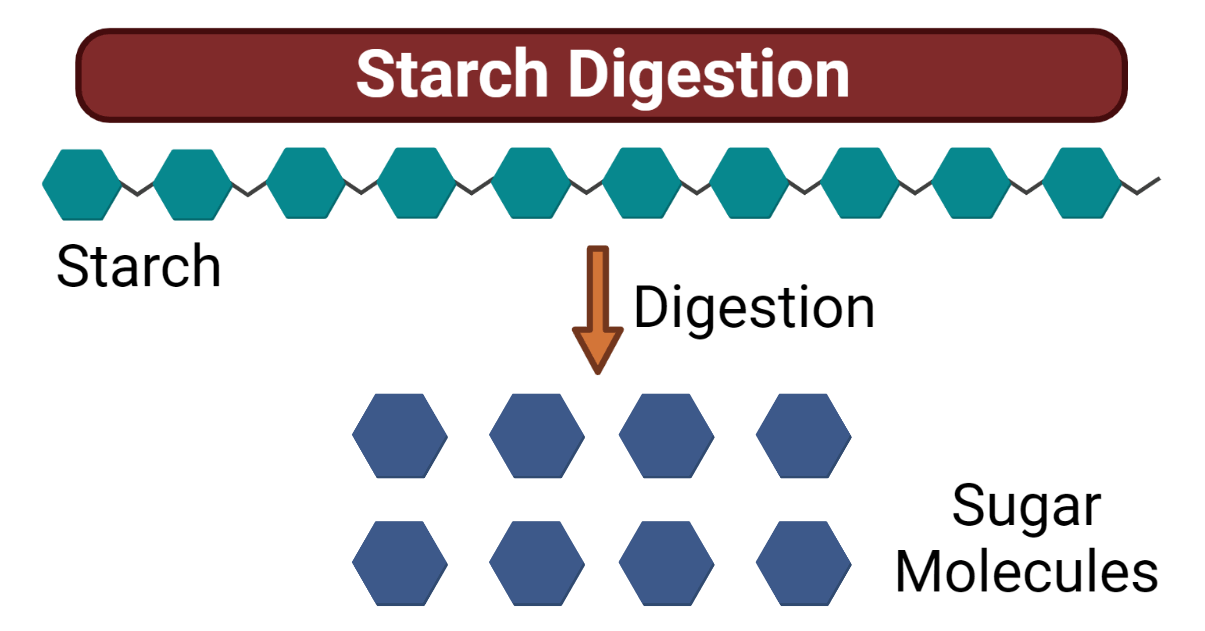
Dietary starches contribute as the largest energy sources by humans to meet their calorie intake. Starch is a complex macromolecule derived from plant-based foods and beverages. Because of their structural complexity, starches seem to take a longer duration to get digested and used for energy needs. The gut employs several processes, such as mechanical and chemical, to degrade the complex conformation of starch into simpler molecules.
Starch digestion is a complex process involving enzymes like salivary and pancreatic amylase that break it down into smaller molecules. These molecules, such as maltose, are further broken down by maltases and sucrases.
The resulting glucose is absorbed in the small intestine and transported to cells for energy or glycogen storage, playing a crucial role in providing a steady glucose supply for vital biological processes.
Interesting Science Videos
Starch and its Structure
Glucose is the functional unit of the starch molecule. Repeating units of glucose molecules joined by glycosidic bonds compose a starch polymer. They undergo hydrolysis reactions to yield glucose molecules. They can be easily derived from the plants as starch granules in semi-crystalline form. Two components of starch include Amylose and Amylopectin.
Amylose
- Makes upto 20-30% of the total starch structure.
- The polysaccharide is composed of alpha-D-glucose monomeric units.
- This component makes up the linear and unbranched polymer of starch.
- The glucose monomers are joined to each other via α-1-4 glycosidic bonds (the 1 and 4 being the carbon number of glucose molecule).
- An amylose polymer may have around 300-3000 units of glucosyl residues.
- It is observed that amylose molecules are resistant to digestion due to a lack of branching.
Amylopectin
- They contribute to about 70-80% of the total starch composition.
- They are constituted by the branching of amylose polymers via α-1-6 glycosidic bonds as the branching point of the glucose units in the amylose.
- They show branching among amylose units after every 24-30 glucose subunits.
- Amylopectin polymers are easily digested due to the presence of branching of glucose units.
- They are responsible for enhancing the gel strength and solubility in the water.
Linkage types (α-1,4 and α-1,6) between glucose molecules
The linkages between glucose molecules in starch involve glycosidic bonds: α-1,4 and α-1,6. The α-1,4 glycosidic bonds connect the glucose units along the linear chains of amylose and Amylopectin.
These bonds are formed between the carbon atom 1 (C1) of one glucose molecule and the carbon atom number 4 (C4) of the adjacent glucose molecule. The α-1,6 glycosidic bonds, found exclusively in Amylopectin, create the branching points in the molecule. They occur between the C1 of a glucose unit and the C6 of another glucose unit, forming a branch.
Granular structure in plants
In plants, starch has a granular structure. These granules are formed inside plastids, specifically in the amyloplasts responsible for starch synthesis and storage. The size and shape of starch granules can vary depending on the plant species.
They can be round, oval, or irregular in shape. Inside the granules, both amylose and Amylopectin are present. Amylose is generally found in the inner region of the granule, forming a helical structure.
Amylopectin is more abundant and forms the outer region of the granule, creating a branched network. This granular structure allows plants to store starch as an energy reserve efficiently.
Enzymes Involved in Starch Digestion
Salivary amylase (ptyalin), pancreatic amylase, and brush border enzymes (maltase, sucrase, and lactase) are all involved in the digestion of starch in the human body. Here’s a brief description of each enzyme:
Salivary amylase (ptyalin)
Salivary amylase is an enzyme secreted by the salivary glands in the mouth. It begins starch digestion by breaking down complex carbohydrates (starch) into smaller polysaccharides and disaccharides like maltose.
Pancreatic amylase
The pancreas generates pancreatic amylase, which is then discharged into the small intestine. It continues starch digestion by breaking down the remaining complex carbohydrates into smaller units like maltose, maltotriose, and alpha-limit dextrins.
Brush border enzymes (maltase, sucrase, and lactase)
Brush border enzymes are located on the surface of the small intestine’s absorptive cells, specifically the microvilli. They further break down the smaller units of carbohydrates into their respective monosaccharides:
- Maltase: Maltase converts maltose into two glucose molecules.
- Sucrase: Sucrase breaks down sucrose into one glucose molecule and one fructose molecule.
- Lactase: Lactase breaks down lactose into one glucose molecule and one galactose molecule.
Mechanism of Starch Digestion
Humans and animals consume starch granules through plant-based food sources such as wheat, potatoes, rice, leaves, and other parts of plants. Also, they are found to be consistent in the staple diet, owing to the major carbohydrate and energy source in humans and animals alike. Starch granules digestion begins in the oral cavity all the way leading to its degradation in the intestine.
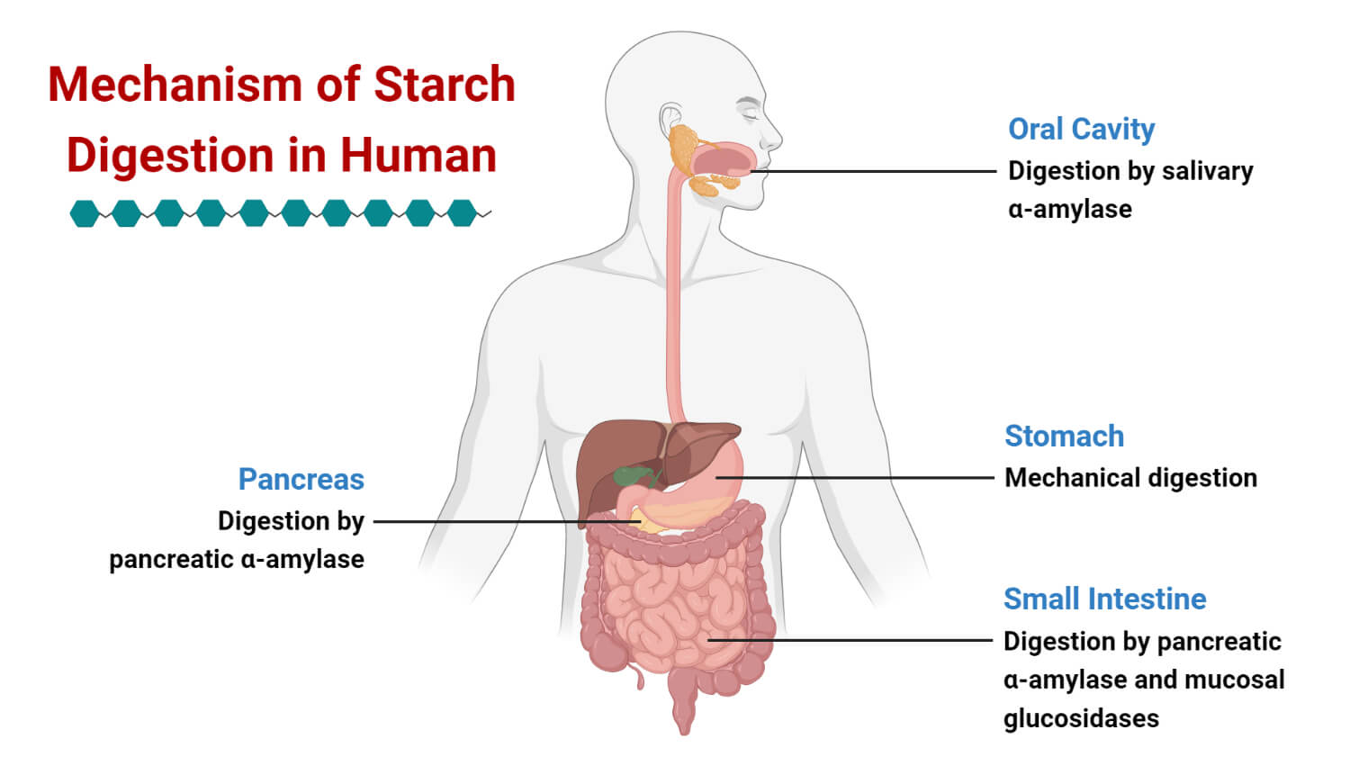
Oral Cavity: Pre-Gastric Processing
As the food is ingested, mechanical and enzymatic processes act on the food. The mastication process is favored for breaking down the macroscopic structure of foods other than starch. Saliva production is commenced before the ingestion of food by sight, smell, or sound of food triggers cephalic digestion in our brain.
- As the food is masticated and turned into a saliva-infused bolus, salivary alpha-amylase enzyme acts on the starch molecule to break it down into short-chain oligopolysachharides such as maltose or isomaltose.
- Salivary amylase’s catalytic activity is enhanced by the presence of chloride ions.
- The gene AMY1 for salivary amylase is located in chromosome 1.
- Individuals or communities that rely on a starchy diet have more number of gene repeats.
- The composition of macromolecules in the food, such as proteins, carbohydrates, and fats, is strongly associated with the release of amylase from acinar cells of salivary glands.
- Tests in rodents have shown an increased salivary amylase production by the consumption of a high-fat diet.
- Mixing of ingested food is also important for lubrication and swallowing as it reduces friction and helps in the easy movement of a food bolus from the oesophagus to the stomach for further degradation.
Stomach-Gastric Processing
Since the pH of the gastric environment is lower than the optimal amylase activity, a minimal digestion process occurs for starch granules. Further homogenization of starch via mechanical contraction of gastric chambers turns the contents into a creamy paste. This paste is allowed to pass through the pyloric sphincter to the duodenum for complete digestion in the small intestine.
Small Intestine- Final Processing
The homogenized mixture enters the duodenum with high hydrogen ion concentration, which leads to the release of cholecystokinin from the hormone enteroendocrine I cell, initiating the process of digestion in the small intestine. To diffuse the acidic influx of gastric juice along with chyme (a liquified mixture of food from the stomach), bicarbonate ions are released from the small intestinal epithelium cells, pancreatic acinar cells and hepatocytes.
- Contractile movement of intestinal walls helps mix the food contents to bile and pancreatic juice.
- Pancreatic juice contains the active pancreatic alpha-amylase enzyme released from acinar cells in the pancreas. It breaks down starch into maltose, isomaltose, sucrose and other simple sugars.
- Pancreatic amylase gene AMY2 and AMY2B is located in chromosome 1.
- Pancreatic alpha-amylase specifically cleaves the 1,4-glycosidic bonds of the amylose component of the starch.
- Another important enzyme secreted by the intestinal epithelium is glucosidases.
- Glucosidases help in the amylolysis of starch, producing glucose, maltose, and maltotriose as the end products.
- Further digestion of disaccharides is achieved by the catalytic actions of enzymes sucrase, maltase, isomaltase, lactase, etc, to convert them into monosaccharides useful in cellular metabolism.
Feedback Mechanism and Starch Digestion
Feedback stimulus, such as food texture, taste perception, the release of enzymes, etc, is very important to upregulate or downregulate the starch digestion mechanism.
| STIMULUS | RESPONSE | MEDIATED BY |
| Food Texture | A conscious decision to keep food in the mouth for processing and swallowing | Mastication and the movement of teeth (periodontal ligaments) for the mechanical breakdown of starch |
| Taste Perception | Enhanced secretion of salivary and gastric enzymes | Taste receptors modulated by CNS (central nervous system) |
| Particle Size | Increased particle size may lead to delayed emptying of gastric contents (chyme); hence further degradation is required | Stretch, and tension receptors in the stomach transmit the signals to CNS |
| Starch Hydroxylates in the Small Intestine | Regulated secretion of enzymes such as amylase, glucosidases, etc, from bile, pancreatic and intestinal juices | Cholecytokinin and ions released from intestinal cells |
Biological Factors controlling Starch Digestion
Biological factors that control starch digestion include
- Mastication – Responsible for the mechanical size reduction of the starch granules.
- Salivation – Breakdown of starch granules and food bolus formation.
- Gastric Peristalsis – Contraction and relaxation of gastric chambers lead to a mechanical reduction of food particle size and mixing food bolus with the gastric juices.
- Gastric Emptying – Rate of delivery and release of food chyme (food bolus mixed with the gastric juice) into the small intestine.
- Intestinal Motility – further size reduction of food particles and fusion of intestinal juices and enzymes to the food chyme.
Conclusion
Starch is a complex carbohydrate and a major dietary component for carbon and energy sources. They are made up of repeating units of glucose molecules as functional units. It has two main components, amylose, and amylopectin, differing in glycosidic bonds. The process of starch digestion begins in the mouth by the action of the enzyme salivary alpha-amylase secreted via salivary glands. Amylases break down the scratch into simpler sugar, such as maltose. The digestion by the intestinal juice further leads to the degradation of starch granules to sugars like glucose by the action of enzymes pancreatic amylases, maltase, sucrase, etc. These monosaccharides are absorbed by the intestinal cells and transported to the liver for cellular metabolism processes and energy requirements.
References
- Brownlee, Iain A., et al. “Starch digestion in the upper gastrointestinal tract of humans.” Starch‐Stärke 70.9-10 (2018): 1700111.
- Lee, Byung-Hoo, et al. “Modulation of starch digestion for slow glucose release through “toggling” of activities of mucosal α-glucosidases.” Journal of Biological Chemistry 287.38 (2012): 31929-31938.
- Nutritional qualities of starch depend on the way it is digested – https://www.sydney.edu.au/science/news-and-events/2021/12/20/nutritional-qualities-of-starch-depend-on-the-way-it-is-digested.html
- Starch – https://alevelbiology.co.uk/notes/starch/#12-degradation-in-animals-
- Li, Cheng, et al. “Biological factors controlling starch digestibility in human digestive system.” Food Science and Human Wellness 12.2 (2023): 351-358.
- https://academic.oup.com/jn/article-abstract/122/1/172/4754868
- https://academic.oup.com/jas/article-abstract/63/5/1624/4662245
- https://pubs.acs.org/doi/abs/10.1021/jf9813900
- https://pubs.acs.org/doi/abs/10.1021/jm800115x
- https://www.sciencedirect.com/science/article/pii/0016508586904361
- https://www.sciencedirect.com/science/article/pii/S0144861714009412
- https://link.springer.com/article/10.1007/BF00451611
- https://pubs.rsc.org/en/content/articlehtml/2014/fo/c4fo00115j
- https://www.sciencedirect.com/science/article/pii/S0268005X21000734
- https://www.sciencedirect.com/science/article/pii/003194227480289X