Interesting Science Videos
Simple columnar epithelium definition
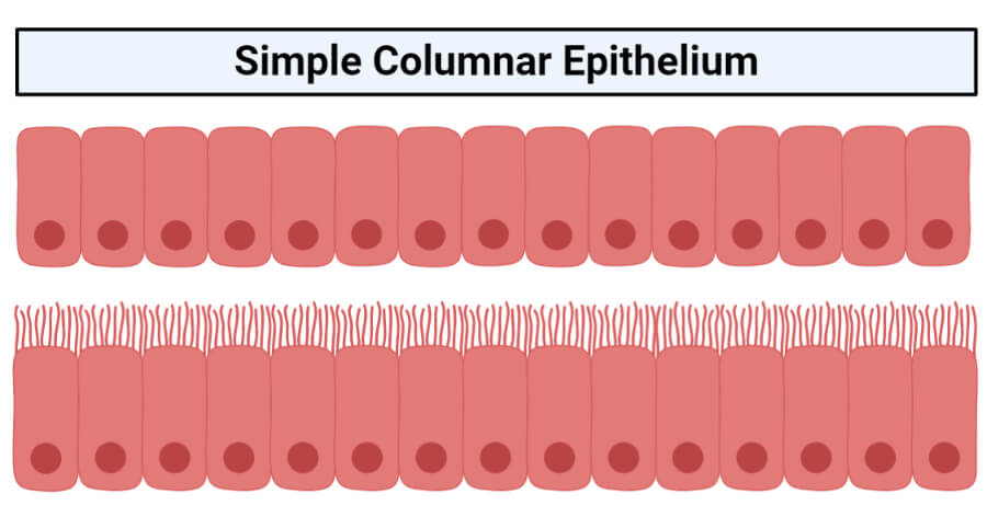
The simple columnar epithelium is a type of epithelium that is formed of a single layer of long, elongated cells mostly in areas where absorption and secretion are the main functions. Like cuboidal epithelium, the cells in the columnar epithelium are also modified to suit the function and structure of the organ better. The simple columnar epithelium is either ciliated or non-ciliated, and microvilli are present on the apical region of the non-ciliated columnar epithelium.
Structure of the simple columnar epithelium
- The simple columnar epithelium is made up of a single layer of elongated cells which are always taller than they are wide and are directly attached to the basement membrane at the distal end.
- The nucleus is oval, large and is present at the base of the cell.
- The cells are arranged tightly adjacent to one other, and goblet cells are present in the layer of non-ciliated columnar epithelium.
- The cells of the columnar epithelium are also provided with a large number of organelles. They thus are capable of a higher level of secretion and absorption than are cuboidal cells.
- As in all other types of epithelial tissues, the apical surface of the cells in the columnar epithelium is located towards the lumen of the organs or ducts while the lateral surface is provided with adhesions and junctions to facilitate the connection between the adjacent cells.
- Complexes of tight and adherent junctions sometimes called “terminal bars”, seen in the microscope are formed at the apical ends of the cells.
- The cells in the simple cuboidal epithelium do not have a direct blood supply. Still, they get their nutrients, water and gases from the underlying tissue via diffusion as they are directly attached to the basement membrane.
- The cells, however, are innervated, having a nerve supply.
Based on the presence and absence of cilia, the simple columnar epithelium is of two types: Non-ciliated columnar epithelium and ciliated columnar epithelium.
1. Non-ciliated columnar epithelium
- The non-ciliated simple columnar epithelium is a single layer of non-ciliated columnlike cells with oval nuclei near the base of cells.
- The layer of cells contains columnar epithelial cells with microvilli at the apical surface and goblet cells.
- Microvilli are tiny finger-like cytoplasmic projections that present on the apical surface, directing towards the lumen of the organ.
- The average microvillus is about 1-μm long and 0.1-μm wide, but these can increase the surface area by 20 or even 30 fold as hundreds to thousands of them are present on the end of each absorptive cell.
- Each microvillus contains bundled actin filaments capped and bound to the surrounding plasma membrane by actin-binding proteins.
- The thick glycocalyx covering microvilli of the intestinal columnar epithelium includes membrane-bound proteins and enzymes for the digestion of specific macromolecules.
- Goblet cells are modified columnar epithelial cells that secrete mucus, a slightly sticky fluid, at their apical surfaces.
- Before release, the mucus accumulates in the upper portion of the cell, causing it to bulge and making the whole-cell resemble a goblet or wine glass.
2. Ciliated columnar epithelium
- The ciliated simple columnar epithelium is a single layer of ciliated column-like cells with oval nuclei near the base of cells, and the goblet cells are usually interspersed between the ciliated cells.
- The cilia are typically 5-10 μm long and 0.2 μm in diameter. Each cilium has a core structure consisting of nine peripheral microtubule doublets arrayed around by two central microtubules.
- Cilia exhibit rapid beating patterns that move a current of fluid and suspended matter in one direction along the epithelium.
- These cilia direct the movement of molecules in a particular direction aids the function of excretion and secretion.
Functions of the simple columnar epithelium
The primary function of the simple columnar epithelium includes secretion, absorption, protection, and transportation of molecules. The non-ciliated columnar epithelium is mainly involved in absorption and secretion. In contrast, the ciliated columnar epithelium aids the transport or movement of molecules and cells from one place to another.
1. Absorption
- As the microvilli present on the apical surface of the non-ciliated columnar epithelium increases the surface area, the function of absorption is greatly enhanced.
- The non-ciliated columnar epithelium present in the gastrointestinal tract, ranging from the stomach to the anus, functions in the absorption of macromolecules and water.
- The cells are also provided with accessory membrane-bound proteins that help in the active absorption of these nutrients.
2. Secretion
- The goblet cells in between the columnar cells in the tissue secrete mucus that lubricates linings of digestive, respiratory, and reproductive tracts, and most of the urinary tract.
- The mucus in the respiratory tract provides protection against foreign particles and prevents their entry through the nasal passage into the inner respiratory tract.
3. Protection
- The simple columnar epithelium also acts as a protective barrier and provides protection against the non-specific movement of luminal substances.
- The complexes of junctions present at the apical region of the cells limit the passage of molecules and ions through the intracellular spaces.
- This also prevents the entry of harmful large macromolecules into the body.
- Similarly, the mucus secreted by the goblet cells protects the lining of the gastrointestinal tract against the harmful effects of the acid produced by the stomach.
4. Transportation
- The ducts of some glands like the gallbladder are lined with simple columnar epithelium which helps in the movement of hormones and enzymes to their site of action.
- In the same way, the cilia present in the columnar epithelium of the respiratory tract beat in unison moving mucus and foreign particles toward the throat, where they can be coughed up and swallowed or spit out.
- The columnar epithelium lining the female reproductive system has cilia which helps move oocytes expelled from ovaries through uterine (fallopian) tubes into the uterus.
Location and examples of simple columnar epithelium
- The simple columnar epithelium is present mostly in areas that perform the function of secretion and absorption.
- The ciliated columnar epithelium lines some bronchioles (small tubes) of the respiratory tract in the respiratory system and the uterine (fallopian) tubes and uterus in the reproductive system.
- In the same way, the columnar epithelium also lines some paranasal sinuses, the central canal of the spinal cord, and the ventricles of the brain.
- The non-ciliated columnar epithelium, however, lines the gastrointestinal tract and the ducts of glands and gallbladder.
- The simple columnar epithelium also lines some parts (collecting ducts) of the kidney.
References and Sources
- Mescher AL (2016). Basic Histology. Fourteenth Edition. McGraw-Hill Education.
- Tortora GJ and Derrickson B (2017). Principles of Physiology and Anatomy. Fifteenth Edition. John Wiley & Sons, Inc.
- Waugh A and Grant A. (2004) Anatomy and Physiology. Ninth Edition. Churchill Livingstone.
- 4% – https://quizlet.com/91194531/nonciliated-simple-columnar-epithelium-flash-cards/
- 3% – https://ibiologia.com/simple-columnar-epithelium/
- 2% – https://quizlet.com/49824769/chapter-3-classification-of-epithelial-tissue-flash-cards/
- 2% – https://quizlet.com/317398032/histology-chapter-4-flash-cards/
- 2% – https://doctor2019.jumedicine.com/wp-content/uploads/sites/10/2020/01/Epithelial-Tissue-1.pdf
- 1% – https://www.sciencedirect.com/topics/agricultural-and-biological-sciences/simple-columnar-epithelium
- 1% – https://www.genetics.org/content/206/1/33
- 1% – https://www.answers.com/Q/Difference_between_simple_ciliated_columnar_and_non_ciliated_columnar
- 1% – https://study.com/academy/answer/what-is-the-function-of-the-microvilli-on-the-apical-surface-of-the-proximal-convoluted-tubule.html
- 1% – https://quizlet.com/520601363/ap-epithelial-tissue-covering-and-lining-flash-cards/
- 1% – https://quizlet.com/262419308/tissues-cells-chapter-4-flash-cards/
- 1% – https://en.wikipedia.org/wiki/Simple_columnar_epithelium
- 1% – https://doku.pub/documents/inderbir-singhs-textbook-of-human-histology-with-colour-atlas-and-practical-guide-united-vrgpdf-z0x292ko9nqn
- 1% – https://biology.homeomagnet.com/cell-junctions/
- <1% – https://www.proprofs.com/discuss/q/64182/describe-simple-columnar
- <1% – https://www.online-sciences.com/biology/tissues-types-epithelial-tissue-features-covering-glandular-epithelium/
- <1% – https://www.lecturio.com/magazine/epithelium/
- <1% – https://www.answers.com/Q/What_is_the_function_of_cilia_and_mucus_in_the_respiratory_system
- <1% – https://quizlet.com/52053286/epithelial-tissues-flash-cards/
- <1% – https://quizlet.com/299843063/ap-1-all-lab-quiz-study-sets-combined-flash-cards/
- <1% – https://en.wikipedia.org/wiki/Respiratory_epithelium
- <1% – https://en.wikipedia.org/wiki/Intestinal_epithelium
- <1% – https://en.wikipedia.org/wiki/Duct_(anatomy)
- <1% – https://courses.lumenlearning.com/wm-biology2/chapter/epithelial-tissues/
- <1% – http://lifesci.dls.rutgers.edu/~babiarz/Frepro.htm