Selective Reabsorption is the process during urine formation that helps retain a large amount of water, salts, and other particular substances from the nephric filtrate by reabsorbing them in the body.
The process of urine formation in our body involves the following steps:
2. Selective Reabsorption
3. Tubular Secretion
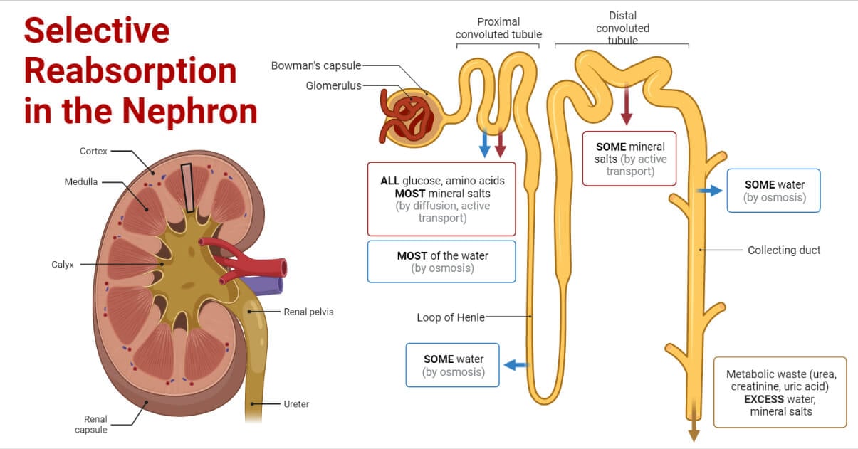
Selective Reabsorption is the second step after the ultrafiltration involved in urine formation. The nephron is surrounded by capillaries into which the osmotic process reabsorbs the chemical components.
It takes place mainly in the following parts of the nephron:
- Proximal Convoluted Tubule
- Loop of Henle
Interesting Science Videos
Proximal Convoluted Tubule (PCT) and its Structure
- It originates from the Bowman’s capsule and carries the filtrate further.
- It is located in the cortex region of the kidney.
- It consists of simple cuboidal epithelial cells.
- The cells of PCT have a large number of mitochondria which aids in providing ATP for the process of active transport.
- PCT is the longest part of the nephron and has the widest lumen.
- The surface of these cells has many microvilli, which are these finger-like extensions into the lumen of PCT. Hence it is also called the brush-bordered epithelium. The brush border aids in incrementing surface area for absorption in the lumen.
- The active transport process in this part entirely absorbs glucose and amino acid.
- The renal threshold for the concentration of glucose absorbed is around 180 mg/dl.
- The chlorides are reabsorbed by the passive transport following the positively charged substances such as ions.
- As the ions are taken up actively, the filtrate becomes less concentrated, and hence to balance the concentration, the water moves into the capillaries surrounding PCT.
- The selective reabsorption is maximum in this part of the nephron.
- The passive transport process selectively reabsorbs nearly eighty percent of the water; hence it is also called “obligatory water reabsorption”.
Loop of Henle and its Structure
- The loop of Henle lies in the medulla region of the kidney.
- It is the part located between PCT and the distal convoluted tubule and is U-shaped.
- It consists of two parts i.e. ascending loop and descending loop of Henle.
- The one that originates from PCT and moves downward is called descending loop of Henle. The descending loop further extends towards the upward direction leading to the Distal Convoluted Tubule and is called ascending loop of Henle.
- Descending loop has a very thin epithelium and comprises squamous epithelium. It has permeability for water and is impermeable to salt.
- The ascending loop comprises cuboidal epithelium. It has both thin and thick segments. It has permeability to salts but is impermeable to water.
- The loop of Henle is surrounded by the capillaries carrying blood called the vasa recta. The vasa recta has two main functions:
- The cells of the medulla region receive the required nutrients through it.
- They carry the reabsorbed water from the filtrate.
Interesting fact: The longest Loop of Henle is found in kangaroo rats which helps them to reabsorb large amounts of water in the body, and hence it is easier for them to adapt to the desert ecosystem.
Process Involved in Selective Reabsorption
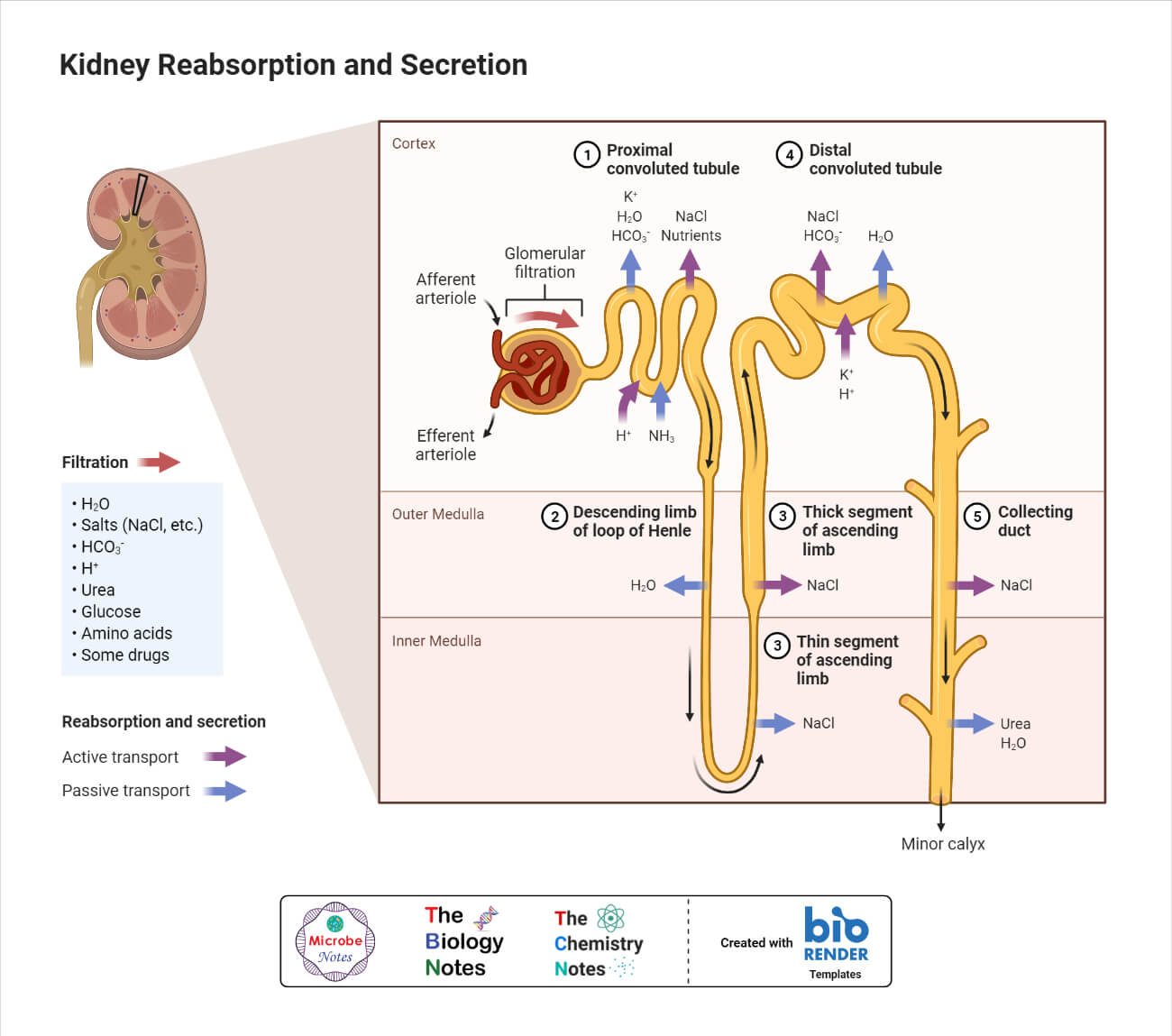
- The filtrate obtained after glomerular filtration is called the nephric filtrate. It contains some valuable substances such as glucose, amino acids, mineral salts, etc., and toxic chemicals such as urea, uric acids, ammonia, and its salts, etc.
- The nephric passes through the lumen of PCT after exiting the Bowman’s Capsule.
- While passing through the PCT, all the valuable substances such as glucose, vitamins, amino acids, hormones, and salts such as sodium, chloride, potassium, and bicarbonates are absorbed from the nephric filtrate into the blood capillaries at various parts of PCT.
- This reabsorption occurs through the process of passive transport and active transport. In active transport, a large amount of ATP is required, provided by the mitochondria present in the epithelial cells of PCT.
- Around eighty percent of the inorganic mineral salts are reabsorbed into the capillaries.
- Besides being useful, toxic chemicals such as urea are also absorbed in the capillaries. The amount of urea reabsorbed is around fifty percent. This process occurs through the diffusion of urea.
- The water in the filtrate is also absorbed back by the process of osmosis. Nearly 80 % of the water in the filtrate is reabsorbed.
| Substance Present in Nephric Filtrate | Amount of reabsorption in PCT | Method of Reabsorption in PCT |
| Glucose | Total amount | Active Transport |
| Vitamins | All | Active Transport |
| Hormones | All | Active Transport |
| Amino acids | Most of the amino acids | Active Transport |
| Sodium ions | 70% | Active Transport |
| Potassium ions | 75% | Active Transport |
| Calcium ions | Larger amount | Active Transport |
| Chlorides | 75% | Passive Diffusion |
| Urea | 50% | Diffusion |
| Water | 80% | Osmosis |
- The further process occurs in the loop of Henle when the filtrate volume reduced in PCT passes through both ascending and descending limbs.
- The wall of descending limb has larger permeability to the water and less to ions of sodium and chloride. A large amount of water is released from this loop to the interstitial space and then moves into the capillaries. The filtrate becomes hypertonic in this due to the excess removal of water.
- The wall of the ascending limb has larger permeability to sodium and chloride ions. In this, the sodium ions are absorbed by active transport and the chlorides by passive transport. These ions move to the interstitial fluid, and the salt concentration rises in the interstitial fluid. To maintain this higher concentration, a large amount of water is drawn from the descending loop. As the salt moves out from the ascending loop, the filtrate becomes hypotonic.
References
- Jones M., Gregory J., McMarty Y. and Jackson N. 2004. Renal Physiology. Biology 2. Cambridge University Press
- Keshari A.K. and Ghimire K.R. 2010. Selective Reabsorption. A textbook of higher secondary biology. Vidyarthi publications. Kathmandu. Pg. 160-161.
- Hall J.E. and Hall M.E. Renal Tubular Reabsorption and Secretion. Guyton and Hall Textbook of Medical Physiology. Pg. 343-363.