pUC plasmids were first developed by Joachim Messing and his co-workers. It is a commonly used cloning vector in the bacteria E. coli.
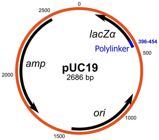
pUC19 is 2686 bp in length. The molecular weight of the pUC19 vector is 1.75×10⁶ Da. It is a small plasmid with a high copy number. It contains the lacz gene and has multiple cloning sites.
Hence, it is widely used as a cloning vector. pUC19 plasmid is similar to pBR322 plasmid in structure.
pUC19 full form
p = plasmid
UC = University of California
19 = numerical designation
Interesting Science Videos
Structure of pUC19
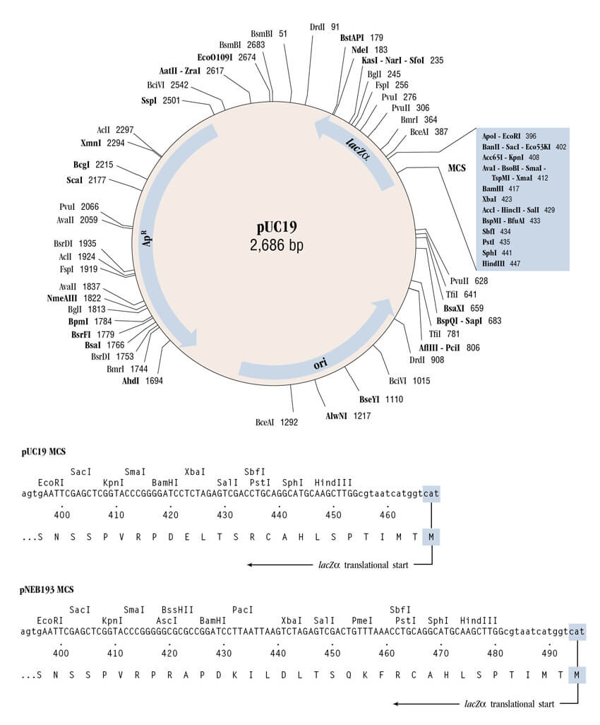
- It is a 2686 bp long plasmid.
- Origin of replication – The origin of replication of the pUC19 plasmid is derived from pMB1.
- Multiple Cloning Sites – There is a short sequence of 2.8 kb which contains sites for various restriction enzymes. This increases the number of potential restriction sites available, enabling the production of the desired fragment for cloning.
- Selectable markers – The pUC19 plasmid contains an Ampicillin resistance gene which can be used to screen the recombinants. The plasmid also contains the E. coli gene lacZ, which encodes for β-galactosidase (β-galactosidase hydrolyses lactose).
- Restriction sites – The pUC19 vector carries a 54 bp long multiple cloning site poly-linker containing 13 different hexanucleotide-specific restriction endonucleases sites.
Some of the restriction sites are EcoR1, HindIII, BamH1, and many more.
Screening of pUC19 vector
The Blue-White screening method is used for pUC19 vectors. The process of screening is as follows:
- This method of screening is based on the fact that the blue pigment is formed when β-galactosidase catalyzes the hydrolysis of a synthetic substance known as X-gal (5-bromo-4-chloro-3-indoyl-β-D-galactopyranoside) in the medium.
- When X-gal is hydrolyzed, it forms galactose and 5-bromo-4-chloro-3-hydroxy indole.
- The later product undergoes dimerization (spontaneous) and oxidation.
- As a result of dimerization and oxidation, a blue pigment is formed.
- The cells which contain the β-galactosidase activity form blue colonies, whereas the cells which do not show β-galactosidase activity form white colonies on the agar medium containing X-gal.
- The recombinant cells, which contain newly inserted DNA fragments, lack the β-galactosidase activity and hence appear white on the agar plates.
- This method of screening the recombinant cells is the easiest and fastest method.
Advantages
- This is a small cloning vector and has large industrial applications.
- It has one step selection process for the recombinants, hence is used on a large scale.
- It has a high copy number.
- The presence of many restriction sites makes it suitable for cloning.
References
- https://www.snapgene.com/resources/plasmid-files/?set=image_consortium_plasmids&plasmid=pUC19
- PRINCIPLES OF GENE MANUPULATION AND GENOMICS BY PRIMROSE AND TWYMAN
- https://enzyquest.com/product/puc19-dna-plasmid/
- Julin, D.A. (2018). Blue/White Selection. In: Wells, R.D., Bond, J.S., Klinman, J., Masters, B.S.S. (eds) Molecular Life Sciences. Springer, New York, NY. https://doi.org/10.1007/978-1-4614-1531-2_94