What is Plasmodium vivax?
Plasmodium vivax is the most common of the human infecting malaria fever parasites. It is an intracellular parasite in man. It lives in the red blood corpuscles and liver cells in man. While extracellular in mosquitoes, living in its alimentary canal and salivary glands. The life cycle of P. vivax is digenetic i.e. it involves 2 hosts man and mosquito. Female Anopheles mosquito is the Primary host or definitive host. The sexual cycle is completed in it. It involves two phases, gametogony, and sporogony. A human is a secondary or intermediate host. The asexual phase of the life cycle is completed in man by schizogony.
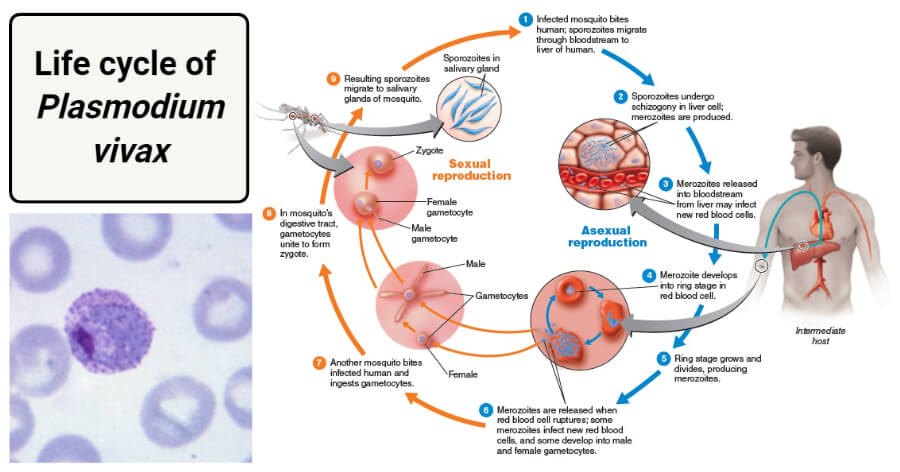
Image Source: Wikipedia and Quizlet.
The life cycle of Plasmodium vivax is divided into:
- Asexual life cycle or schizogony in man
- Sexual life cycle or sporogony in female Anopheles mosquito
1. Asexual cycle or Schizogony in man
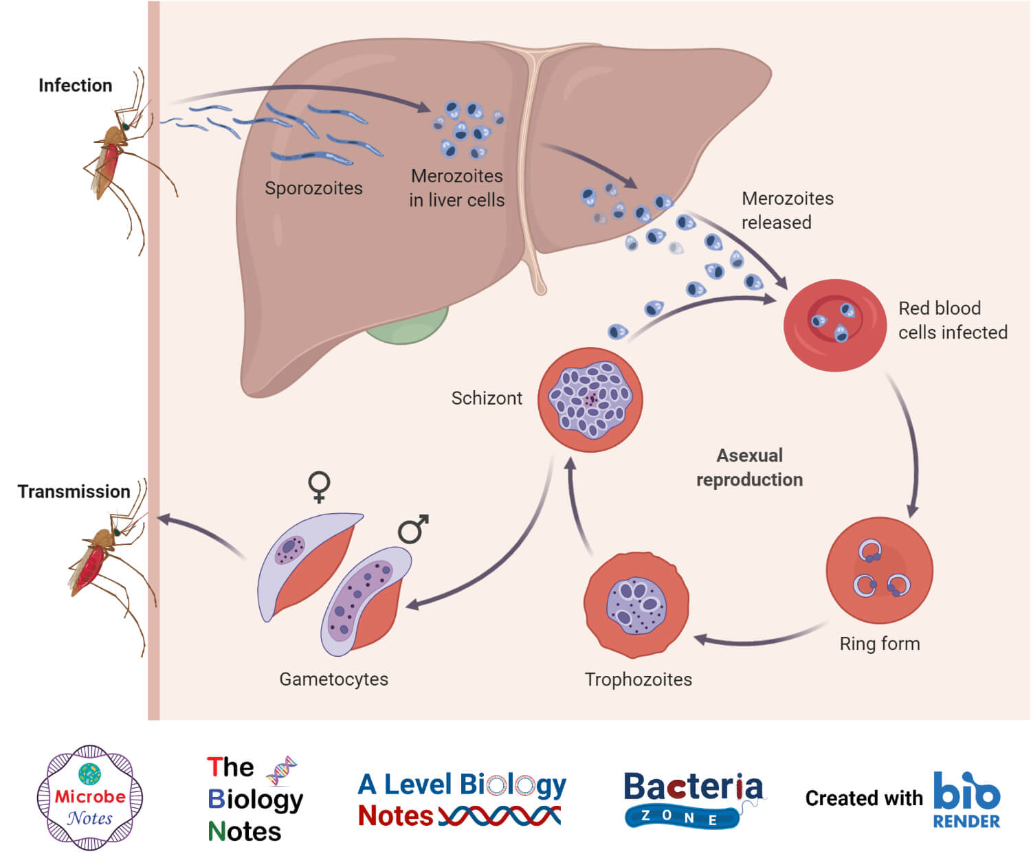
a. Infection
- Schizogony is the process of asexual reproduction by which Plasmodium undergoes asexual multiplication in liver cells and RBCs of man.
- It occurs in the human liver cells (liver schizogony) and in RBC (erythrocytic schizogony).
- A healthy person acquires the infection when an infected female Anopheles, containing infective stage of parasites (sporozoites) in its salivary glands bites him for sucking blood.
- The mosquito punctures the host’s skin with proboscis and introduced some saliva in the bloodstream first.
- Along with saliva, it inoculates thousands of sporozoites into the bloodstream.
b. Sporozoites
- These are infective forms of parasites.
- It is minutes measures about 11 to 12 µ in length and 0.5-1 µ in width.
- It is a spindle-shaped, slightly curved or sickle-shaped, and uninucleated organism.
- The electron microscopy of the sporozoites has shown that they are covered externally by an elastic, firm pellicle having longitudinally arranged contractile microtubules.
- The microtubules help in their wriggling movements.
- Its anterior end is the apical cap, made of 3 or more concentric rings, into which opens the secretory paired organelles.
- These secretory organelles are supposed to secrete some secretion which helps its penetration into the liver cell.
- It contains a single and vesicular nucleus having a nucleolus in its center.
- It contains a single mitochondrion with tubular cristae.
c. Liver schizogony
- The sporozoites are capable of slight gliding movements.
- In about a half hour sporozoites disappear from bloodstreams and enter the parenchymatous cells of the liver where they multiply asexually by schizogony.
- Inside the liver and RBC different forms of sporozoite cause infection.
- Liver schizogony has 2 phases, pre-erythrocytic and exo-erythrocytic.
i) Pre-erythrocytic phase
- In the liver cells, Sporozoites grow in size and become large and spherical in shape called schizonts.
- The nucleus of schizont multiply asexually (multiple fission) and forms thousands of merozoites.
- The schizonts ruptures and merozoites are liberated into the sinusoids or venous passage of the liver in the form of Cryptozoites or cryptomerozoites.
- These cryptozoites are immune to medicines and the resistance of the host.
- It is completed in 8-10 days.
- The process of formation of many cryptozoites from single sporozoites in the liver cells is called pre-erythrocytic schizogony.
- During this stage, the blood remains sterile and inoculation does not produce infection.
ii) Exo-erythrocytic phase
- The second phase of asexual multiplication known as the exo-erythrocytic phase in which cryptomerozoites enter fresh liver cells and grow into schizonts, the schizonts divide to form merozoites, and merozoites of 2nd generation are termed as metacryptozoites or phanerozoites.
- The same process is repeated several times in liver cells to form a reservoir of merozoites and each time new liver cells are infected.
- It has been reported that metacryptozoites which are smaller in size and numerous called micro metacryptozoites and some are larger and less numerous in size called macro metacryptozoites.
- The micro metacryptozoites enter the red blood cells to start the erythrocytic phase while the macro metacryptozoites infect the fresh liver cells to continue the exo-erythrocytic phase.
- The merozoites attack only the young and immature corpuscles (the merozoites of P. malariae attack only old corpuscles, while P. falciparum attacks all kinds of corpuscles indiscriminately).
- The exo-erythrocytic phase of parasites remain immune to the resistance of the host and parasites are not susceptible to the action of any kind of anti-malarial drug.
iii) pre-patent and incubation periods
- The duration between initial sporozoite infection and 1st appearance of parasites in blood is termed as pre-patent period.
- It is for about 8 days in P. vivax.
- The period between infection and appearance of 1st malarial symptoms is the incubation period which is about 10-17 days (average 14days) in P. vivax.
d. Erythrocytic schizogony
- 3rd multiplication phase of schizogony occurs in the erythrocytes known as erythrocytic schizogony.
- This cycle starts when the micro metacryptozoites enter into erythrocytes.
- Single metacryptozoites enter into single RBC and pass through the trophozoite stage, signet ring stage, amoeboid stage, and schizont stage.
- When metacryptozoites invade the RBC, it becomes rounded with a large nucleus and grows in size by ingesting hemoglobin of corpuscles. This stage of the parasite is called the trophozoite stage.
- As the trophozoites grow in a size, a large non-contractile vacuole appears which pushes the nucleus towards the periphery and forms a ring-like structure known as the signet ring stage.
- The signet ring stage is 1/3 to ½ the size of an erythrocyte.
- The signet ring trophozoites ingest a large portion of the cytoplasm of RBC forming a food vacuole into which it secretes digestive enzymes.
- The enzyme brings about the proteolysis of blood hemoglobin, which breaks down the protein component into hematin.
- Protein is used as food by the trophozoite, while hematin is deposited in the form of hemozoin (toxic malarial pigment).
- Trophozoites enlarge and vacuole starts disappearing and develops pseudopodial processes in the cytoplasm and changed into the amoeboid stage. This stage is called the amoeboid stage.
- The amoeboid trophozoites after feeding becomes rounded grow in size and become erythrocytic schizont.
- Asexual multiplication takes place in schizont to form 12 to 24 oval-shaped merozoites this phase is known as erythrocytic schizogony.
- The much-weakened erythrocyte burst and merozoites are liberated into the plasma in the form of erythrocytic merozoites.
- The merozoites enter new erythrocytes, then they repeat the erythrocytic schizogony once every 48 hours.
- The merozoites are arranged towards the periphery due to the presence of hemozoin at the center. The arrangement is just like the arrangement of petals in rose flowers. So, this stage is called the rosette stage.
- Numerous yellowish eosinophilic granules appear in the cytoplasm of the host corpuscles which are called schuffner’s granules. These dots are believed to be the antigen excreted by the parasites.
e. Post-erythrocytic schizogony
- Sometimes, some merozoites which are produced in erythrocytic schizogony reach the liver cell and undergo schizogonic development in liver cells. This is known as pre-erythrocytic schizogony.
f. Formation of gametocytes
- After many generations of schizogony in the blood, some of the merozoites invade the new RBC and do not change into schizonts but they grow and transformed into 2 types of gametocytes called macrogametocytes and microgametocytes.
- Gametocytes appear in the peripheral blood at various intervals after the onset of fever, they remain inactive while in the human blood.
- Macrogametocytes or female gametocytes are large (10-12µ) and numerous in number. They have a small compact peripheral nucleus. They have reserved food materials and the cytoplasm is dark in color.
- Microgametocytes or male gametocytes are smaller (9-10 µ) motile and few in number. They have large centrally placed nuclei. They lack reserved food and stains faintly hence the cytoplasm is light in color and clear.
- Both of the gametocytes contain a large amount of hemozoin; they enlarge the erythrocytes.
- The gametocytes do not divide but remain in human blood corpuscles for several weeks.
- It is necessary for them to be taken into the body of Anopheles for further development, if this does not happen, they will either degenerate or die.
2. Life cycle in mosquito or Sexual Cycle in mosquito
a. Ingestion by mosquito
- When female Anopheles mosquito sucks the blood of the infected persons, containing the gametocytes and other stages of the erythrocytic cycle (e.g. erythrocytic merozoite). They reach the stomach where all the stages along with RBCs are digested except gametocytes. Now, the life cycle is continued towards the completion by the following processes.
b. Gametogenesis/ Gametogony (Formation of gametes)
- The process of the formation of gametes from the gametocytes is called gametogenesis or gametogony.
- Like gametocytes, the gametes are also of 2 types i.e. microgametes and macro-gametes.
- Microgametocytes undergo the ex-flagellation process in the mid-gut of the mosquito.
- The cold-bloodness of the mosquito is said to stimulate the process of ex-flagellation.
- The nucleus of microgametocytes divides to form 6-8 daughter nuclei, the first division is meiotic.
- These nuclei move to the periphery along with the cytoplasm, forming flagella like structure. Thus 6-8 flagella-like male gametes are formed from each microgametocyte. The elongated structure is called microgametes or sperms.
- These microgametes measures about 20-25 microns in length.
- The movement of flagella causes the gametes to separate and move actively in the stomach of the mosquito in search of female gametes.
- Macrogametocyte undergoes a maturation process, thereby 2 polar bodies are pushed out to become female gametes or macrogametes or megagametes.
- The female gamete is non-motile and develops a cytoplasmic projection called the cone of reception or fertilization cone on one side.
c. Fertilization
- The nucleus of the female gamete comes to lie near its receptive cone.
- If microgametes happen to reach the macrogametes, then it enters into the female gamete at the point of the cytoplasmic cone and finally completes the fusion of nucleus and cytoplasm of 2 gametes occurs, resulting in the formation of a diploid zygote or synkaryon.
- The fertilization takes place known as syngamy.
- Syngamy is anisogamous as the uniting male and female gametes are dissimilar.
- Zygotes form in the stomach of mosquitoes about 9 to 10 days after the blood meal.
d. Ookinete
- For some period of time, about 24 hours zygote remains rounded and motionless but soon it becomes elongated to become worm-like having pointed ends and motile.
- The zygotes are now called ookinetes or vermicules.
- It measures about 15-22 µ in length and 3 µ in width.
- It penetrates the wall of the stomach with the help of lytic secretion. It settles into the inner portion of the stomach wall.
- Electron microscopy of ookinetes has shown the presence of a central irregular nucleus, dense cytoplasm, brown pigment granules, many mitochondria, and ribosomes.
- This suggests that very rapid synthesis of protein takes place within this stage of the parasite.
- The ookinetes are motile due to the presence of ectoplasmic contractile fibrils.
e. Encystment
- Ookinetes penetrate the wall of the midgut to settle down just under the thin membranes that separate the midgut from the hemocoel.
- The ookinete then changes into a spherical shape, takes nutrition from the wall of the stomach, and gets enclosed in a thin, elastic, and permeable cyst wall, such stage is called the oocyst stage.
- The oocyst grows in size and sometimes called sporont.
- The cyst wall is secreted partly by ookinete and partly derived from the stomach tissue of the mosquito. Many oocysts (<500) are seen on the stomach wall of an infected mosquito. The ookinetes fail to penetrate the stomach wall pass out from the mosquito’s body with fecal matter.
f. Sporogony
- Each oocyst enters a phase of asexual multiplication know as sporogony.
- It is the process of the formation of sporozoites from the zygote nucleus by asexual multiple fission.
- Oocysts mature and develop. The nucleus of oocyst divides first by meiosis and then by mitosis, forming a large number of haploid nuclei (2-3 days) and forms sporozoites forming cells known as sporoblasts.
- The nuclei of sporoblast again multiply and the cytoplasm gets constricted around them.
- Thus, the resultant structures in the sporoblasts elongate to form slender or sickle-shaped sporozoites
- Each oocyst may have 10 thousand sporozoites, and a group of sporozoites gets arranged around the vacuoles.
- The process of formation of sporozoites is known as sporogony which is completed in 10-20 days from the time the gametocytes are taken by the mosquito, the time depending on the temperature.
- Therefore, each oocyst fills with numerous sporozoites. Now, these give pressure to the oocyst, and due to which the oocyst burst or rupture, and thousands of sporozoites are released in the body cavity (hemocoel) of the mosquito.
- The sporozoites are very active and motile, then they reach the salivary glands of the mosquito and enter the duct of the hypopharynx.
- Then the sporozoites are ready to infect the healthy person when the mosquito bites each bite. And the life cycle is repeated again.
References and Sources
- 21% – https://microbiologynotes.com/life-cycle-of-plasmodium-vivax/
- 4% – https://www.biologydiscussion.com/invertebrate-zoology/protozoa/plasmodium-vivax-p-vivax-habitat-structure-and-life-cycle/28254
- 1% – https://www.shareyouressays.com/knowledge/biology-question-bank-57-mcqs-on-biological-classification-answered/114624
- 1% – https://www.modernagespirituality.com/what-is-life-cycle-and-how-to-break-the-life-cycle/
- 1% – https://fremonthypnosiscenter.com/site/article.php?id=01339e-life-cycle-of-plasmodium-notes-pdf
- <1% – https://www.youtube.com/watch?v=Dt9VEbxUAKQ
- <1% – https://www.sciencedirect.com/science/article/pii/S1369527409000794
- <1% – https://www.notesonzoology.com/protozoa/plasmodium/life-cycle-of-plasmodium-protozoa-microorganisms-zoology/9172
- <1% – https://www.biologydiscussion.com/animals-2/phylum-protozoa/plasmodium-host-habitat-and-life-history/32509
- <1% – https://quizlet.com/236408551/fluid-electrolyte-acid-base-flash-cards/
- <1% – https://pediaa.com/what-is-the-difference-between-endoplasm-and-ectoplasm/
- <1% – https://en.wikipedia.org/wiki/Zygote
- <1% – https://en.wikipedia.org/wiki/Malaria
- <1% – https://edurev.in/course/quiz/attempt/-1_Test-Plasmodium-Lower-Animal/f0d4deef-e9bd-4bc1-bff7-61903b2d76e7
- <1% – https://ccforum.biomedcentral.com/articles/10.1186/cc2183
- <1% – https://biologydictionary.net/fertilization/
- <1% – http://www.vdci.net/mosquito-biology-101-life-cycle
This website is very beneficial for students.I feel it quite good🙃♥️