- The primary cell and middle lamella never occur in the form of a continuous layer, but many minute apertures through the cells of a tissue maintain cytoplasmic relation with each other. Such cytoplasmic junctions or bridges between the adjacent cells are known as plasmodesmata.
- Plasmodesmata are found only in plant cells and algal cells. They are intracellular organelles. In animal cells, similar structures are presently called gap junctions.
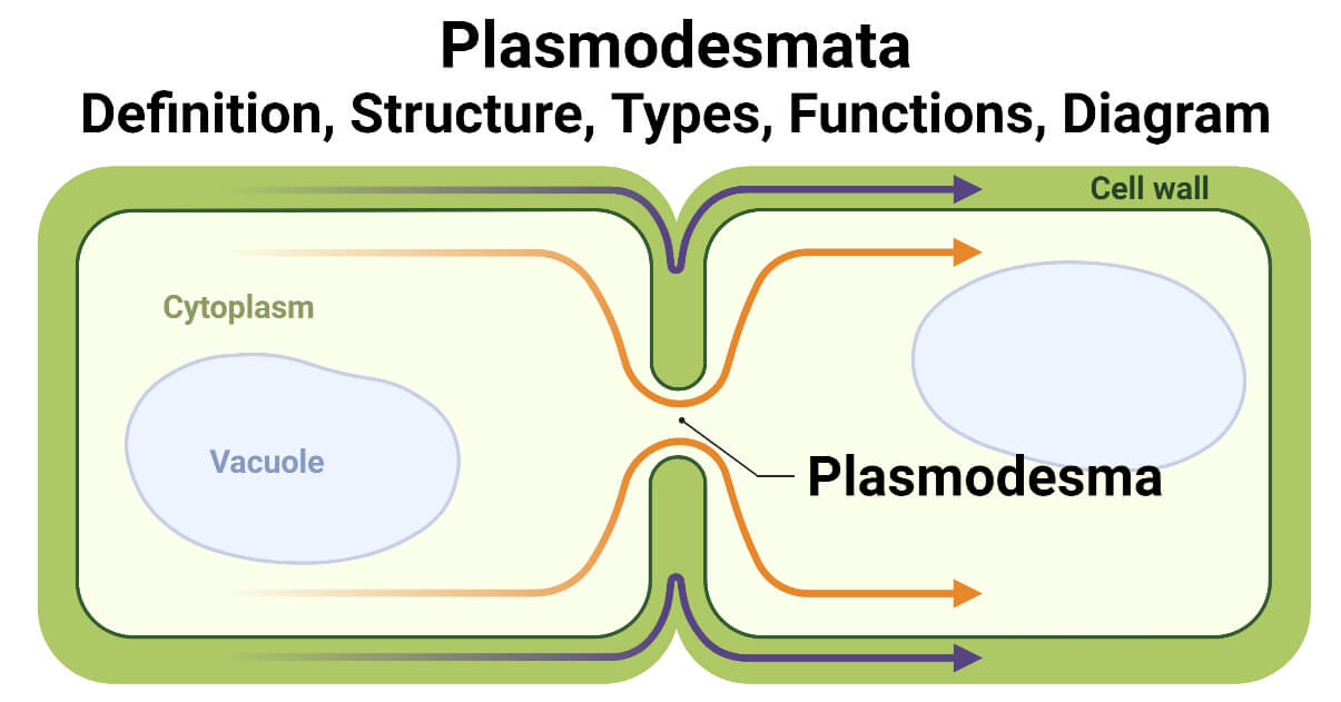
- In a higher plant, every living cell is linked to its living neighbor’s cell by fine cytoplasmic channels. This is called a plasmodesma.
- Plasmodesmata permit to pass a molecule directly from one cell to another and are important in cellular communication.
- The plasmodesmata (singular, plasmodesma) were first reported by Strasburger in 1901 A.D.
- The word plasmodesma derives from the Latin word ‘plasmo’ meaning fluid and the Greek ‘desma’ meaning bond.
- They are essential for plant life because they serve as a channel for conveying water, fluid, protein, small RNAs hormone, and transport of metabolites during developmental and defense signaling.
- They permit the passage of molecules weighing less than 800 daltons.
- Transport through the plasmodesmata is also found under complex regulation which may involve Ca2+ and protein phosphorylation.
- The number of plasmodesmata may vary from one place to another.
- For example, in the wall of a column of cells, the number of plasmodesmata may be 15 or greater per square micrometer of the wall surface.
- In the other cell walls, plasmodesmata are fewer than 1 per square micrometer.
- 103 to 105 plasmodesmata are present in a typical cell which equates to 1-10 per µm2.
- They are easily seen in the endosperm of seeds such as phoenix, diospyros, Aesculus, and cotyledons of some cells.
- Callose, a β-1,3-glucan polymer, appears to serve as both a structural and a regulatory element of plasmodesmata.
Interesting Science Videos
Plasmodesmata Structure
- Different structures of plasmodesmata are identified, which range from simple (characterized by a single sheath) to complex (characterized by branched), H-shaped, and twinned structures.
- Usually, young tissue has simple plasmodesmata and complex plasmodesmata developing later, after cell expansion.
- A plasmodesma measures about 20 to 40 nm in diameter and it is a roughly cylindrical membrane-lined channel.
- They are assembled in three main compartments:
- The plasma membrane
- The cytoplasmic sleeve
- Desmotubule
- Plasmodesmata have their plasma membrane or plasmalemma which is the extension from the membrane of the cell. Its structure is similar to having the phospholipid bilayer.
- A fluid-filled space surrounded by the cell membrane (plasmalemma) is called a cytoplasmic sleeve and is a continuous extension of the cytosol.
- Myosin-like proteins and actin filament are localized within the cytoplasmic sleeve, and proteinaceous spike-like projections that are regularly positioned within the cytoplasmic sleeve are thought to create nanochannels of varying size.
- The cytoplasmic sleeve helps to traffic the ions and molecules through plasmodesmata.
- Through the diffusion, smaller molecules and amino acids can pass through it.
- The desmotubule runs from cell to cell through the center of plasmodesmata in most cases.
- The desmotubule is a dense rod or narrower cylindrical structure, that is connected to the smooth endoplasmic reticulum of adjacent cells.
- Desmotubules are derived from the smooth endoplasmic reticulum of the connected cells.
- Now it is called desmotubules but initially, it was called the axial component.
- The annulus of the cytosol is present between the outside and inside of the desmotubule and cylindrical plasma membrane respectively.
- At each end of plasmodesmata, it appears to be constructed.
- The space between desmotubules and plasma membrane contains 8-10 microchannels.
- Electron microscopic images of plasmodesmata are, that the plasma membrane shows up as a tripartite structure that is 7.2nm wide, and the dense central rod is 1.4nm in radius.
- Width of the pale ring: 2.2 nm. It surrounds the dense central rod.
Types of Plasmodesmata
Two types of plasmodesmata are formed, it is defined by their origin.
Primary plasmodesmata
- Primary plasmodesmata are those plasmodesmata that are formed during cell division.
- Chara zelanica produces both primary and secondary plasmodesmata.
Secondary plasmodesmata
- They are developed totally de novo in the existing cell wall. Hormone cytokinin has been shown to increase secondary plasmodesmata.
- Chara corallina produces only secondary plasmodesmata. Both primary and secondary plasmodesmata are the initial structure that is simple but can form complex structures by forming branch and/or fusion of exiting plasmodesmata or the fusion of established and newly formed plasmodesmata.
Plasmodesmata Functions
- Usually, plant cells have tough, rigid cell walls. Due to its nature, larger molecules could not pass easily to the cell wall of the plant The plasmodesmata present in the plant cells facilitate the entry of these entities inside of the cell wall.
- Communication between one cell to another cell is predominant for plant growth and plant survival, plasmodesmata play an important role in both cellular communication and molecular translocation.
- Passive and active pores are present in plasmodesmata. Nutrients and water pass from the passive pores.
- Actin structure which is present in plasmodesmata helps to move transcription factors like messenger RNA, viroids, short interfering RNA, and plant viruses.
- Plasmodesmata located protein 5(PDLP5), which is discovered by a researcher, can produce salicylic acid. It enhances defenses against plant pathogenic attacks. It also protects from pathogenic bacteria.
- The cells present in the phloem also use plasmodesmata.
- Plasmodesmata are involved in the short-distance movement of viruses.
References
- Verma, P. S., & Agrawal, V. K. (2006). Cell Biology, Genetics, Molecular Biology, Evolution & Ecology (First edition.). S Chand and Company Ltd.
- Alberts, B. (2004). Essential cell biology. New York, NY: Garland Science Pub.
- Wayne, R. ((2009). Plant Cell Biology, from Astronomy to Zoology. Academic Press in an imprint of Elsevier.
- Keshari, A.K(2020). A textbook of higher secondary biology. 13th edition. Vidyarthi pustak bhandar, Kathmandu, Nepal.
- Maule, Andrew (December 2008). “Plasmodesmata: structure, function and biogenesis”. Current Opinion in Plant Biology. 11 (6): 680–686.
- https://journals.biologists.com/jcs/article/131/11/jcs209346/56923/Plasmodesmata-at-a-glance
- https://www.vedantu.com/biology/plasmodesmata
- https://www.sciencedirect.com/science/article/pii/S0960982210003738
- https://www.thoughtco.com/plasmodesmata-the-bridge-to-somewhere-419216