Pinocytosis is a type of endocytosis in which small particles suspended in the extracellular fluid are moved into the cell through pores formed on the cell membrane.
- The term pinocytosis is formed of two words “pino” and cytosis” where ‘pino’ means “to drink” while ‘cytosis’ means relating to the cell.
- It is a continuous process in most cells and is a non-specific way fo internalizing fluid and dissolved nutrients.
- The process of pinocytosis deals with the movement of a large number of tiny molecules through a fluid, which is why it is also called the fluid endocytosis or bulk-phase endocytosis.
- The molecules once inside the cells form vesicles which are then fused with the endosomes for the metabolic processes.
Interesting Science Videos
Process/ Steps of Pinocytosis
- The process of pinocytosis is initiated by the presence of the desired molecule in the extracellular fluid.
- The molecules, which acts as an inducer at this point, binds to the cell membrane triggering the initiation of the pinocytosis process.
- The molecules can be proteins, sugar, ions, or other molecules. In humans, pinocytosis is mostly associated with the absorption of fat.
- Although pinocytosis involves the binding of molecules to receptors in the cell membrane, it is different from other receptor-mediated endocytosis processes as the receptor is not specific for one molecule.
The following is the generalized description of the steps involved in pinocytosis:
- The process of pinocytosis begins with initiation. The molecules bind to the receptor on the cell membrane sending signals to the cell membrane for the further process.
- The cell membrane then creates small open-ended pockets or folds around the extracellular fluid to be absorbed. The molecules in the extracellular fluid are also captured into the pockets.
- The cell membrane begins to connect at the open end of the pocket to form a complete invagination.
- The pocket part of the cell membrane then pinches off of the cell membrane, which is now called a vesicle. The vesicle is an organelle originating from the cell membrane that surrounds the fluid containing the desired molecules. The vesicles allow the movement of movement through the cytoplasm without disturbing the normal functioning of the cell.
- Depending on the purpose of the molecules, the vesicles either fuse with the endosome or move towards the other end of the cell membrane for exocytosis.
- In the first cases, the vesicles fuse with the endosomes in the cells in order to hydrolyze the particles into smaller molecules so that they can be utilized by the cell.
- Alternatively, the vesicles might simply move towards the other end of the cell membrane to remove the extracellular fluid out of the cell by exocytosis. This process is essential to maintain the size of the cell as membrane materials are returned back to the cell membrane.
- In some cases, the molecules might be dumped into the cytoplasm immediately after the vesicle formation.
Types of Pinocytosis
- Pinocytosis is divided into categories on the basis of the size of the molecules to be transferred or on the basis of the mechanism of vesicle formation.
- On the basis of the size of the molecules, pinocytosis is of two types:
Macropinocytosis
- In macropinocytosis, the molecules to be moved are rather large in size.
- The size of the vesicles formed might be about 1-2 µm in length.
- In macropinocytosis, large invaginations or pockets are formed for the entry of the molecules.
- Besides, ruffles are formed in the cell membrane during macropinocytosis. These ruffles are generated as cytoskeleton of the cell rearranges the actin filaments in the cell membrane.
- The vesicles formed in this process are termed macropinosomes that mature in the cytoplasm and either fuse with the lysosomes or migrate towards the cell membrane for recycling.
- This process is common with white blood cells like macrophages and dendritic cells
Micropinocytosis
- In micropinocytosis, the molecules to be moved are smaller in size.
- The size of the vesicles formed is about 0.1 µm in length.
- For the entry of the molecules, tiny indentations are formed on the cell membrane.
- Caveolin-mediated pinocytosis is a common example of micropinocytosis that are formed in the epithelium of the blood vessels.
On the basis of the receptor involved in the process and the mechanism of vesicle formation, pinocytosis is divided into :
- Clathrin-mediated pinocytosis
- Caveolin-mediated pinocytosis
- Clathrin- and caveolin- independent pinocytosis
Functions of Pinocytosis
- Pinocytosis is a form of active transport that plays a vital role in cellular processes like nutrients uptake, excretion of waste materials, and signal transduction.
- Pinocytosis is utilized by various unicellular organisms for the uptake of nutrients such as certain sugars, most amino acids, organic acids, and many inorganic ions.
- In higher organisms, pinocytosis plays an important role for the bulk transport of dissolved molecules like fats and vitamins.
- Because pinocytosis is a non-specific absorption of molecules, it allows the transport of a large number of different molecules at the same time.
- Pinocytosis is also involved in the removal of waste materials out of the cells like in the cells of the kidneys removing water and waste products into the urine.
- Pinocytosis is employed by cells of the immune system like the macrophages and dendritic cells as a means of testing the extracellular fluid for the presence of antigens.
Examples of Pinocytosis
- An example of pinocytosis is observed in the microvilli of the small intestine to absorb nutrients from the lumen of the gastrointestinal tract.
- Similarly, it is also observed in cells in the ducts of the kidneys during the formation of urine.
- Besides, the human egg in the female reproductive system uses pinocytosis to absorb nutrients prior to fertilization.
- Pinocytosis is seen in unicellular organisms like an amoeba for the uptake of water and dissolved nutrients.
- Pinocytosis is also seen in most of the cells in the body to recycle the components of the cell membrane and maintain the size of the cell.
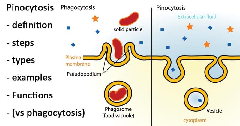
Image Source: Wikipedia (Mariana Ruiz Villarreal).
Pinocytosis vs Phagocytosis
Basis of comparison |
Phagocytosis |
Pinocytosis |
| Definition | Phagocytosis is a type of endocytosis involved in the transport of particles sized >0.5 µm. | Pinocytosis is a type of endocytosis involved in the transport of particles sized 0.5 µm or less. |
| Process of intake | Phagocytosis involves the formation of pseudopodia (false feet) for the intake of molecules. | Pinocytosis intakes the particles via invagination and formation of pockets in the cell membrane. |
| Nature of particles ingested | Phagocytosis intakes particles which are solid and larger in size. | Pinocytosis intakes particles that are dissolved in liquid and more comparatively smaller in size. |
| Specificity | Phagocytosis is specific and only moves a particular molecule at a time. | Pinocytosis is not molecule-specific and moves a large number of molecules at one time. |
| ATP utilization | Phagocytosis utilizes or requires more ATP and in turn, more amount of energy. | Pinocytosis requires a comparatively lesser number of ATPs. |
| Vesicles formed | Vesicles formed by phagocytosis are termed phagosomes. | Vesicles formed by pinocytosis are termed pinosomes. |
| Purpose | Phagocytosis is mostly employed by immune cells for the defensive purpose of engulfing the foreign invaders. | Pinocytosis is mostly employed for the intake of nutrients, |
| Lysosome involvement | Phagosomes formed fuse with lysosomes for the formation of food vacuole. | The pinosomes do not fuse with lysosomes. |
| Types of particles ingested | Phagocytosis engulfs bacteria, viruses, and other foreign invaders. | Pinocytosis intakes nutrients like sugar, amino acids, vitamins, and ions. |
| Site of the process | Phagocytosis takes place in immune cells like macrophages and neutrophils. | Pinocytosis takes place in almost all cells, including the secretory cells and epithelial cells of the blood vessels. |
References
- Friedman, M. (2008). Principles and models of biological transport. Springer.
- https://biodifferences.com/difference-between-pinocytosis-and-phagocytosis.html
Sources
- 1% – https://www.thoughtco.com/pinocytosis-definition-4143229
- 1% – https://www.sciencedirect.com/topics/pharmacology-toxicology-and-pharmaceutical-science/pinocytosis
- 1% – https://www.answers.com/Q/Which_transport_process_does_the_cell_use_to_direct_the_contents_of_vesicles_out_the_cell_membrane
- 1% – https://quizlet.com/98649618/chapter-7-flash-cards/
- 1% – https://quizlet.com/94006737/chapter-3-flash-cards/
- 1% – https://openstax.org/books/anatomy-and-physiology/pages/3-1-the-cell-membrane
- 1% – https://en.wikipedia.org/wiki/Dendritic_cell
- 1% – https://brainly.com/question/16598655
- 1% – https://biologydictionary.net/pinocytosis/
- <1% – https://www.thoughtco.com/what-is-endocytosis-4163670
- <1% – https://www.quora.com/Which-system-is-an-action-for-removal-of-the-waste-material-produced-in-human-body
- <1% – https://quizlet.com/7810575/biology-cells-flash-cards/
- <1% – https://courses.lumenlearning.com/wmopen-biology1/chapter/endocytosis-and-exocytosis/
- <1% – https://chem.libretexts.org/Bookshelves/General_Chemistry/Map%3A_Chemistry_-_The_Central_Science_(Brown_et_al.)/11%3A_Liquids_and_Intermolecular_Forces/11.1%3A_A_Molecular_Comparison_of_Gases%2C_Liquids%2C_and_Solids
- <1% – http://www.differencebetween.net/science/difference-between-pinocytosis-and-receptor-mediated-endocytosis/