Interesting Science Videos
Porifera Definition
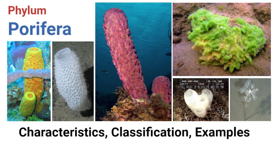
The Porifera may be defined as an asymmetrical or radially symmetrical multicellular organism with a cellular grade of an organization without well- definite tissues and organs; exclusively aquatic; mostly marine, sedentary, solitary or conical animals with body perforated by pores, canals, and cambers through which water flows; with one or more internal cavities lined with choanocytes; and with a characteristic skeleton made of calcareous spicules, siliceous spicules or horny fibers of spongin.
Phylum Porifera Characteristics
- Porifera are all aquatic, mostly marine except one family Spongillidae which lives in freshwater.
- They are sessile and sedentary and grow like plants.
- The body shape is vase or cylinder-like, asymmetrical, or radially symmetrical.
- The body surface is perforated by numerous pores, the Ostia through which water enters the body and one or more large openings, the oscula by which the water exists.
- The multicellular organism with the cellular level of body organization. No distinct tissues or organs.
- They consist of outer ectoderm and inner endoderm with an intermediate layer of mesenchyme, therefore, diploblastic
- The interior space of the body is either hollow or permeated by numerous canals lined with choanocytes. The interior space of the sponge body is called spongocoel.
- Characteristic skeleton consisting of either fine flexible spongin fibers, siliceous spicules, or calcareous spicules.
- Mouth absent, digestion intracellular.
- Excretory and respiratory organs are absent.
- Contractile vacuoles are present in some freshwater forms.
- The nervous and sensory cells are probably not differentiated.
- The primitive nervous system of neurons arranged in a definite network of bipolar or multipolar cells in some, but is of doubtful status.
- The sponges are monoecious.
- Reproduction occurs by both sexual and asexual methods.
- Asexual reproduction occurs by buds and gemmules.
- The sponge possesses a high power of regeneration.
- Sexual reproduction occurs via ova and sperms.
- All sponges are hermaphrodite.
- Fertilization is internal but cross-fertilization can occur.
- Cleavage holoblastic.
- Development is indirect through a free-swimming ciliated larva called amphiblastula or parenchymula.
- The organization of sponges are grouped into three types which are ascon type, sycon type, and leuconoid type, due to simple and complex forms.
- Examples: Clathrina, Sycon, Grantia, Euplectella, Hyalonema, Oscarella, Plakina, Thenea, Cliona, Halichondria, Cladorhiza, Spongilla, Euspondia, etc.

Image Source: Wikipedia.
Phylum Porifera Classification
The phylum includes about 5,000 species of sponges, grouped into 3 classes depending mainly upon the types of skeleton found in them. The classification here is based on Storer and Usinger (1971) which appears to be a modification from Hyman’s classification.
Class 1. Calcarea (L., calx=lime) or Calcispongiae (L., calcis= lime+ spongia= sponge)
- Small-sized calcareous sponges, below 10 cm in height.
- Solitary or conical; body shape vase-like or cylindrical.
- They may show asconoid, Syconoid, or leuconoid structures.
- A skeleton of separate one or three or four-rayed calcareous spicules.
- Exclusively marine.
Order 1. Homocoela (=Asconosa)
- Asconoid sponges with cylindrical and radially symmetrical bodies.
- Body wall thin, not folded. Choanocytes line the Spongocoel.
- Often conical.
- Examples: Leucosolenia, Clathrina.
Order 2. Heterocoela (=Syconosa)
- Syconoid and leuconoid sponges having a vase-like body.
- The body wall is thick, folded. Choanocytes line the flagellated chambers (radial canals) only.
- Spongocoel is a line by flattened endoderm cells.
- Solitary or conical
- Examples: Sycon or Scypha, Grantia.
Class 2. Hexactinellida (Gr., hex=six + actin=ray) or Hyalospongiae (Gr., hyalos=glass+ spongos= sponge)
- Moderate -sized. Some reach 1 meter in length.
- Called glass sponges.
- Body shape cup, urn, or vase-like.
- Skeleton is of siliceous spicules which are triaxon with 6 rays. In some, the spicules are fused to form a lattice-like skeleton.
- No epidermal epithelium.
- Choanocytes line finger-shaped chambers.
- Cylindrical or funnel-shaped
- Found in deep tropical seas.
Order 1. Hexasterophora
- Spicules are hexasters i.e. star-like in shape with axes branching into rays at their ends.
- Flagellated chambers regularly and radially arranged.
- Usually attached to substratum directly.
- Examples: Euplectella (Venus’ flower basket), Farnera.
Order 2. Amphidiscophora
- Spicules are amphidiscs i.e. with a convex disc, bearing backwardly directed marginal teeth at both ends.
- Flagellated chambers are slightly different from the typical type.
- Attached to the substratum by root tufts.
- Examples: Hyalonema, Pheronema.
Class 3. Demospongiae (Gr., dermos= frame+ spongos= sponge)
- Contains the largest number of sponge species.
- Small to large-sized.
- Conical or solitary.
- The body shape is a vase, cup, or cushion.
- Skeleton of siliceous spicules or spongin fibers, or both, or absent.
- Spicules are never 6-rayed, they are monaxon or tetraxon and are differentiated into large megascleres and small microscleres.
- The body canal system is leucon type.
- Choanocytes restricted to small rounded chambers.
- Generally marine, few freshwater forms.
Subclass I. Tetractinellida
- Sponges are mostly solid and simple rounded cushion-like flattened in shape usually without branches. Dull to brightly colored.
- Skeleton comprised mainly of tetraxon siliceous spicules but absent in order Myxospongida.
- The Canal system is a leuconoid type.
- Mostly in shallow water.
Order 1. Myxospongida
- Simple structure.
- Spicules absent.
- Examples: Oscarella, Halisarca.
Order 2. Carnosa
- Structure simple.
- Spicules are not differentiated into megascleres and microscleres.
- Asters may be present.
- Examples: Plakina, Chondrilla.
Order 3. Choristida
- Both large and small spicules present.
- Examples: Geodia, Thenea.
Subclass II. Monaxonida
- Occurs in a variety of shapes from rounded mass to branching types or elongated or stalked with funnel or fan-shaped.
- Spicules monaxon. Spongin present or absent.
- Spicules are distinguished into megascleres and microscleres.
- Found abundant throughout the world.
- Mostly in shallow waters, some in the deep sea, some in freshwater.
Order 1. Hadromerina
- Monaxon megascleres in the form of tylostyles.
- Microscleres when present in the form of asters.
- Spongia absent.
- Examples: Cliona, Tethya.
Order 2. Halichondrina
- Monaxon megascleres are often of 2 types i.e. monactines and diactines.
- Microscleres are absent.
- Spongia present and scanty.
- Example: Halichondria (crumb-of-bread sponge).
Order 3. Poecilosclerina
- Monaxon megascleres are of 2 types, one type in the ectoderm and another type in the choanocyte layer.
- Microscleres are typically chelas, sigmas, and toxas.
- Example: Cladorhiza.
Order 4. Haplosclerida
- Monaxon megascleres are of only one type i.e. diactinal.
- No microscleres.
- Spongia fibers are generally present.
- Examples: Chalina, Pachychalina, Spongilla.
Subclass III. Keratosa
- The body is rounded and massive with a number of conspicuous oscula.
- Horny sponges with the skeleton of spongin fibers.
- No spicules.
- Found in shallow and warm waters of tropical and subtropical regions.
- Examples: Euspongia, Hippospongia.
References
- Kotpal RL. 2017. Modern Text Book of Zoology- Invertebrates. 11th Edition. Rastogi Publications.
- Jordan EL and Verma PS. 2018. Invertebrate Zoology. 14th Edition. S Chand Publishing.
