
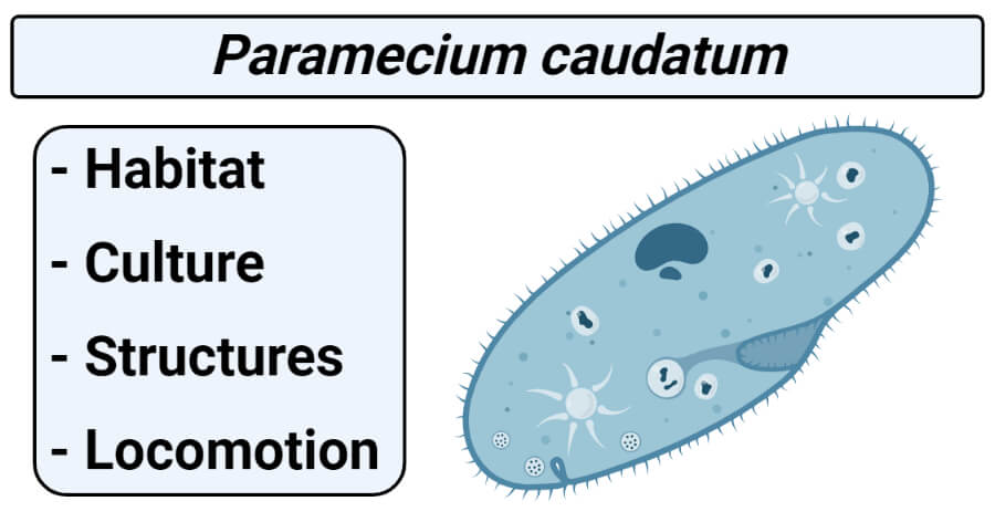
Habit and Habitat
- Paramecium caudatum (Gr., paramekes=oblong+ L., caudata=tail) is a free-living organism which is one of the most common species of Paramecium having worldwide distribution.
- It is commonly found in freshwater, ponds, pools, ditches, streams, lakes, reservoirs, and rivers.
- It is usually found abundant in water containing decaying organic matters, in organic infusion, and in the sewage water.
- It thrives well in ponds or slowly running streams containing aquatic plants.
- It is also found to gather near the surface in scum.
Culture
- Paramecium is easily grown in wide mouth jars with glass covers, 3 quarters filled with boiled pond water or chalkey’s medium dissolved in 1 liter of water, and with 7-12 drops of skimmed milk added weekly.
- The jars are kept away from direct light to allow bacteria to flourish which serves as food for the multiplying bacteria.
External structures
1. Size and shape
- It is a microscopic, elongated organism which are visible to the naked eyes.
- It is light gray or white in color.
- P. caudatum measures between 170µm and 330 µm long.
- It looks like the sole of a slipper or shoe-shaped, cigar-shaped, or spindle-shaped, hence the animal is commonly called a slipper animalcule.
- Its shape is usually constant and in general asymmetrical.
- It is 4 times as long as broad and somewhat cylindrical with different ends.
- Its body is elongated, blunt and rounded at the anterior end and somewhat pointed at posterior ends.
- Its anterior half of the body is slightly twisted.
- Its body is distinguished into an oral or ventral surface and an aboral or dorsal surface.
2. Oral groove
- Situated at the ventral surface of the body.
- Is a large, oblique, and shallow depression.
- It originates from the middle of the body and extends to the left side of the anterior end.
- It drives food materials.
- Oral groove leads into a v-shaped cavity called a vestibule. The vestibule leads into an oval-shaped opening called cytostome. Cytostome leads into funnel-shaped cytopharynx.
- Cytopharynx the turn sharply towards the center of the body is the wide tapering esophagus.
- Esophagus turns again towards the center of the anima to lead into the forming food vacuole.
NOTE: Due to the oblique position of the oral groove the organism has lost its symmetry.
3. Cytoproct /cytopyge
- Is a small opening present on the ventral surface just behind the cytopharynx.
- From this opening, undigested food materials are passed outside the body. So, it is called cell anus.
4. Pellicle
- It is the covering of paramecium.
- It is a thin, double-layered, tough, elastic, and colorless membrane.
- It holds the shape of the animal but elastic enough to permit contractions.
- It has a double membrane; the outer membrane is continuous with cilia and the inner membrane with the ectoplasm.
- It shows rectangular or hexagonal depression on its surface under the high magnification of the microscope.
- It protects the internal organelles from mechanical injuries.
5. Cilia
- They are numerous, tiny, hair-like projections distributed throughout the body.
- They measure 10-12 µm in length and 0.27 µm in diameter.
- They occur in longitudinal rows all over the body, this condition is known as holotrichous in which body cilia are equal.
- Paramecium contains 10000-14000 cilia.
- There are two types of cilia present in paramecium. They are oral cilia and body cilia.
- Oral cilia are present in the vestibule and oral groove. They help in the collection of food materials.
- Body cilia are found on the body surface and help in locomotion. They are of moderate and fairly uniform in length but at the posterior end, cilia are quite longer called caudal tuft hence the species name caudatum.
Ultrastructure of cilia
- Same fundamental structures as of flagellum.
- At the base, the cilium has a diameter of about 0.2 microns or 2,000 Aº which may be up to 10 microns above the cell surface.
- The cilia are bounded by a unit membrane of 90 Aº thickness which resembles and remains continuous with the plasma membrane.
- The bounded space of cilium consists of a fluid, called
- In the matrix, there remain embedded 11 longitudinal fibrils or microtubules.
- Out of the 11 fibrils, two are located in the center, while the remaining 9 fibrils remain arranged peripherally around the central fibrils.
- Each of the 9 outer fibrils is 360Aºin diameter and composed of two sub-fibrils of 180 to 250Aº diameter.
- These sub-fibrils are designated as sub-fibril A and sub-fibril B.
- The sub-fibril A is slightly larger than the sub-fibril B.
- The sub-fibril A gives out two thick projections or arms from its one side. The arms of the sub-fibril A of all the outer fibrils remain directed in a clockwise direction.
- Further, the sub-fibril A occurs more closely to the center of the cilium than the sub-fibril B. Both the sub-fibrils have a common wall of 50Aº thickness.
- Each central fibril has a diameter of about 250Aºand is composed of a 60Aº thick wall.
- Both the central fibrils remain separated by a space of 350Aº and remain enveloped in a common sheath.
- The high-resolution electron microscopy has revealed that each of the peripheral and central fibrils of the cilia and flagella is composed of ten to twelve filaments of 40Aº thickness.
- Each filament is beaded. Each bead remains arranged in the lattices of 40 by 50Aºin the plane of the wall of the tubule. These beads are considered as the basic subunit of the tubule structure.
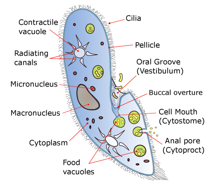
Figure: Paramecium caudatum Structure. Image Source: Deuterostome.
Internal structures
Internal structures of Paramecium consist of cytoplasm, trichocysts, food vacuoles contractile vacuoles, etc.
Cytoplasm: within pellicle, it is differentiated into two types i.e. ectoplasm and endoplasm.
A. Ectoplasm
- It is a permanent part of the body, strikingly delimited from the endoplasm.
- It is a narrow, peripheral thin clear transparent, and dense zone.
- It includes the structure of trichocysts, infraciliary systems.
1. Trichocysts
- These are small oval or spindle-shaped structures situated in the ectoplasm alternating with basal bodies and oriented at right angles to the body surface.
- These are numerous and tiny structures.
- These were first seen in paramecium by Elis which were measuring about 4µm in length.
- Each trichocyst consists of an elongated shaft and a terminal pointed tip, called spike or barb covered by a cap.
- The matrix of the shaft consists of a dense mass of fibrous protein called trichinin.
- These are opened outside through a minute pore on the pellicle surface.
- The trichocysts are discharged on mechanical, chemical, or electric stimuli. After they are discharged, new ones are generated from kinetosomes.
- Functions are uncertain but it is believed that it helps in the offense, defense, and adhesions. Also helps to form the endoskeleton or supports the body.
2. Infraciliary system
It is situated just below the pellicular alveoli. It consists of the kinetosome or basal body and kinetodesma.
a. Basal bodies
- They are tube-like structures produced at the base of each cilium.
- Their wall is formed by the fusion of 9 pairs of peripheral fibrils. The central fibers do not enter into it.
- They are self-duplicating units and progenitors of new cilia.
- Each basal body is either a centriole or its derivatives.
b. Kinetodesma
- Associated closely with basal bodies of cilia and lying in the ectoplasm is a system of specialized striated fibrils, called kinetodesmal fibrils.
- Kinetodesmos or a single fibril arise from the kinetosome or basal body of each cilium. And runs anteriorly somewhat tapering along the course.
- It joins its counterparts from the posterior kinetosomes, forming a bundle of overlapping longitudinal fibrils, called kinetodesma (pleural, Kinetodesmata)
- In each Kinetodesmata the numbers of fibrils constantly remain 5.
- A longitudinal row of kinetosomes with their Kinetodesmata forms a longitudinal unit called a kinety.
- A Kinety is apparently characteristics of all ciliates.
- All the kineties or kinetia makes the infraciliary system of ciliate.
- The kinetia lie in the cortex below the pellicle, their number remains constant for each ciliate.
- The function of the infraciliary system: control and coordinates the movements of the cilia, it brings about the formation of organelles in cell division. e.g. in paramecium, one set of kinety, is solely responsible for the formation of mouth.
B. Endoplasm
- It is a large, central, granular, voluminous, and semi-fluid zone of the cytoplasm.
- It contains many cytoplasmic granules as well as other inclusions and structures of specialized nature.
- The cytoplasmic inclusions are mitochondria, Golgi apparatuses, vacuole, crystals, granules, and chromidia, etc.
- Other structures include nuclei, numerous food vacuoles, and contractile vacuoles.
1. Nucleus
- Paramecium is heterokaryotic as it possesses 2 types of nuclei.
- In P. caudatum there are two nuclei, one large macronucleus or mega nucleus and another small micronucleus.
- These nuclei are not only differing in shape and size but also function.
a. Mega nucleus or macronucleus
- It is a conspicuous, ellipsoidal, or roughly kidney-shaped body.
- Located in the middle dorsal part of the body opposite to cytopharynx.
- It is of compact types containing fine threads and tightly packed discrete chromatin granules of variable size and embedded in an achromatic matrix.
- It has many nucleoli and much more chromatin material (DNA).
- The nuclear membrane is absent in it.
- It divides asexually during reproduction. During transverse binary fission, it divides amitotically.
- It controls the vegetative functions (metabolic activities) of the animals.
b. Micronucleus
- It is small, compact, and spherical or rounded in structure.
- It is lodged in a depression on the surface of the macronucleus.
- Fine chromatin granules and threads are uniformly distributed throughout the structures.
- It divides mitotically during transverse binary fission. During conjugation reproduction, it divides first meiotically then mitotically.
- It controls the process of sexual reproduction but fails to reproduce.
NOTE: Paramecium also survives without micronucleus but fails to reproduce.
2. Contractile vacuoles
- There are two types of contractile vacuoles present in paramecium, occupying a somewhat fixed position in the endoplasm.
- It is located on the dorsal surface of the anterior and posterior end.
- Each contractile vacuole is surrounded by a circlet of 6 to 10 long, narrow, spindle-shaped radial canals extending far into the cytoplasm.
- They open to the outside through a permanent pore in the pellicle of the dorsal side of the body.
- The 2 contractile vacuoles do not lose their identity when water is expelled.
- The two contractile vacuoles work alternatively, but the posterior vacuole contracts more rapidly than the anterior contractile vacuole because it is near to cytopharynx and more water comes into it.
- The main function hydrostatic, helps in the removal excess of water from the protoplasm, the water is partly absorbed and partly taken in while feeding, helps in the removal of carbon dioxide and other metabolic waste products outside. And also maintains the osmoregulation.
3. Food vacuoles
- Numerous non- contractile vacuoles present in paramecium called food vacuoles
- Volkonsky (1934) proposed name gastrioles for these vacuoles, can be seen moving with streaming endoplasm (cyclosis).
- They differ in shape and size according to the quality of food particles but mostly they are rounded in shape.
Locomotion
Paramecium performs locomotion by 2 methods, i.e. metaboly or body contortions and by ciliary beats.
1. Metaboly or body contortions
- The body of Paramecium Caudatum possesses elasticity, it can squeeze itself through a passage narrower than its body, after which the body assumes its normal shape.
- Metaboly is a temporary change in the body, which is brought about in Paramecium by the protoplasm.
2. Ciliary beats
- This is the main method of locomotion.
- During movements, a cilium oscillates like a pendulum. Each oscillation comprises a fast-effective stroke and a slow recovery stroke.
- The cilia can beat forwards or backward enabling the animal to swim anteriorly or posteriorly.
- During effective strokes or strong backward lash, the cilium becomes slightly curved and rigid and strikes the water like an oar, so that body propelled forward in the opposite direction of stroke.
- The cilia become limp and return slowly to the original vertical position, this is, called recovery stroke.
- All the cilia of the body do not move simultaneously and independently but progressively in a characteristics wave-like manner, called metachronal rhythm.
- Cilia of the same transverse row beat together and those of the same longitudinal row beat one after the other from the anterior to the posterior end.
- Metachronal rhythm is due to the infra-ciliary system; this causes swimming forward by the animal.
- But when the body cilia are beating obliquely backward, then at the same time the longer cilia of the oral groove beat more vigorously which causes the anterior end to swerve to the left.
- The action of the cilia of the body and oral groove makes the animal rotate on its long axis.
- This rotation is always to the left (except in P. calkinsi which rotates in a right-hand spiral).
- This combination of forward motion, swerving, and rotation make the animal move forwards in a counterclockwise spiral path.
- This path has a straight axis, and the same body surface of the animal remains towards the axis of the spiral path.
- But in swimming backward, all species rotate to the right.
- By ciliary action, Paramecium moves with a speed of 1500 microns or even more per second.
References and Sources
- Kotpal RL. 2017. Modern Text Book of Zoology- Invertebrates. 11th Edition. Rastogi Publications.
- Jordan EL and Verma PS. 2018. Invertebrate Zoology. 14th Edition. S Chand Publishing.
- 20% – https://androbose.in/paramecium-caudatum-habitat-structure-and-locomotion/
- 5% – https://microbiologynotes.com/external-and-internal-features-of-paramecium-caudatum/
- 4% – https://www.biologydiscussion.com/invertebrate-zoology/protozoa/paramecium-caudatum-habitat-structure-and-locomotion/28315
- 1% – https://www.shareyouressays.com/knowledge/305-words-short-essay-on-the-locomotion-of-paramecium/92346
- 1% – https://www.notesonzoology.com/protozoa/paramecium-protozoa/morphology-of-paramecium-protozoa-microorganisms-zoology/9166
- 1% – https://www.101diagrams.com/paramecium-diagrams-to-print/
- <1% – https://www.sparknotes.com/physics/rotationalmotion/rotationaldynamics/section3/
- <1% – https://www.liveabout.com/what-is-the-sole-of-a-shoe-2987688
- <1% – https://www.amazon.com/Ball-Wide-Mouth-Quart-Bands/dp/B00CNHCDR6
- <1% – https://sciencing.com/left-side-body-human-anatomy-7363170.html
- <1% – https://quizlet.com/120287413/combo-with-ap-ch-3-and-27-others-flash-cards/
- <1% – https://quizlet.com/11328551/facehead-embryology-flash-cards/
- <1% – https://jcs.biologists.org/content/joces/11/3/899.full.pdf
- <1% – http://shodhganga.inflibnet.ac.in/bitstream/10603/78652/9/09_chapter%204.pdf