Vertebrates are the only group that has well-developed functional kidneys to conserve water in terrestrial environments. Primitive vertebrates like fish excrete ammonia (NH4) as the byproduct of protein metabolism. Birds and Reptiles excrete a more concentrated form of ammonia, uric acid. Ammonia is toxic to mammalian cells. Mammals have nephrons with an extended ‘loop of Henle’, which reabsorb water and excrete urea, hence concentrating the urine with urea to a high extent. This reabsorption of water helps the mammal to survive in hot and dry environments.
Interesting Science Videos
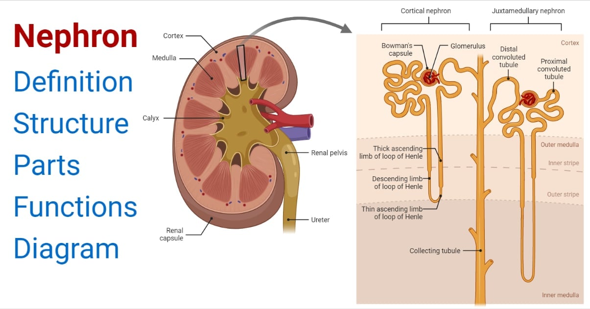
What is a Nephron?
Nephron is a basic microscopic structural and functional unit of the Kidney. They are used by the kidney to separate substances from bodily fluids like ions, small molecules, and water, filtering out toxins and wastes from the blood and returning substances of interest back to the blood.
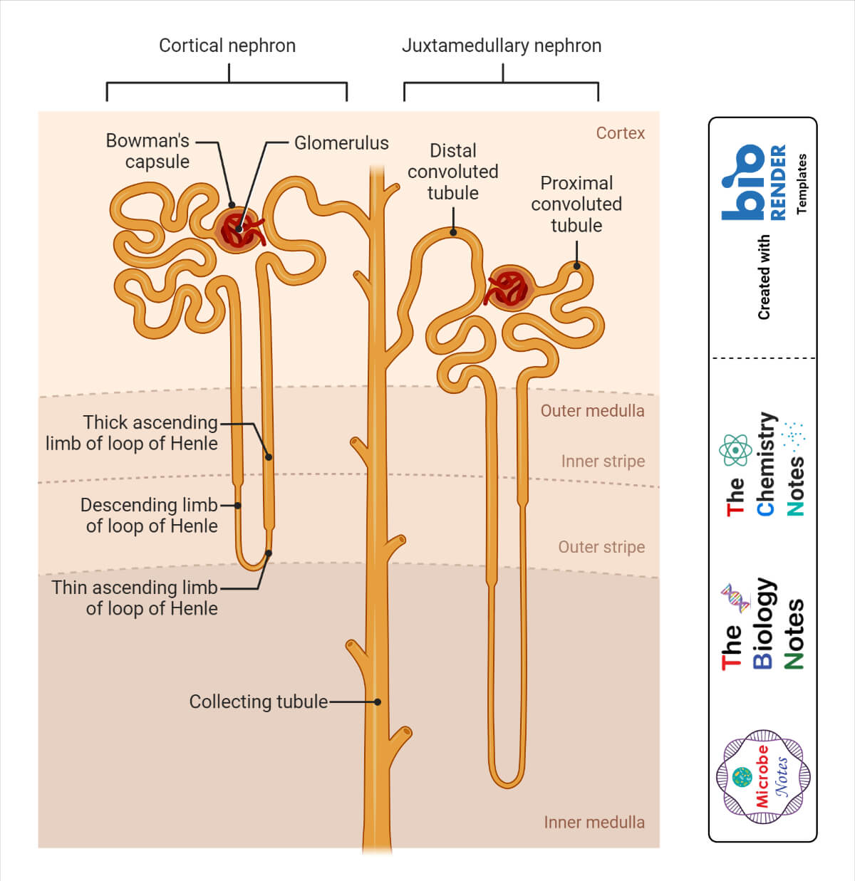
- Cortical Nephrons – They are present in the renal cortex with shorter loops of Henle.
- Juxtamedullary Nephrons – Present in the medullar region with longer Loops of Henle.
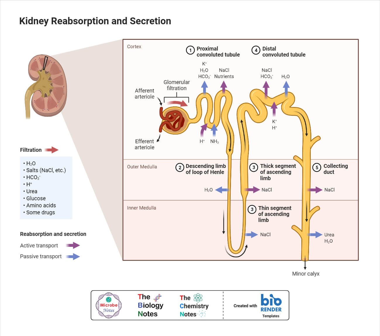
Nephrons work on the principle of ultrafiltration for extracting and reabsorption of molecules. Filtration occurs passively in the glomerulus with the aid of intracapillary blood pressure. Blood plasma constituents which include water and other substances are filtered as the plasma traverses through the tiny gaps in glomerular capillaries. The components of the blood that are excluded from filtration into Bowman’s capsule are RBCs, WBCs, platelets, and blood proteins.
Physiology of Nephron
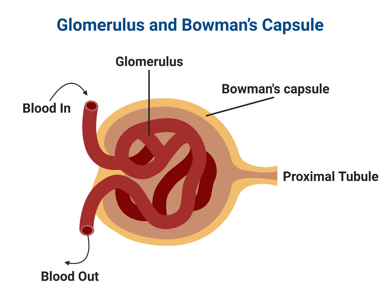
Glomerulus and Bowman’s Capsule
- It is a specialized network of blood capillaries present in the nephron forming a tuft-like structure.
- Located at the vascular pole of Bowman’s Capsule which surrounds the glomerulus.
- Glomerulus and the Bowman’s Capsule form the Renal Corpuscle.
- The visceral inner layer of the capsule is made up of specialized podocyte cells and the outer layer is composed of cells of simple squamous epithelium.
- Renal blood circulation is responsible for the blood supply in the kidneys.
- Glomerulus collects the blood supply from the afferent arterioles which generate the high pressure necessary to force solutes and water out of the blood plasma as a filtrate.
- About one-fifth of the blood plasma is filtered into the Bowman’s capsule.
- The rest of the blood containing RBCs, WBCs, platelets, and plasma proteins are passed into the efferent arteriole.
- The difference between the afferent and the efferent arteriole diameter with the efferent arteriole being smaller leads to an increase in the hydrostatic pressure in the glomerulus.
- The filtrate in the Bowman’s capsule then passes through the rest of the nephron tube which consists of PCT (proximal convoluted tubule), the loop of Henle, and DCT (distal convoluted tubule), before finally exiting the nephron into the ‘Collecting Duct’, commonly shared by many nephrons.
Renal Tubule
The long pipe-like structure that arises from the Bowman’s capsule and contains the filtrate (also referred to as tubular fluid) passed from the glomerulus. This consists of the following components:
Proximal Convoluted Tubule (PCT)
- They lie in the cortex region of the kidney.
- Divided into the initial convoluted portion and the descending straight portion.
- They are lined by simple cuboidal epithelial cells with prominent microvilli forming a ‘brush border’ on the luminal surface.
- Microvilli perform the essential function in PCT by increasing the surface area for reabsorption and secretion of solutes like glucose, sodium ions, etc into the filtrate.
- The filtrate is also reabsorbed by the peritubular capillaries.
- The cuboidal epithelial cells of PCT actively transport ions and solutes across the membrane, suggesting the high amount of ATP required to carry out the transportation, hence possessing a large number of mitochondria.
Loop of Henle
- The U-shaped tube extending from the descending PCT is called as Loop of Henle.
- It consists of ascending and descending limbs running adjacent and parallel to one another.
- The ascending limb receives filtrate from PCT in the cortex region, extending into the medullar region as the descending limb, and ascending back to the renal cortex as ascending limb emptying filtrate into DCT (distal convoluted tubule).
- The descending limb has an initial short thick portion made up of simple cuboidal epithelial cells and a long thin portion.
- The ascending limb has an initial short thin portion made up of simple squamous epithelium and a long thick portion.
- The primary role it performs is to generate concentrated urine by making the interstitial fluid hypotonic.
- The ascending and descending limb is water impermeable and less permeable to salts.
- As the filtrate moves down deeper into the renal medullar interstitium, the water passes freely out of descending limb via osmosis to equilibrate the tonicity interstitium and the filtrate.
- The ascending limbs pump out sodium ions from the filtrate via active transport, causing hypotonic filtrate solution due to the loss of sodium ions.
- The hypotonic filtrate is moved to DCT in the renal cortex.
Distal Convoluted Tubule (DCT)
- Lined by simple cuboidal epithelium cells.
- They are shorter in length than PCT with fewer microvilli.
- There is an active pumping of ions against the concentration gradient, so a large number of mitochondrial organelles are present in the epithelial cells to produce sufficient ATP.
- DCT is regulated by endocrine hormones like aldosterone.
- The presence of the parathyroid hormone in the DCT causes more reabsorption of calcium ions and secretion of potassium ions
- Aldosterone in the DCT causes sodium reabsorption and potassium secretion.
- The atrial natriuretic peptide in the DCT causes sodium secretion.
Collecting Duct
- The collecting tubule is continuous with the distal convoluted tubule and is not technically part of the nephron due to a different developmental origin than reproductive and urinary organs.
- The collecting duct is joined by several nephrons, hence collecting filtrate from each one of them.
- They are made up of simple cuboidal epithelium cells.
- The duct begins at the renal cortex and extends deeper into the medullary region.
- Urine passes the duct all the way through the medullary interstitium with a high sodium ion concentration.
- The collecting duct membrane is impermeable to water molecules but in the presence of ADH (antidiuretic hormone), it becomes permeable due to the opening of aquaporins.
- Levels of ADH in the urine help determine if the urine is diluted or concentrated. Diluted urine has low levels of ADH while concentrated urine has high levels of ADH indicating dehydration condition.
Juxtaglomerular apparatus (JGA)
- A specialized region that separately associates with nephrons to carry out important functions.
- Located at the juncture of the afferent arteriole and thick ascending limb of PCT.
- It consists of components like macula densa, juxtaglomerular cells, etc.
- Macula densa is present adjacent to the DCT made up of cuboidal epithelial cells.
- The cells of macula densa release paracrine signals like adenosine or ATP in response to the flow rate of the filtrate and the sodium ion concentration.
- The juxtaglomerular (JG) cells are located at the junction of the afferent arteriole.
- JG cells are a type of smooth muscle cell that contracts and relax in response to ATP release by the cells of macula densa.
- The contraction and relaxation of JG cells help regulate the flow of blood to the glomerulus.
- JG cells produce and secrete the Renin enzyme also known as Angiotensinogenase. This enzyme cleaves Angiotensinogen forming Angiotensin-1, which is further converted to Angiotensin-2 via ACE (angiotensin-converting enzyme).
- Angiotensin-2 is a potent vasoconstrictor.
Nephron Functions
- They are the functional unit of kidneys.
- They cleanse the blood from the toxic wastes present and help in excreting out of the body via urine.
- Filter and reabsorb water molecules and other important solutes present in the blood.
- Helps in balancing the homeostatic pressure via the process of filtration, reabsorption, and secretion of ions, water, and other solutes.
- Regulates the blood pressure with the aid of renin production which causes fluctuations in vasodilation and vasoconstriction.
- Stimulate RBC production in the bone marrow via the production of erythropoietin hormone released from the peritubular cells of the kidney.
- Regulates the concentration of phosphate and calcium in the bloodstream via the conversion of calcidiol to calcitriol (the biologically active form of Vit. D), promoting remodelling and growth of bones.
Conclusion
Nephrons are the basic functional unit of the Renal (Kidney) system. They have the necessary function of filtering blood leading to the production of urine. They maintain the balance of body fluids and electrolyte concentration in the body. Also helps in regulating pH levels and blood pressure. Nephrons have many components like glomerulus, bowman’s capsule, loop of Henle, etc. Glomerulus is a network of blood capillaries surrounded by the bowman’s capsule which filters blood out of toxins and waste products along with other ions and solutes. The filtered fluid is passed down to the tubule with regions of PCT, loop of Henle, DCT, and collecting tubule. As the filtrate flows through each region, it is processed and refined with water and nutrients being reabsorbed into the blood and waste material being concentrated into urine. The final urine is collected in the collecting duct which is further transported to the renal pelvis and expelled out via the urethra.
References
- Nephron – https://biologydictionary.net/nephron/
- Microscopic Anatomy of the Kidney: Anatomy of the Nephron – https://open.oregonstate.education/aandp/chapter/25-2-microscopic-anatomy-of-the-kidney-anatomy-of-the-nephron/
- Stern, Rachel A., and Paul E. Mozdziak. “Differential ammonia metabolism and toxicity between avian and mammalian species, and effect of ammonia on skeletal muscle: A comparative review.” Journal of animal physiology and animal nutrition 103.3 (2019): 774-785.
- Renin – https://my.clevelandclinic.org/health/body/22506-renin
- Erythropoietin Stimulating Agents – https://www.ncbi.nlm.nih.gov/books/NBK536997/
- Bone and Calcium Homeostasis – https://med.libretexts.org/Bookshelves/Anatomy_and_Physiology/Anatomy_and_Physiology_(Boundless)/6%3A_Skeletal_System/6.5%3A_Bone_and_Calcium/6.5A%3A_Bone_and_Calcium_Homeostasis