Mitochondria are oxygen-consuming ribbon-shaped cellular organelles of immense importance floating free throughout the cell.
They are known as the “powerhouse of the cell” since these organelles supply all the necessary biological energy to the cell by oxidizing the substrates available.
The enzymatic oxidation of chemical compounds in the mitochondria releases energy.
Since mitochondria act as the power-houses, they are abundantly found on those sites where energy is earnestly required such as sperm tail, muscle cell, liver cell (up to 1600 mitochondria), microvilli, oocyte (more than 300,000 mitochondria), etc.
Typically, there are about 2000 mitochondria per cell, representing around 25% of the cell volume.
In 1890, mitochondria were first described by Richard Altmann and he called them bioblasts. Benda in the year 1897 coined the term ‘mitochondrion’.
- Mitochondria is the cell organelle which is filamentous and granular structure.
- It is present in higher plants, animals, and some microorganisms.
- It is absent in bacteria but is found in algae, protozoa, and fungi.
- To carry out the energy metabolism, mitochondria have got the lipoprotein framework.
- It consists of the different enzymes and coenzymes.
- Mitochondria also consist of specific DNA and ribosomes.
- Ribosomes are involved in protein synthesis whereas specific DNA is involved in the cytoplasmic inheritance.
- Mitochondria were first observed in 1850 by Kolliker.
- In the striated muscles, it was observed as the granular structure.
- Later in 1888, he isolated them from the insect muscles.
- It was named fila by Flemming in 1882.
- In the early days, mitochondria were given the different names such as
- Fuchsinophilic granules
- parabasal bodies
- plasmosomes
- plastosomes
- fila
- vermicules
- bioblasts
- chondriosomes
- In the cytoplasm, mitochondria are uniformly distributed.
- But it is also found that their distribution is restricted in many cells.
- Depending on the function of the cell, the mitochondria are distributed accordingly.
- The distribution of mitochondria varies according to species and cell type.
- But some cell may contain a large number of mitochondria as:
- 50,000 in Chaos chaos
- 140,000 to 150,000 in eggs of sea urchin
- 300,000 in oocytes of amphibians.
- It is found that only 500 to 1600 mitochondria are present in the liver cells of the rat.
- As compared to the animal cell, the number of mitochondria is less in green plants.
- It is because in the green plants there is the presence of chloroplast which carries out the function as of mitochondria.
- In the animal cell, sarcosomes are the mitochondria present in the myocardial muscle cell. They are numerous and large.
- The shape of the mitochondria can be filamentous or granular.
- Depending upon the physiological condition of the cell, their shape may change from one form into another.
- So the shapes can be of club, racket, vesicular, ring or round-shape.
- In the rat or primary spermatocyte, mitochondria are granular.
- In the liver cell mitochondria are club-shaped.
- Mitochondria may fuse and separate the cell which causes the changes in shape.
- During the day the mitochondria may fuse into the reticulate structure in certain cells of euglenoid.
- Then it separates or dissociates during the darkness.
- In the case of yeast depending on the different cultural condition changes occurs in it.
- The size of the mitochondria is 0.5 to 2.0 µm, so it cannot be seen clearly under the light microscope.
- It is also found sometimes the length reaches 7 µm.
Interesting Science Videos
Structure of Mitochondria
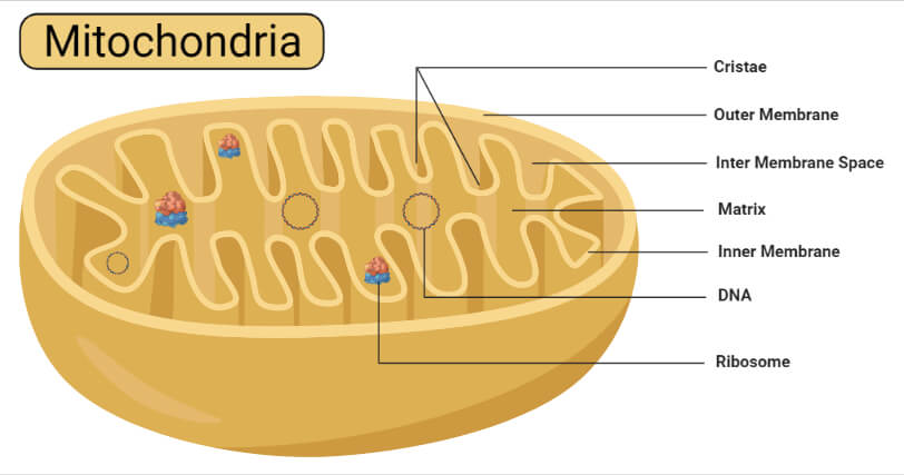
Mitochondria are mobile, plastic organelles that have a double-membrane structure. It ranges from 0.5 to 1.0 micrometer in diameter. It has four distinct domains: the outer membrane, the inner membrane, the intermembrane space, and the matrix.
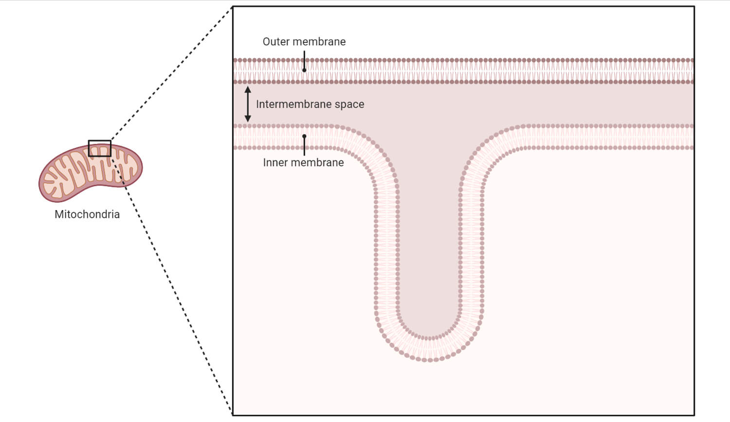
- The organelle is enclosed by two membranes—a smooth outer membrane and a markedly folded or tubular inner mitochondrial membrane, which has a large surface and encloses the matrix space.
- The intermembrane space is located between the inner and outer membranes.
- The number and shape of the mitochondria, as well as the numbers of cristae they have, can differ widely from cell type to cell type.
- Tissues with intensive oxidative metabolism— e. g., heart muscle—have mitochondria with particularly large numbers of cristae.
- Even within one type of tissue, the shape of the mitochondria can vary depending on their functional status.
- Both mitochondrial membranes are very rich in proteins.
Mitochondria consists of a mitochondrial membrane and a mitochondrial chamber.
1. Mitochondrial membrane
It consists of two membranes. They are:
a. Outer membrane
- It is a smooth membrane.
- It is made up of 40% lipids and 60% proteins.
- Due to the presence of the pores or porins, it is permeable.
b. Inner membrane
- It is made up of 20% lipids and 80% proteins.
- It is the selectively permeable membrane.
- Since the membrane is folded inwards and there is the presence of cristae, it is said as the rough membrane.
- Cristae are the numerous and finger-like projections.
- Tennis racket-like particles are present in each crista.
- The particles were previously named as the inner membrane subunits, F0-F1 particles, elementary particles, or oxysomes.
- It was called the electron transport particles (ETP)by Parsons in 1963.
- In each mitochondrion about 104-105 particles are present.
- Base, stalk, and head are present in each elementary particle.
- Base and head are also called the F0 and F1 particles respectively.
- Stalk acts as the link which connects the base and head.
- The base is made up of hydrophobic proteins and is embedded in the lipid molecules of the membrane.
- The Head is made up of five types of the polypeptide.
- ATPase or ATP synthetase is the enzyme present in it.
- The ADP aids in the formation of ATP.
- Similarly, inorganic phosphate is also formed. It is due to oxidative phosphorylation.
- The stalk also consists of the coupling factors which connect the respiratory chain with the elementary particle.
2. Mitochondrial Chambers
Two chambers are present in the mitochondria i.e Outer and inner chamber.
a. Outer chamber (peri-mitochondrion space)
- Between the outer and the inner mitochondrial membrane, a space is present in between them which is known as the peri-mitochondrion space.
- Few enzymes are also present in the fluid present in it.
b. Inner chamber
- It is present in the inner part of the inner membrane.
- A semi-fluid matrix is present in it which consists of:
Mitochondrial Matrix
- The mitochondrial matrix which is the liquid (colloidal) area encircled by the inner membrane, contains the soluble enzymes of the Krebs cycle which completely oxidize the acetyl-CoA to produce CO2, H2O and hydrogen ions. Hydrogen ions reduce the molecules of NAD and FAD, both of which pass on hydrogen ions to respiratory or electron transport chain where oxidative phosphorylation takes place to generate energy-rich ATP molecules.
- Mitochondria also contain in their matrix single or double circular and double-stranded DNA molecules called mt DNA and also the 55S ribosomes, called mitoribosomes. Since mitochondria can synthesize 10 percent of their proteins in their own protein-synthetic machinery, they are considered as semi-autonomous organelles.
Functions of Mitochondria
- Mitochondria stores and releases energy in the form of ATP ( Adenosine triphosphate ). It occurs by the oxidation of carbohydrates, proteins, and fats. It will be further utilized in the different metabolic activities. So, mitochondria are known as the powerhouse of the cell or storage batteries of the cell.
- Mitochondria help in the formation of the heme of hemoglobin.
- During cellular respiration, mitochondria form the different intermediate products. They are utilized for the synthesis of cytochromes, chlorophyll, ferredoxin, steroids, alkaloids, pyrimidines, etc.
- Calcium can be stored and released by the mitochondria.
- It helps in the formation of amino acids.
- In the matrix of the mitochondria, several fatty acids can be synthesized.
- During the process of oogenesis, they help in the formation of the yolk.
- During the process of spermatogenesis, they help in the formation of the middle part of the sperms.
- By the process of maternal inheritance, traits are directly transferred by mitochondria from the mothers to the offsprings.
- Mitochondria are also present in the liver cell. They help in the detoxification of ammonia using their enzymes.
- Mitochondria are the site of heat generation which is known as thermogenesis.
- Sometimes there can be the abnormal death of the cell. It might be due to the dysfunctioning of the mitochondria. It can affect the function of the organ.
- It helps in the formation of some parts of the hormone of testosterone and estrogen.
References and Sources
- Shakya, M., Mehata, K.R., Gautam, M.K., Pokhrel, K.R., and Khanal, K. (2020 ) “ Principles of Biology”, Asmita Books Publisher and Distributors Ltd, Bhotahity, Nepal
- Verma, P. S., and Agrawal, V. K. (2006). Cell Biology, Genetics, Molecular Biology, Evolution & Ecology (1 ed.). S.Chand and Company Ltd.
- https://kullabs.com/class-11/biology11/cell-organelles/mitochondria
- https://alevelbiology.co.uk/notes/mitochondria-structure-and-functions/
- https://www.biologydiscussion.com/cell/plant-cell/structure-of-a-plant-cell-with-diagram/68806 – 12%
- https://biologyteach.com/power-house-of-the-cell/- 11%
- https://www.sciencedirect.com/topics/agricultural-and-biological-sciences/pavement-cells- 4%
- https://www.sciencefacts.net/mitochondria.html- 2%
- https://biologywise.com/mitochondria-structure-functions- 1%
- https://www.sciencedirect.com/science/article/pii/S0005272810000058- 1%