Interesting Science Videos
Systematic position
Phylum: Protozoa
Subphylum: Sacromastigophora
Superclass: Mastigophora
Class: Zoomastigophora
Order: Kinetoplastida
Genus: Leishmania
Species: donovani
Discovery and Distribution
- The species of Leishmania donovani was reported simultaneously by Leishmania from London and Donovan from Mardas in (1903) hence the name Leishmania donovani.
- L. donovani causes a malaria-like fever-oriental disease in the man called kala-azar, Dumdum fever, or Black fever.
- L. donovani, the causative organism of Visceral leishmaniasis (kala-azar) in Africa, the Middle East, Mediterranean coasts, Asia, and South America.
- It is endemic in Asia, Africa, the Americas, and the Mediterranean region.
Habits and Habitat of Leishmania donovani
- It is an intracellular parasite of man and other mammalian hosts.
- In man, intracellular amastigote forms are found in reticuloendothelial cells of the spleen, bone marrow, leucocytes or cells of the liver, intestinal mucosa, and mesenteric lymph nodes.
- The promastigote form is found in the midgut of sandfly or in artificial culture.
Morphology of Leishmania donovani
The parasite occurs in two forms or stages, leishmanial or amastigote and leptomonad or promastigote, which alternate between a vertebrate (man) and an invertebrate (sandfly) host.
1. Leishmanial or Amastigote stage
- This stage occurs intracellularly in blood cells or reticuloendothelial cells of the vertebrate hosts or man.
- It is microscopic, rounded, or oval in shape measuring 2-4 micrometer in length.
- There is no free flagellum, it is greatly reduced, fibril-like, and lies embedded in the cytoplasm.
- A flagellar stage of amastigote is known as LD bodies.
- The nucleus is central or eccentric.
- The cell membrane is delicate and can be demonstrated only in a fresh specimen.
- Kinetoplast is rod-shaped or dot-like and lies at the right angle to the nucleus.
- The axoneme(rhizoplat) is a delicate filament extending from the kinetoplast to the margin of the body. It represents the foot of the flagellum.
- They are stained well with Giemsa or Wright stain.
- In a Giemsa stained preparation, the cytoplasm surrounded by a limiting membrane appears pale blue. The nucleus relatively is larger and stained red. The kinetoplast stained deep red.
- Amastigote divides by binary fission at 37°C.
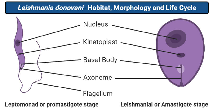
2. Leptomonad or promastigote stage
- It is found in the midgut of the invertebrates host or sandfly.
- It is elongated, slender, and spindle-shaped measuring 15-20µ in length and 1-2µ in width.
- A flagellum is long measures 15-28µ and free and arises from a minute basal body or blepharoplast situated near the anterior end.
- The flagellum does not curve around the body of the parasite and therefore there is no undulating membrane.
- The nucleus is centrally placed.
- The kinetoplast lies transversely near the anterior end.
- A vacuole is present near the root of the flagellum
- With Leishman stain, the cytoplasm appears blue, the nucleus pink or violet, and the kinetoplast bright red.
- Promastigote multiplies by binary fission at 27°C.
Life cycle of Leishmania donovani
Hosts
- Leishmania is also a digenetic parasite that requires 2 hosts for completion of its life cycle.
- The primary host is a vertebrate or man, in which the parasite feeds and multiplies asexually.
- The secondary host or vector is invertebrates or blood-sucking insects or sand-fly, belonging to the genus Phlebotomus.
- Some mammals like dogs, jackals, gerbils, and squirrels also serve as reservoir hosts in which the parasite does not undergo any change but simply waits for its introduction into the human host.
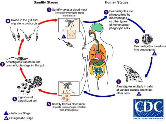
Image Source: DPDx – Laboratory Identification of Parasites of Public Health Concern.
(I) Life cycle in Man
- The parasite has two stages in its life cycle:
- Amastigote form occurs in humans and mammals.
- Promastigote form occurs in sandfly.
- L. donovani is transmitted to humans or other vertebrates by the bite of blood-sucking sandfly Phlebotomus argentipes
- The parasites introduced by sandfly into the human body are in the promastigote form.
- Some of the promastigote entering the blood circulation directly become destroyed.
- while those entering the reticuloendothelial system(liver, spleen, bone marrow, and lymph nodes ) change into amastigote or leishmanial forms.
- The amastigotes multiply by simple binary fusion inside the Reticuloendothelial system to form a large number of amastigotes.
- When the number of parasite reaches 50 to 200 or even more, the host cell rupture.
- The liberated parasites are taken up by new host cells and the multiplication cycle is repeated so that the reticuloendothelial system becomes progressively infected.
- Some of the free amastigotes are phagocytosed by the neutrophils and monocytes(macrophages) in the bloodstream.
- These heavily parasitized cells wander through the general blood circulation leading to a general infection.
(II) Life cycle in sandfly
- When the sandfly sucks the blood of an infected person, it obtains free amastigotes as well as the parasitized neutrophils and monocytes along with the blood-meal.
- The parasite begins a process of transformation and the amastigotes change to procyclic promastigotes and then to metacyclic promastigotes in the midgut of the sandfly.
- These promastigotes multiply by longitudinal binary fusion and produce large numbers of promastigotes completely filling the lumen of the gut.
- In 6 to 9 days, the number of parasites becomes enormous and heavily spread into the pharynx and buccal cavity. The salivary glands are not infected.
- Transmission into a new host occurs when such a heavily infected sandfly bites the host.
References and Sources
- Kotpal RL. 2017. Modern Text Book of Zoology- Invertebrates. 11th Edition. Rastogi Publications.
- Jordan EL and Verma PS. 2018. Invertebrate Zoology. 14th Edition. S Chand Publishing.
- Torres-Guerrero, E., Quintanilla-Cedillo, M. R., Ruiz-Esmenjaud, J., & Arenas, R. (2017). Leishmaniasis: a review. F1000Research, 6, 750. https://doi.org/10.12688/f1000research.11120.1
- 8% – https://www.notesonzoology.com/term-paper/leishmania/term-paper-on-leishmania-protozoa-microorganisms-zoology/9106
- 1% – https://www.yourarticlelibrary.com/zoology/parasite-leishmania-donovani-life-cycle-mode-of-infection-and-treatment/24263
- 1% – https://www.who.int/neglected_diseases/news/NTD_course_now_accessible_via_eLearning/en/
- 1% – https://www.slideshare.net/PakRose1/leishmania-31612660
- 1% – https://en.wikipedia.org/wiki/Leishmania_mexicana
- 1% – https://biologydictionary.net/salivary-glands/
- <1% – https://www.toppr.com/ask/question/plasmodium-multiplies-by-binary-fission/
- <1% – https://www.sciencedirect.com/topics/immunology-and-microbiology/trematoda
- <1% – https://www.sciencedirect.com/science/article/pii/S1367578818302116
- <1% – https://parasitesandvectors.biomedcentral.com/articles/10.1186/1756-3305-4-82
Good notes,
Easy to understand, brief and precise
Thanks so
much
Amazing 👌 notes really it’s play a great role 👌 in my study
Good notes ! It helps me a lot to understand