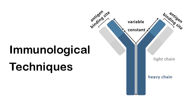
Most of the immunological techniques are based on antigen-antibody reactions. Precipitation reactions are one of the important reactions that occur when antigen and antibody come to contact. When a soluble antigen reacts with its antibody in the presence of NaCl at optimal temperature and pH, the antigen-antibody complex forms an insoluble precipitate. Generally, liquid media and gels such as agar, agarose, and polyacrylamide are used for this kind of reaction.

Interesting Science Videos
Immunodiffusion tests
This is an immunological technique used to find out different antigens and antibodies in clinical samples. The tests are performed in 1% agar. There are some advantages of using immunodiffusion tests in a clinical set up such as:
- The band formed after the reaction is easily visible, stable, and can be stained for preservation.
- Different antigens can be used to observe the reaction. As each antigen-antibody reaction gives a specific precipitation line, therefore, it helps to identify specific antigens.
- Identical, partial identical and non-identical antigens can be observed.
Single Diffusion in One Dimension (Oudin Procedure)
- The antibody is mixed with agar in a test tube and the antigen solution is added over it.
- As a result, antigen diffuses downward through agar gel and a line of precipitation is formed.
- The number of different precipitate bands will indicate different types of antigens.
Double Diffusion in One Dimension (Oakley- Fulthrope Procedure)
- The antibody is mixed with agar in a test tube.
- A column of plain agar is added on top of the antibody solution.
- The antigen is poured on the plain agar column.
- The antigen and antibody move toward each other through the intervening column of plain agar and a precipitate band will form when at the optimum concentration of the antigen and antibody.
Single diffusion in two dimensions (Radial immunodiffusion)
- The antibody is mixed with agar gel and a layer of this mixture is formed on a glass slide.
- Wells are cut on the surface of the gel.
- The antigen solution will be added to the wells. As a result, it diffuses and a ring-shaped precipitation band is formed.
- The diameter of the band is proportional to the concentration of the antigen.
- This technique was used for the estimation of IgG, IgM, and IgA in sera and for the screening of antibodies of influenza virus.
Double diffusion in Two Dimension (Ouchterlony Procedure)
- A layer of agar gel is formed on a Petri plate. Then wells are formed by using the template.
- Antibody solution is added to the central well and different antigens are added to the surrounding wells.
- If two adjacent antigens are identical, the precipitation lines will fuse.
- If two adjacent antigens are unrelated, the precipitation lines will cross.
- In the case of partially related antigens, spur formation will be observed.
- This technique is used for the toxicity test of C. diphtheria (Elck’s Test).
Immunoelectrophoresis
- Immunoelectrophoresis is a combination of electrophoresis and immune diffusion.
- A glass slide is used which is layered with semisolid agar.
- A well will be formed on the surface of the agar and antigen solution will be added to the well.
- Electrophoresis will be performed for 1 hour.
- Then a rectangular trough will be cut parallel to the direction of migration of antigen. Antibody solution will be added to the trough and it will be left for 18-24 hours for diffusions.
- As a result precipitation band will be formed based on each separated compound.
- This technique is used for the detection of different antigens in human serum and normal and abnormal serum proteins such as Myeloma proteins.
Counter Immunoelectrophoresis
- It is a one-dimensional double elctro-immunodiffusion test.
- The test is based on the movement of antigen and antibody in the opposite direction.
- This test is also performed on a glass slide which will be layered with agar.
- Two different wells will be formed on the surface of the agar. In one well antigen will be added and another well will contain antibodies.
- The electricity will pass through this which accelerates the movement of antigen and antibody towards each other.
- A precipitation line will be formed at a specific point between the two wells.
- This test is a sensitive, standard technique and requires around 30 minutes to perform.
- This technique is used for clinical detection of hepatitis B antigens and antibodies, antigens of Cryptococcus in cerebrospinal fluid.
Rocket Electrophoresis
- It is a one-dimensional single electroimmunodiffusion test.
- It is mostly used for the quantitation of antigens.
- In this case, the antibody is mixed with the agarose gel and this mixture will be used to form a layer on the glass slide.
- Wells will be formed on the surface of the gel and antigens are added to those wells in increasing concentration.
- Electrophoresis will be performed.
- As a result, cone-like precipitation bands (rocket-like structures) will be observed.
- The length of the rocket-like structure is directly connected with the concentration of antigens.
Radioimmunoassay (RIA)
- This technique was first described by Berson and Yallow.
- It is mostly used for the quantitation of hormones, drugs, hepatitis B surface antigen, IgE, and viral antigens.
- The test is based on the competition for a fixed amount of specific antibodies between a known radiolabelled antigen and an unknown test antigen.
- The competition is determined by the level of test antigen present in the reacting system.
- At the end of the antigen-antibody reaction, the antigen will be found in free and bound fractions and their radioactivity will be measured.
- The concentration of the test antigen is calculated from the ratio of bound and total antigen levels using a reference curve.
Enzyme-Linked Immunosorbent Assay (ELISA)
ELISA is a simple and sensitive test used for the detection of different antibodies and antigens. It requires only microliter quantities of test reagents. The principle of ELISA is based on an enzyme that acts on its specific substrate to produce a color. The color will indicate a positive result. Based on this principle there are different types of ELISA such as
Sandwich ELISA
- Microtitre plates are used to perform this test. The wells of the plate are coated with a specific antibody against the antigen to be detected.
- The clinical specimen is then added to the wells.
- If the antigen is present in the specimen, it binds with the coated antibody.
- This antigen-antibody reaction is detected by using antiserum conjugated with an enzyme.
- This antibody attaches to the antigen which is already bound with its specific antibody.
- A substrate is then added to the reaction mixture which will bind with the conjugated enzyme.
- In case of a positive result, the enzyme-substrate complex will produce color and the intensity of the color is then further read by the ELISA reader.
- Positive and negative controls should be performed along with the test.
Indirect ELISA
- This test is used for the detection of antibodies.
- The wells of the microtitre plate are coated with antigen.
- A clinical sample or Sera is added to these wells.
- If the specific antibody is present in the sample, it will form a complex with the antigen-coated in the wells.
- This antigen-antibody complex is detected by adding enzyme-conjugated antihuman immunoglobulin which will bind with the antibodies of the specimen.
- A substrate is then added which forms a complex with the enzyme and produces color as a positive result.
- Positive and Negative controls are equally important to perform this test.
- The enzymes which are generally used are horseradish peroxidase, alkaline phosphatase, etc. The substrate used for horseradish peroxidase is o-phenyl-diamine-dihydrochloride and the substrate for alkaline phosphatase is p-nitrophenyl phosphate.
Competitive ELISA
- It is used for the detection of HIV antibodies.
- It is different than the previous two types of ELISA. In this case, the appearance of color indicates a negative test while no color change indicates positive results.
- In this case, the competition occurs between an enzyme-linked antibody and a test antibody that is present in the clinical sample. These two antibodies compete for the same antigen.
- The wells of the microtitre plate are coated with the antigen of HIV. The sera (test sample) is added to these wells and incubated at room temperature and then washed.
- If the specific antibody is present in the test sample, the antigen-antibody reaction will occur.
- Enzyme labeled antibodies are then added to the reaction mixture to detect the antigen-antibody complex. The plate is further washed after incubation.
- If the test sample contains the antibodies of HIV then, these enzyme-linked antibodies will not be able to form a complex with the antigen and will leave the reaction mixture after washing.
- Then, the substrate is added and as there are no enzyme-linked antibodies present in the system, no color change will be observed as a positive test.

Very good
Thanks
Greeting my dear, sir, madam, Dr. please share this notes by adding more corroborate similar support articles in the form of PDF through my email address if it is possible.
Thank u so much!