Hydra are inconspicuous freshwater relatives of corals, sea anemones, and jellyfish that are classified under the phylum Cnidaria, Class Hydrozoa, Order Anthomedusae/Anthoathecata, and Family Hydroidea. It is essentially a sedentary organism living attached to stones, pebbles, and water plants, but can get released from the substratum and swim. The name Hydra was given by scientist Linnaeus because of its special power to regenerate its lost part like Hydre ( a nine-headed dragon serpentine of the Greek mythology).
Systematic position
Phylum: Coelenterata
Class: Hydrozoa
Order: Hydroida
Suborder: Anthomedusae
Genus: Hydra
Habit and Habitat
- Hydras are solitary, sessile, freshwater animals, cosmopolitan in distribution.
- They can be found in a large variety of freshwater habitats mostly occur in ponds, lakes, and slow-flowing parts of streams and rivers.
- They may be found attached to and hanging downwards from the underside of solid objects in the water such as leaves, sticks, stones, weeds, etc.
- The tentacles and body of Hydra are stretched to the maximum limit in water in order to capture any prey that comes in contact with them. When disturbed the body at once contracts into a minute jelly-like knob.
- They are carnivorous in habit and feeds on insect larvae and, especially, small crustaceans, such as water fleas, seed shrimps, and copepods.
Collection and preservation
- Hydra can be collected from shallow water of ponds, lakes, and streams during early winter.
- As these animals live habitually clinging to aquatic vegetation, quantities of vegetation may be picked and put in a jar filled with pond water.
- In the laboratory, hydras may be dislodged from vegetation by squirting them with a jet of water from a pipette.
- They are then transferred to a dish with a pipette or small aquarium net.
- When fully expanded, hydras may be fixed in Bouin’s fixative for 30 minutes, given several washes with 50% alcohol, kept in 50% alcohol for 10-15 minutes, and finally preserved in 70% alcohol.
Interesting Science Videos
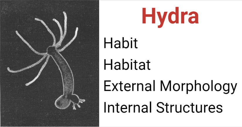
External morphology
1. Shape and size
- Hydra is a polyp-like or polypoid coelenterate with a tubular or cylindrical body.
- It becomes elongated and slender and measures about 1 cm in length when fully extended.
- When retracted, the body becomes shortened and somewhat globular and measures only a few millimeters.
- The symmetry of the body is typically radial, comprising an oral-aboral axis, with different parts arranged concentrically around it.
2. Coloration
- H. vulgaris is colorless, H. gangetica is white or pink in color, H. oligactis and H. fusca which is brown in color, and H.viridissima which is green in color (green hydra). *The green coloration of H. viridissima is due to the presence of zoochlorellae, a unicellular green alga, that lives in symbiosis with the hydra.
3. Pedal Disc
- The proximal or aboral end is termed as a pedal disc is flattened and plays an important role in temporary attachment.
- It consists of the glandular zone involved in the secretion of adhesives that make it possible for the organism to attach to the substratum and also produce gas bubbles that allow Hydra to float.
4. Hyptostome, Mouth, and Tentacles
- The distal or oral end of the body column consists of a conical hypostome.
- Hyptostome contains a circular aperture or mouth at its apex which opens into the gastrovascular cavity or enteron.
- Hypostome is surrounded by 6–10 slender, contractile, and tubular thread-like processes, called tentacles(L. tentare, to feel) which can be stretched to several millimeters to capture prey.
- These tentacles are controlled by a simple neural net. Tiny stinging cells, called nematocysts, cover the tentacles and are very specialized, and engage in offense and defense, playing a role in feeding by paralyzing the prey.
- Tentacles assist organisms in feeding and locomotion. *Stinging cells or nematocytes of cnidarians represents one of the most poisonous and sophisticated cellular inventions in animal evolution.
5. Buds
- The external surface of the Hydra in some individuals may bear proximally lateral buds in various stages of development.
- Hydras reproduce asexually, budding.
- Budding results in the rapid production of a large number of genetically identical Hydras.
6. Gonads
- Gonads may be found as projections from the external surface of the body during the breeding season.
- When present, the male gonads or testes are found at the oral end as conical projection, whereas the single female gonad or ovary occurs near the basal disc as oval projection.
Internal structures
The internal or histological structures of Hydra are best seen in its longitudinal and transverse sections.
Gastrovascular cavity
- The internal structures reveal the presence of a body wall and a central cavity or coelenteron (Or., Koilos, hollow+ enteron, gut), functionally referred to as the gastrovascular cavity.
- It is surrounded by the body wall. The mouth leads into this cavity which extends into tentacles as their lumen. There is no anus and no excretory pore.
Body wall (Histology)
- Hydra is a diploblastic animal, i.e., it is derived from 2 germ layers, the ectoderm, and endoderm.
- These germ layers from 2 distinct cellular layers, the outer epidermis, and the inner gastrodermis, receptively of body wall and tentacles.
- The body is a hollow tube consisting of two layers of cells, separated by a thin, delicate, transparent, and non-cellular mesoglea.
A. Epidermis
- It is composed of small, more or less cuboidal cells.
- It forms a thin layer, about one-third of the thickness of the body.
- It acts as the protective and sensory layer and is enveloped by a thin coating of the cuticle.
- It’s composed of epithelio-muscle cells, gland cells, interstitial cells, Cnidoblasts, sensory cells, nerve cells, and germ cells.
1. Epithelial muscle cells
- Roughly conical or pear-shaped epithelio-muscle cells have both epithelial and muscular parts in the same cells.
- The outer epithelial part extends up to the body surface, while the inner or basal muscular part is drawn out into two muscle processes along the longitudinal axis of the body.
- The muscle processes contain a contractile fibril myoneme or unstriped muscle fiber. The ectodermal myoneme run parallels to the long axis of the body and tentacles, they form longitudinal muscle which brings about contraction of the body.
- These cells have a large nucleus, the row of granules along the border which secretes the cuticle.
- The epidermal cells of the basal disc are granular and they secrete mucus for the attachment of Hydra; the basal epidermal cells also can form pseudopodia by which the animal glides on its attachment. Some granular epidermal cells of the basal disc secret a gas to form a bubble by which Hydra breaks from its attachment and lifted up.
- Electron microscopy has revealed the detailed structures of epithelio-muscle cells. In addition to the presence of usual intracellular organelles, like the nucleus, Golgi bodies, mitochondria, endoplasmic reticulum, ribosomes, lysosomes, and vacuoles, there are some other organelles as well.
- The cell membrane, at the outer free surface, has a few outward projections or microvilli. At the periphery below this membrane are present a few mucous bodies. These secrete a finely granular material to form a thick, protective mucous layer on the cell surface.
- The base region of the cell lies above the mesogloea and has many muscular processes, which run parallel to the long axis of the animal body. These muscular processes are filled with myofilaments which constitute myoneme.
Functions
- They form a protective covering of the body.
- They help in contraction, shortening, and bending of the body.
- They help in locomotion.
- They help in attachment with the solid object, and
- They help in respiration through the mucous layer at the cell surface.
2. Gland cells
- These are tall cells found abundant in tentacles, oral region, and pedal disc.
Functions
- These secrete a mucus-like sticky material that serves for attachment, protection, and entanglement of prey.
- Sometimes they secrete a gas bubble by which Hydra can rise and fasten onto the surface of the water to float.
3. Interstitial cells
- These cells lie between narrow basal ends of epithelio-muscle cells hence known as interstitial cells.
- These are small, rounded, undifferentiated embryonic cells that measure about 5µ in diameter.
- Each cell contains a clear cytoplasm and a large nucleus with one nucleolus.
- Interstitial cells form a growth zone just below the tentacles, from this zone all kinds of new cells arise which push out the old worn-out cells, which are shed at the proximal and distal ends.
- These cells are capable of developing into any kind of cells such as reproductive, glandular, stinging and buds, etc., as required. They are thus totipotent or reserve cells.
- All the cells of Hydra are replaced by interstitial cells every 45 days according to (Brein, 1955).
- Electron microscopy also reveals interstitial cells are small, rounded, or oval, about 5µ in diameter. The cytoplasm is filled with the smooth endoplasmic reticulum, scattered ribosome, and a few small mitochondria. The central nucleus contains scattered granules; the nucleolus is smaller or absent.
Functions
- These cells are the main agent in rebuilding tissues during growth, budding, and regeneration.
- They form gonads during the breeding season to give rise to germ cells.
- They are capable of forming cnidocytes.
4. Cnidoblasts
- Cnidocytes (Cnidoblasts) are weapons of offense and food capture.
- Many of the interstitial cells of the epidermis, become specialized to form stinging cells, called cnidoblast (Gr., knide, nettle+ blastos, germ).
- They are found mostly on the tentacles and arranged in groups or clusters (batteries).
- A cnidoblast is somewhat oval with a basal nucleus and contains the sac-like organoid the nematocyst or stinging cell. nematocyst are characteristic of cnidaria.
- The stinging cage contains a capsule with a coiled tube or thread. A sensitive hair “looks” out on the cell surface.
- When the victims of the hydra swim by and touch the hairs, a stinging thread shoots out of the cage. In some stinging cells, the threads pierce the cover of the arthropod, in others, they inject poison inside, in others, they stick to the victim.
Functions
- These form organs of offense and defense of Hydra.
- They also help in food-captures, locomotion, and anchorage.
Nematocysts
- One of the most characteristics structures of all coelenterates are the stinging cells, called nematocysts.
- Nematocyst, minute, elongated, or spherical capsule produced exclusively by members of the phylum Cnidaria.
- These are organelles found in specialized cells called cnidocytes or cnidoblast.
- These are not a cell because it is chitinous and non-living.
- These cells develop only from modified interstitial cells of the epidermis and are not found in the gastrodermis.
- When fully developed, cnidoblasts containing developing nematocysts migrate to tentacles through mesogloea by means of amoeboid movement.
- Projecting in between the epithelio-muscle cells (host cells), the cnidoblasts act as organs of offense and defense. They also serve locomotion, food capture, and anchorage.
I. structures of a cnidoblast
- It is an oval or rounded cell with a conspicuous basal nucleus lying on one side. The nucleus contains a small inconspicuous nucleolus.
- the interior of the cnidoblast is filled by a peculiar oval or pyriform sac or bladder, the stinging capsules, or nematocyst (Gr., nema, thread+ Kystis, bladder).
- Nematocysts consist of a tiny bulb or capsule,5µ to 5oµ in length, and composed of collagen-like protein.
- The capsule is filled with a poisonous fluid, or hypnotoxin, which is a chemical mixture of proteins and phenols.
- The narrow outer end of the capsule is invaginated into along, hollow, tubular filament or thread tube, coiled like a watch-spring inside the sac itself.
- The base of the threaded tube is swollen to form the butt or shaft.
- Inside the butt are 3 large spines, called barbs or stylets, and three spiral rows of minute spines, called barbules, or spines.
- The butt is covered externally by a little lid-like structure, operculum.
- The outer narrow edge of the cnidocyte projects freely beyond the epidermal surface, as a tiny, pointed and hair-like process, the cnidocil(Gr., Knide, nettle+ Cilium, hair) or trigger. Upon contact of the cnidocil with some small animals causes sudden contraction of the cytoplasm, the nematocyst are discharged firing a dart-like thread containing neurotoxins.
- The cytoplasm of the cnidoblast may contain contractile muscle fibrils.
- In some, a restraining thread, called a lasso, is attached to the base of the cnidoblast, It prevents the nematocyst from being thrown out of it.
- Electron microscopic studies have shown the presence of endoplasmic reticulum, free ribosomes, basally located Golgi bodies, mitochondria, and multivesicular bodies in the cytoplasm.
- A bundle of small myofilaments(probably lasso) is present in the basal region of the cnidoblast extending from the capsule of the nematocyst.
- The cnidocil is composed of a central core surrounded by large 20-21 hollow 1rods. The core is structurally like a cilium as it contains fibers in 9+2 pattern, Fine microtubules are attached to, the base of the capsule which is double-walled.
II. Occurrence of Nematocysts
- Nematocysts scattered, mostly singly, throughout the epidermis of the body but remain absent on the basal disc.
- They are especially abundant in the oral region and on tentacles where they form nematocyst batteries. A battery consists of 1 or 2 large central nematocysts, surrounded by 10-12 smaller ones, all enclosed within a single large epithelio-muscle cell.
- Cnidoblasts are never formed in tentacles where they literally migrate from their seat of origin in the epidermis of the body, or they may enter the body cavity. hence they are passively transported in large quantities to the tentacles where they encyst in the cluster to become batteries.
- None seems to encyst in the gastrovascular cavity.
III. Mechanism of Discharge
- When the cnidocil is stimulated by food, prey, or enemy the explosion or discharge of nematocyst takes place. Neither touch alone, nor the presence of food together causes discharge, but touch and the presence of food together cause it.
- Both mechanical (such as contact with prey) and chemical stimulation(emanating from an approaching prey) are involved in the mechanism of discharge.
- The exact nature of discharge remains unknown. The response is wholly local and not under the control of the nervous system.
- The enzymes involved in discharge also remain unknown.
- It is not known why the well-fed Hydra fails to discharge nematocysts in the usual manner.
- The wall on nematocysts remains impermeable to water except at discharge.
- On the stimulation, the capsule wall suddenly increases its permeability. This causes a rapid intake of water and greatly increases osmotic pressure inside the capsule. As a result, the lid of an operculum is forced to open, the coiled thread tube turns inside out, and the entire nematocyst explodes to the outside.
- The contraction of contractile microtubules, surrounding the capsule help in the discharge of nematocyst as attributed by some workers.
- Probably the neuronal connections bring about the coordinated discharge of nematocysts. As the threaded tube everts, the barbs and spines present inside the butt unfold to the outside.
- Thread tube once discharged can not be withdrawn, so that exploded nematocysts cannot be used again. Their cnidoblast migrates to the gastrovascular cavity and are digested.
- The discharged nematocyst are replaced within 48 hours.
IV. Types of Nematocysts
Hydra has 4 types of nematocysts serving different functions as follows:
a. Penetrants(Stenoteles)
- It is the largest and complex nematocysts which are about 16 µm,
- It is spherical occupying almost the entire inner space of the cnidoblast in which it lies.
- Its thread is long and the hollow tube remains coiled in a transverse plane.
- The thread bears three pairs of large stylets and three rows of small thorns or nettles at its stout base.
- When discharged, thread pierces the animal body and injects the poisonous fluid hypnotoxin, which paralyzes the victim or kills it outright. Hydra then seizes its prey with tentacles and draws it into its mouth.
b. Volvents (desmonemes)
- It is small 9 µm in size and pear-shaped nematocyst resembling a miniature bola.
- It has no butt.
- It contains a thick, spineless, smooth, and elastic thread tube forming a single loop and closed at the far end.
- When discharged, the thread coils round small projections such as hairs or bristles of the prey, thus impeding its movements.
- They are also useful in capturing the prey.
c. Steroline glutinant or atrichus isorhize(small glutinants)
- It is oval or elongated nematocysts of about 7µm in size.
- It has no butt.
- Its thread is open at the tip and has no spines.
- They discharge a straight and unarmed thread tube pen at the tip and used for attachment.
d. streptoline glutinant or holotrichous isorhizas (large glutinants)
B. Gastrodermis
- The gastrodermis is the inner cell layer of the body wall which lines the hollow and bag-like gastrovascular cavity.
- It constitutes nearly two-thirds of the entire thickness of the body wall.
- It is formed chiefly of large, typical columnar epithelial cells with irregular flat bases.
- The gastrodermis is largely involved in digestion and consists of the following cells.
1. Endothelio-muscle or nutritive muscle or digestive cells
- These are long, club-shaped, numerous, and conspicuous cells forming the bulk of the gastrodermis.
- These cells resemble the epithelo-muscle cells of the epidermis in all respects except that the basal end is provided with two contractile extensions containing a myoneme.
- This myoneme are oriented at the right angle to the long axis of the body next to the mesogloea, thus forming a circular muscle layer by which animals contract and slowly expand the body.
- The contractile processes are highly developed around the mouth and bases of tentacles to form sphincters.
- The free end of a nutritive muscle cell, projecting into the gastrovascular cavity, bears long, whip-like flagella, usually 2 in number, by means of which the liquid food inside body-cavity is kept in motion.
- The gastrodermal cells in the green Hydra (Chlorohydra) bear green algae (Zoochlorella) which give the hydra its color.
- Nutritive muscular cells may also secrete digestive enzymes into the coelenteron for the digestion of foods.
- Besides flagella, blunt pseudopodia may also be put out from the free end to engulf the particles of food.
- The protoplasm of these cells remains much vacuolated in starving Hydra. After a meal, however, the cells become gorged with nutritive particles.
- Electron microscopic studies have shown that the free end of nutritive cells produces microvilli and 2 or more flagella that contain the usual 9+2 pattern of fibers.
- The apical cytoplasm contains a large number of mitochondria, glycogen granules, secretory granules, and food vacuoles.
- Both smooth and rough endoplasmic reticulum, free ribosome, and lipid droplets are in abundance in the cytoplasm.
- Centrally or basally lie nucleus which includes a single nucleolus.
- At the base of the cell occurs circularly oriented muscle processes containing myofilaments.
- Pinocytotic invaginations are common at the base of microvilli and membranous vesicles, and channels are present immediately below the plasma membrane.
2. Endothelio-gland cells
- These cells are smaller than nutritive cells and occur interspersed singly among them.
- These cells are club-shaped, with the larger end facing the coelenteron.
- They lack muscle tails at their tapering basal ends but bear one or two flagella at their free ends.
- Endothelio-gland cells are of 2 types: mucous gland cells and enzymatic gland cells.
- The enzymatic gland cells are found in the stomach, they secrete digestive enzymes into the gastrovascular cavity for extracellular digestion.
- Mucous gland cells are found in the region of the hypostome and mouth, they secrete a slimy fluid serving as a lubricant and also for entangling and paralyzing the prey.
- The gastrodermis of stalk and tentacles lack gland cells.
- Gland cells are not under the control of the nervous system, they are independent effectors.
- Electron microscopy has revealed the presence of many Golgi bodies and a large number of secretory granules, which are functionally divisible into the mucus and enzyme-secreting types. The nucleus is basal, with or without nucleolus and rough endoplasmic reticulum.
3. Interstitial cells
- A few interstitial cells occur between the bases of endothelio-nutritive muscle cells.
- They may transform into other types of cells when the need arises, i.e., totipotent in nature.
4. Sensory cells
- Large sensory cells are also found in the gastrodermis.
- These cells are supposed to be stimulated by the entry of prey into the gastrovascular cavity.
5. Nerve cells
- These are similar to that of the epidermis but occurs in far fewer numbers.
- They form a separate(gastrodermal) nerve net on the mesogloea.
- Nematocysts are absent in the gastrodermis.
C. Mesogloea
- Mesogloea (Gr., Mesos, middle+ glea, glue) of Hydra is a non-cellular, thin layer, sandwiched between epidermis and gastrodermis.
- It is a continuous layer that extends over both body and tentacles, thickest in the stalk portion and thinnest on the tentacles. This arrangement allows the pedal region to withstand the great mechanical strain and gives the tentacles more flexibility.
- It consists of a proteinaceous matrix devoid of cellular elements.
- It serves for the attachment of cellular layers, thus serving as the supporting lamella or elastic framework of the body.
References and Sources
- Murugadas A., Zeeshan M., Akbarsha M.A. (2019) Futuristic Approach to Alternative Model Organisms: Hydra Stakes Its Claim. In: Kojima H., Seidle T., Spielmann H. (eds) Alternatives to Animal Testing. Springer, Singapore. https://doi.org/10.1007/978-981-13-2447-5_14
- Boumis, Robert. “The Anatomy of the Hydra” sciencing.com, https://sciencing.com/anatomy-hydra-19470.html. 15 January 2021.
- 6% – https://www.biologydiscussion.com/invertebrate-zoology/phylum-coelenterata/internal-structure-of-hydra-with-diagram/28580
- 1% – https://www.researchgate.net/publication/221888435_A_view_to_kill
- 1% – https://www.biologydiscussion.com/invertebrate-zoology/phylum-coelenterata/hydra-history-habitat-and-locomotion-with-diagram/28686
- 1% – https://link.springer.com/chapter/10.1007%2F978-981-13-2447-5_14
- 1% – http://www.iaszoology.com/nematocysts/
- 1% – http://lifeinfreshwater.net/hydra/
- <1% – https://www.zigya.com/previous-year-papers/NEET/12/Biology/2007/NEET2007001/BIENNT11134248/30
- <1% – https://www.typesof.com/types-of-epidermal-cells/
- <1% – https://www.thoughtco.com/integumentary-system-373580
- <1% – https://www.slideshare.net/DrDineshCSharma/coelenterates-the-obelia
- <1% – https://www.reference.com/science/four-functions-nervous-system-78ef29631e4be78b
- <1% – https://www.prevention.com/health/a20449619/your-mucus-and-your-health/
- <1% – https://www.newhealthadvisor.org/Types-of-White-Blood-Cells.html
- <1% – https://www.microscopemaster.com/endoplasmic-reticulum.html
- <1% – https://www.dictionary.com/browse/gastrodermis
- <1% – https://www.deepdyve.com/lp/wiley/primary-cutaneous-extragenital-canine-transmissible-venereal-tumour-WVHhD7pRMk
- <1% – https://www.britannica.com/science/smooth-endoplasmic-reticulum
- <1% – https://www.britannica.com/science/nematocyst
- <1% – https://www.biologydiscussion.com/zoology/hydra/hydra-habitat-locomotion-and-reproduction-zoology/49465
- <1% – https://www.biologydiscussion.com/animals-2/phylum-cnidaria/phylum-cnidaria-characters-and-classification-animal-kingdom/69876
- <1% – https://woelen.homescience.net/science/chem/exps/BaCl2_2H2O/index.html
- <1% – https://science.sciencemag.org/content/131/3394/160
- <1% – https://quizlet.com/87237073/kingdom-animalia-flash-cards/
- <1% – https://quizlet.com/22483119/anatomy-digestive-system-flash-cards/
- <1% – https://quizlet.com/22415017/21-cell-theory-flash-cards/
- <1% – https://quizlet.com/19720093/human-anatomy-flash-cards/
- <1% – https://opentextbc.ca/psychologyopenstax/chapter/cells-of-the-nervous-system/
- <1% – https://medical-dictionary.thefreedictionary.com/carcinoma
- <1% – https://en.wikipedia.org/wiki/Tentacles
- <1% – https://en.wikipedia.org/wiki/Male_reproductive_system
- <1% – https://en.wikipedia.org/wiki/Jelly_fish
- <1% – https://en.wikipedia.org/wiki/Hydra_(genus)
- <1% – https://en.wikipedia.org/wiki/Goblet_cell
- <1% – https://en.wikipedia.org/wiki/Germ_layer
- <1% – https://en.wikipedia.org/wiki/Cnidocyte
- <1% – https://brainly.in/question/14240899
- <1% – http://cronodon.com/BioTech/Ctenophores.html