- The heart is a muscular organ located in the middle mediastinum that pumps blood through the circulatory system.
- It is one of the earliest differentiating and functioning organs in the human body.
- In human embryos, the heart begins to beat at about 22-23 days, with blood flow beginning in the 4th week.
- It begins very early in mesoderm within the trilaminar embryonic disc.
- The heart forms initially in the embryonic disc as a simple paired tube inside the forming pericardial cavity, which when the disc folds, get carried into the correct anatomical position in the chest cavity.
- Heart development is also known as cardiogenesis refers to the prenatal development of the human heart.
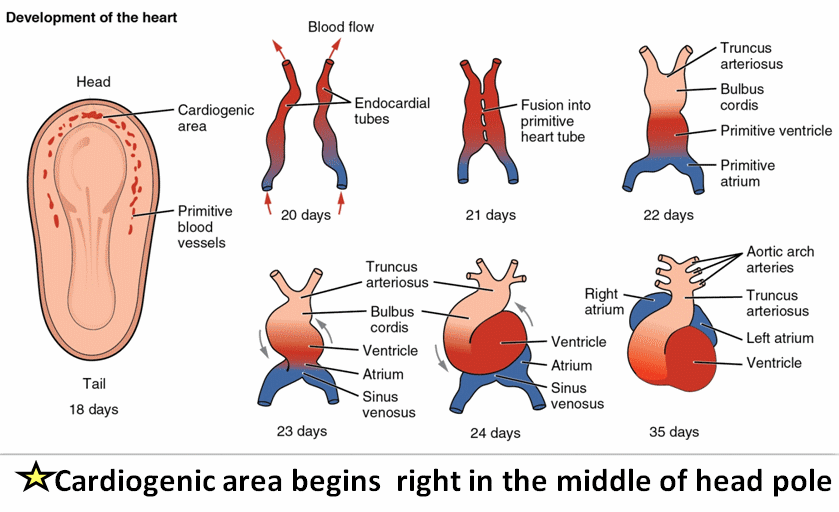
Image Source: drsvenkatesan.com
Development of the Heart (Heart Embryology)
- Cardiogenesis begins with the formation of two endocardial tubes which merge to form the tubular heart, also called the primitive heart tube, that loops and separates into the four chambers and paired arterial trunks that form the adult heart.
- The tubular heart quickly differentiates into the truncus arteriosus, bulbus cordis, primitive ventricle, primitive atrium, and the sinus venosus.
- The truncus arteriosus splits into the ascending aorta and pulmonary artery.
- The bulbus cordis forms part of the ventricles.
- The sinus venosus connects to the fetal circulation.
- The heart tube elongates on the right side, looping and becoming the first visual sign of the left-right asymmetry of the body.
- Septa form within the atria and ventricles to separate the left and right sides of the heart.
The development of the heart can be studied under the following headings:
1. Generation and fusion of the developing heart tubes
- The heart fields are of mesodermal origin and established in the cranial-most end of the embryo.
- The heart fields are patterned into primary and secondary heart fields:
- Primary heart field will develop into left and right atria and the left ventricle
- Secondary heart field will become the right ventricle and outflow tract
2. Repositioning the cardiogenic fields
- Lateral folding brings the forming heart tubes to the midline to fuse into a single tube
- Cranio-caudal folding swings the heart tube into a position just ventral to the foregut pocket in the neck of the embryo with the inflow oriented toward the tail of the embryo and outflow oriented toward the head.
- The heart tube is suspended from the body wall by a sling of connective tissue called the dorsal mesocardium.
- Differential growth of the embryo causes the heart to be displaced toward the tail of the embryo such that the heart ends up in the chest.
3. Folding of the developing heart
- Degeneration of the central portion of the dorsal mesocardium leaves the primitive heart attached at the outflow and inflow ends (this is how the transverse pericardial sinus forms).
- The heart grows rapidly, but it is still fixed at both ends so it loops.
4. Partitioning the Atrio-Ventricular Canal
- The atrioventricular canal is divided by the fusion of the dorsal and ventral AV cushions.
- The AV cushions are formed by the conversion of endogenous heart tissue in the endocardium of the AV canal into mesenchyme that proliferates to form the two “cushions” or swellings that grow toward each other.
- The cushions fuse in the middle of the AV canal to form a left and right AV canal.
- However, the two canals initially empty into the future left ventricle –with additional growth and remodeling, the canals shift toward the middle such that the left AV canal lines up with the left ventricle and the right AV canal lines up with the right ventricle.
5. Partitioning the Atria
- A septum, the septum primum (“first” septum) grows down from the roof of the primitive atrium toward the AV cushions that have fused into a block of tissue dividing the left and right AV canals.
- The foramen primum is the space between the free edge of the septum primum and AV cushions –it becomes progressively smaller as the septum primum grows toward the AV cushions and eventually closes off completely when the septum primum fuses with the AV cushions.
- As the foramen primum is closed off, programmed cell death in the wall of the septum primum near the roof of the atrium opens up a second foramen, the foramen secundum.
- A second, more rigid, septum called the septum secundum grows down on the right side of the septum primum from the roof of the atrium toward the AV cushions.
- However, the septum secundum does not grow all the way down to the cushions but leaves an oval opening, the foramen ovale, located toward the lower back wall of the right atrium (near the opening of the inferior vena cava).
- Over time, the tissues of the two septa grow together with such that they typically become anatomically fused. However, in about 25% of the population, this fusion is not complete and said to be “probe patent” (meaning a probe pushed into the foramen ovale would push open the septum secundum and pass into the left atrium).
6. Partitioning the ventricles
- A ridge of muscular tissue from the wall of ventricles proliferates at the transition from the future left ventricle and future right ventricle to form the “muscular interventricular septum” that almost completely separates the right and left ventricles.
- The upper portions of the left and right ventricles (near the AV valves and semilunar valves) are divided by a much thinner septum known as the “membranous interventricular septum.”
7. Partitioning the Outflow Tract
- Neural crest cells associated with pharyngeal arches 4 and 6 migrate into the truncus arterosus (undivided outflow tract) and conus cordis (aka bulbus cordis, which is the conical-shaped outflow portion of the primitive right ventricle) and transform into mesenchymal tissue that proliferates to form two so-called truncoconal or truncobulbar cushions (or ridges).
- The two truncoconal ridges grow toward each other and fuse first at the truncoconal transition and then “zip” distally (toward the outflow tract) and proximally (toward the ventricles.
- As the ridges zip together, they spiral in a right-handed twist such that the pulmonary trunk ends up anterior to the aorta.
- As the truncoconal ridges grow toward the ventricles, they also contribute a portion of the membranous interventricular septum.
8. Formation of valves in the heart
- Semilunar (pulmonary and aortic) valves are formed via cavitation of truncoconal ridge tissue to form three triangular valve leaflets in each of the outflow vessels in a highly stereotypical pattern:
- the pulmonary semilunar valve develops three cusps: left, right, and anterior
- the aortic semilunar valve also develops three cusps: left, right and posterior
- Atrioventricular (tricuspid and mitral, or bicuspid) valves are formed via cavitation of atrioventricular cushion tissue and ventricular walls to form valve leaflets attached via chordae tendinae to the myocardium (i.e. papillary muscles).
References
- Larsen’s human embryology (5th ed.). New York; Edinburgh: Churchill Livingstone.
- Sadler, T. W., & Langman, J. (2004). Langman’s medical embryology. Philadelphia, Pa: Lippincott Williams & Wilkins.
- Moore, K. L., Persaud, T. V. N., & Torchia, M. G. (2008). The developing human: Clinically oriented embryology. Philadelphia, PA: Saunders/Elsevier
- https://embryology.med.unsw.edu.au/embryology/index.php/CardiovascularSystem-HeartDevelopment
- https://www.amboss.com/us/knowledge/Heart
- https://web.duke.edu/anatomy/embryology/cardiovascular/cardiovascular.html
- https://step1.medbullets.com/embryology/103009/cardiac-development