Disclaimer: The information and images presented on this page are for academic, study, and educational purposes only. The terms, keywords, and images used on this page may not be suitable for some users, but they are regular terms for the human female reproductive system used only for educational purposes as a part of study information.
The group of organs, cells, and hormones working together from the formation of the female gamete, ova, to the parturition is the female reproductive system. The organs in the female reproductive system are divided into external and internal organs.
Interesting Science Videos
External organs of the female reproductive system
- Labia majors
- Labia minora
- Clitoris
- Vaginal orifice
- Vestibular gland (Bartholin’s gland)
- Hymen
Internal organs of the female reproductive system
- Vagina
- Uterus
- Uterine tubes (Fallopian tubes)
- Ovaries
External Genitalia (Vulva)
The external organs in the female reproductive system are collectively termed, vulva consisting of labia majora, labia minora, clitoris, vaginal orifice, vestibular gland, and hymen.
Labia majors
- These are large muscular folds surrounding the vulva, comprised of akin, fibrous tissue, fat, and a large number of sebaceous glands.
- Anteriorly, the folds join together in front of the pubis whereas anteriorly, they merge with the perineum.
Labia minora
- A smaller fold present beneath the labia majora is the labia minora. It is also provided with a large number of sebaceous glands.
- The cleft between the two folds is called the vestibule, which is the opening for the vagina, vestibular glands, and urethra.
Clitoris
- Clitoris has numerous sensory nerve endings and corresponds to the penis in Male. They have no reproductive significance.
Hymen
- The hymen is a thin mucosal membrane present in front of the opening of the vagina.
Vestibular glands
- Vestibular glands (Bartholin’s gland) are small pea-sized glands with ducts opening into the vestibule. These are present on either side of the vaginal opening
- The secretion of these glands helps to keep the vagina moist.
Internal Genitalia
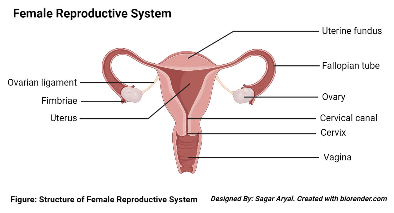
The internal genitalia is present in the pelvic cavity of a female and consists of a uterus, a vagina, two uterine tubes, and two ovaries.
Ovaries
An ovary is the gonad of the female reproductive system which produces an ovum that develops into a zygote after fertilization.
Structure of Ovaries
- Ovaries are female gonads or glands that exist in pairs in humans and each lie on the ovarian fossa present on the lateral wall of the pelvis. Birds have only one functional ovary with the other being vestigial.
- They are 2-3 cm long, 2 cm wide, and 1 cm in thickness. The ovaries are attached to the upper part of the uterus by the ovarian ligament called mesovarium and are connected to the fallopian tubes by a deep tissue called an infundibulopelvic ligament.
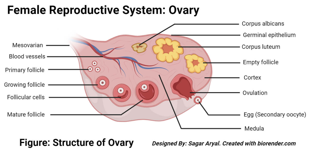
- An ovary is surrounded by a capsule which encloses an outer cortex and an inner medulla. The capsule is a fibrous connective tissue called tunica albuginea.
- Medulla: It lies in the middle of the ovary and consists of blood vessels, fibrous tissue, and nerves. Amphibians and reptiles have no ovarian medulla where the inside is a lymph filled space.
- Cortex: Cortex is present around the Medusa and has a framework of fibrous tissue or stroma surrounded by germinal epithelium. It contains ovarian follicles in different stages of development with an ovum. The follicular cells are flattened epithelial cells derived from the germinal epithelium surrounding the ovary.
Function of ovaries
The ovaries have two distinct functions.
- Gamete production: Ovaries are the site of periodic development and release of the egg cell or female gamete by the process of ovulation. The developing egg cell mature in the fluid-filled follicles. At once, only one egg cell is released. However, different cells can also mature simultaneously.
- Hormone secretion: Ovaries secrete a set of hormones including the estrogen, testosterone, progesterone, and inhibin which are involved in the development of secondary s*xual characteristics and the maintenance of the reproductive function of the ovaries.
Uterine tubes (Fallopian tubes)
Uterine tubes, also known as oviducts, are the part of the female reproductive system that carry ova from the ovary to the uterus each month. After fertilization of the ovum and sperm, these tubes transport the fertilized egg to the uterus for implantation.
Structure of uterine tubes
Each uterine tube is 10-13 cm long with a 1-2 cm diameter that extends laterally from the superior part of the uterus.
A Uterine tube consists of four parts:
- The infundibulum is a funnel-shaped structure that opens into the peritoneum of abdominal Ostia. It is found with associated fimbriae which are finger-like mucosal projections. These fimbriae projects over the ovaries with the longest of the fimbriae called ovarian fimbriae that attach to the superior aspect of the ovum.
- The ampulla is the longest part of the tubes with a length of 5cm. It has a thin wall with the folded luminal surface, and the fertilization occurs in this lumen.
- The isthmus is the narrower part of the tube that extends towards the uterus.
- The intramural (interstitial) part that transverses the myometrium of the uterus.
- The channel is surrounded by a layer of mucous membrane folded into papillae. The inner layer is of simple columnar epithelium that has cells with minute hair-like structures called cilia.
- IN between the ciliated cells are peg cells that secrete a fluid that provides nutrients for spermatozoa, oocytes, and zygotes.
Functions of uterine tubes
- The primary purpose of uterine tubes is to facilitate the transport of ovum from the ovary to the uterus. This transport is aided by the peristaltic movement of the muscles and the rhythmic movement of the ciliated cells.
- During fertilization, the spermatozoa travel through the tube towards the ovum and fertilization usually occurs in the ampulla. After fertilization, the secretions of the tubes provide nutrients to the zygote.
Uterus
The uterus is a hollow muscular pear-shaped organ of the female reproductive system which is narrow both anteriorly and posteriorly.
Structure of uterus
- It lies in between the urinary bladder and the rectum. In an adult female, the uterus weighs about 30-40 grams and is approximately 7.5cm long, 5cm wide with 2.5cm thick walls.
- The wall of the uterus is comprised of three layers viz—Perimetrium myometrium and endometrium.
- Perimetrium is the outermost layer of the uterus which is folded anteriorly to form the vesicouterine pouch. Posteriorly, it extends over the uterus to the rectum to form the rectouterine pouch.
- Myometrium is the middle layer which is also the thickest layer in the uterine wall, which is composed of smooth muscles, connective tissues, blood vessels, and nerves.
- The endometrium is the innermost layer that is composed of columnar epithelium provided with a large number of mucus-secreting tubular glands.
- The endometrium is further divided into their layers with the upper layer that thickens during the first half of the menstrual cycle. The basal layer is present towards the myometrium. The upper layer of the endometrium is shed during the menstrual cycle, whereas the basal layer is not lost.
- Externally, the uterus is divided into three parts, the fundus, the body, and the cervix. The fundus is the dome-shaped part present above the opening for the uterine tubes. The body is the main part of the uterus, which is connected to the cervix. The cervix or ‘the neck of the uterus protrudes out through the anterior wall of the vagina.
Functions of uterus
- The primary purpose of the uterus is to provide a feasible environment for a newly developing baby.
- Uterine secretions provide nourishment to the ovum before its implantation on the wall and, later after fertilization, the rapidly expanding ball of cells is nourished by the endometrial cells.
- The uterus undergoes a periodic shedding after puberty in the female, which is called the menstrual cycle. This shedding prepares the uterus to nourish and protect a fertilized ovum.
Vagina
The vagina is a fibromuscular tube that is lined with stratified squamous epithelium that connects the internal and external organs of the female reproductive system. The vagina is present obliquely upwards bent at an angle of 45° with the bladder in front and rectum and anus behind.
Structure of the vagina
- The vagina is surrounded by three layers, the outer layer of areolar tissue, a middle layer of smooth muscle, and the inner layer of stratified squamous epithelium that forms ridges or rugae.
- Vagina has no secretory cells; however, the surface is kept moist by the cervical secretions.
- Between puberty and menopause, Lactobacillus acidophilus is found abundantly in the vagina. This organism secretes lactic acid that maintains the pH of the vagina between 4.9-3.7. This acidity inhibits the growth of other microorganisms that may reach the vagina through the perineum.
Functions of vagina
- The vagina is the receptor for the penis during coitus or s*xual reproduction.
- Vagina also provides an elastic passage for the baby during childbirth.
Hormones involved in the female reproductive system
The hypothalamic releasing hormone, gonadotropic hormone (GnRH): This hormone stimulates the anterior pituitary to release s*x hormones, Follicle Stimulating Hormone (FSH) and Luteinizing Hormone (LH).
FSH and LH: These hormones stimulate the production of ovarian hormones estrogen and progesterone and also support the proliferation of follicles on the wall of the ovary.
Estrogen hormone: This hormone supports the proliferation of egg follicles and helps in the maintenance and development of female characteristics in the human body.
Progesterone hormone: Progesterone hormone regulates the inner lining (endometrium) of the uterus and also functions in the release of the egg during ovulation.
References and Sources
- Hall JE and Guyton AC. (2011) Textbook of Medical Physiology. Twelfth Edition. Elsevier Saunders.
- Waugh A and Grant A. (2004) Anatomy and Physiology. Ninth Edition. Churchill Livingstone.
- 3% – https://basicmedicalkey.com/the-reproductive-systems/
- 2% – https://www.kenhub.com/en/library/anatomy/fallopian-tubes
- 1% – https://www.medicalnewstoday.com/articles/inside-the-vagina
- 1% – https://www.innerbody.com/image_repfov/repo11-new.html
- 1% – https://www.dummies.com/education/science/anatomy/organs-female-reproductive-system/
- 1% – https://www.differencebetween.com/difference-between-endometrium-and-vs-myometrium/
- 1% – https://www.britannica.com/science/progesterone
- 1% – https://wikimili.com/en/Ovary
- 1% – https://quizlet.com/248765778/reproductive-system-flash-cards/
- 1% – https://quizlet.com/16386285/chapter-10-ap-of-the-female-reproductive-system-flash-cards/
- 1% – https://medhyaherbals.com/female-reproductive-system-functioning-organs-diseases/
- 1% – https://healthfully.com/94700-main-organs-reproductive-system.html
- 1% – https://en.wikipedia.org/wiki/Ovaries
- 1% – https://en.wikipedia.org/wiki/Follicle-stimulating_hormone
- 1% – https://courses.lumenlearning.com/boundless-ap/chapter/the-female-reproductive-system/