Euglena viridis (Gr.,eu, true+ glene, eyeball or eye pupil+L., Viridis, green) is a unicellular green organism with an eye-like photoreceptive structure. It is phytoflagellate as it possesses both chloroplasts as well as flagella. It is autotrophic in sunlight but becomes heterotrophic in dark.
Systematic position
Phylum: Protozoa
Subphylum: Sarcomastigophora
Superclass: Mastigophora
Class: Phytomastigophorea
Order: Euglenida
Genus: Euglena
Species: viridis
Habits and Habitat of Euglena viridis
- It is a solitary and free-living freshwater flagellate.
- It is found in great numbers in stagnant freshwater ponds, pools, ditches, and slowly-running streams, etc., containing a considerable amount of vegetation.
- It is fairly active and often found at various depths below the surface of the water.
- Ponds in well-maintained gardens, containing decaying nitrogenous organic matter, such as feces of animals, leaves, twigs, etc., are a good source of this organism.
- It multiplies rapidly and forms green scum on the water surface (like algal blooms)under favorable conditions.
Culture of Euglena viridis
- Euglena can be cultured and studied in the laboratory.
- For the culture, the clean and wide-mouth large bottles are filled with boiled tap or pond water and boiled wheat grains are added to each bottle. Then, the bottles are kept in a sunny place for a week. The wheat grain is added monthly to maintain the culture.
- Another method, a culture of Euglena in manure solution (horse or cow manure) is boiled in the pond or distilled water in a jar. These manure solutions are allowed to cool for 2 days and some weeds from the pond containing Euglena were put in a jar. Then, the jar was kept near the well-lightened window. In few days, Euglena will appear in this nitrogenous infusion.
Structure of Euglena viridis
1. Shape and size
- It is a small microscopic, unicellular organism measuring about 40-60µ in length and 14-20µ in breadth at the thickest part of the body.
- It is elongated and spindle-shaped in appearance.
- The anterior end is blunt, the middle part is wider and the posterior part is pointed.
- The whip-like flagellum arises from the anterior end.
2. Pellicle
- The body is covered by distinct, thin, elastic, and tough, and strong pellicle or periplast which lies beneath the plasma membrane.
- It is made of protein, not cellulose so that it is not homologous to the plant cell wall.
- It is flexible enough to permit movements.
- It has spiral or parallel striations, called myonemes all around.
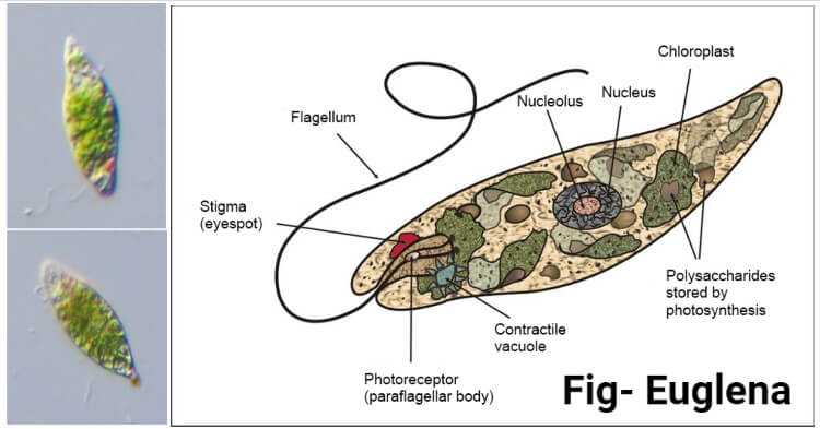
Figure: Euglena diagram. Image Source: Wikipedia.
Electron structures of pellicle
- Electron microscopy of pellicle shows that it is made of thin, firm, elastic, and helically disposed of strips.
- This pellicular strips fuse at both the ends of the cell body and bear a groove along one edge and a ring along with the other.
- The edges of adjacent strips overlap and articulate along their entire length in such a way that the ridge of one fits into the groove of the other.
- Under the light microscope, these articulating edges appear as striations, called myonemes. BEneath the pellicle are present arow of mucus-secreting muciferous bodies and bundles of microtubules.
3. Cytoplasm and cytopharynx
The cytoplasm is divisible into two zones: The ectoplasm is a clear, dense, and outer or peripheral zone, and the endoplasm is a fluid-like granular central zone. which contains:
(a) Nucleus
- A single, large, spherical, or oval nucleus lies near the middle of the endoplasm or towards the posterior end of the body.
- It has a distinct double-layer nuclear membrane that is perforated by pores.
- It contains a large solid central body or nucleolus of chromatin, called endosome or karyosome.
- Chromatin forms the small granules in the space between the nuclear membrane and the endosome.
- Electron microscopy has revealed that the nucleoplasm contains several nucleoli and a large number of granular and thread-like chromosomes.
(b) Contractile vacuole
- It is a large osmoregulatory body and is found close to the reservoir.
- It includes a large central contractile vacuole.
- It remains surrounded by several minutes, accessory contractile vacuoles which later on fuse together to form the large vacuole
- It discharges its watery contents along with some waste products of metabolism to outside via the reservoir, cytopharynx, and cytostome.
(c) Stigma
- It lies near the reservoir, on the side opposite to the contractile vacuole, is a small discoid or shallow cup-shaped red pigment spot, the stigma or the eye-spot which function as a lens.
- According to Leedale (1966), it is composed of a plate of oil droplets, each charged with red pigment, the carotenoid.
- The stigma and the paraflagellar body ( a small swelling lies on either one root or at the junction of 2 roots of the flagellum which is sensitive to light) together form a photoreceptor apparatus.
- It serves as a cover or shield over the light-sensitive paraflagellar body.
(d) Chloroplasts or chromatophores
- There are a large number of elongated rod-like bodies called chloroplasts or chromatophores.
- They are green bodies that give Euglena its green color because they contain green pigments, chlorophyll a and b, along with ß-carotene.
- The chloroplast of E. viridis are slender and radiate from a central point so as to form a star- shape grouping.
- The central body of each chloroplast is a single proteinaceous pyrenoid which is surrounded by a number of small granules of paramylum (a polysaccharide).
- Paramylum is a polysaccharide (ß-1, 3 glucans) similar to starch but not identical with it, s it is not colored blue with iodine solution. It is the refractile body and the reserve food of Euglena and is formed by the products of photosynthesis. These paramylum bodies may be so numerous that they interfere with the observation of the chloroplast. If the Euglena, is kept in dark for several days, the granules for paramylum become less and the chloroplasts are more easily seen.
- A chloroplast contains a group of chlorophyll-bearing lamellae or thylakoids, each with 3 lamellae. These are placed in the matrix or stroma, also containing ribosomes and fat globules.
- Each chloroplast is bounded by a triple membrane envelope ( Sleigh, 1973).
(e) Other cytoplasmic content
- The cytoplasm also contains Golgi bodies, endoplasmic reticulum, mitochondria whose number is more near the reservoir, and the ribosomes which are found scattered in the endoplasm, on the endoplasmic reticulum, and in the chloroplast.
4. Reservoir
- It is a permanent flask-shaped invagination at the blunt anterior end of the body.
- It consists of a wide reservoir or flagellar sac which leads through a short tubular canal, the cell gullet or Cytopharynx, to the outside.
- Its external opening 1s called the cell mouth or cytostome.
- It is lined by a plasma membrane without a pellicle beneath it.
5. Flagellum
- At the anterior end of the body, a single, long,thread-like flagellum arises through the cytostome.
- It is typically made of an axial elastic filament or axoneme, covered by a protoplasmic sheath.
- The length of flagellum differs in different species of Euglena but in E. viridis it is as long as the body of the animalcule.
- Electron microscopy has revealed that the flagellum is not just one but paired, the other being smaller and confined within the reservoir. Each flagellum originates from a tiny granule, the blepharoplasty (Gr., blepharon, eyelid +plastos, formed), or kinetosomes, located in the cytoplasm near the base of the reservoir.
- The long flagellum bears a lateral swelling, the paraflagellar body near its base within the reservoir which acts as a photoreceptor and probably contains lactoflavin as a sensitizer.
- The animal moves with the help of a long flagellum only. The short flagellum does not come out of the reservoir.
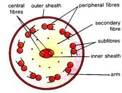
Figure: T.S. of a Euglena flagellum. Image Source: Biology Discussion.
Electron structure of a flagellum
- Each flagellum consists of 2 central and 9 peripheral fibers.
- The central fibers are enclosed in an inner membranous sheath.
- Each central fiber is single, whereas the peripheral fibers are paired, which is each made of 2 sub-fibers.
- One of the 2 sub-fibers of each peripheral bears a double row of short projections or arms, all pointing in the same direction.
- The whole flagellum is covered by an outer protoplasmic sheath which is continuous with the plasma membrane.
- Nine secondary fibers are present in the space between the peripheral and central fibers which are somewhat inconspicuous.
- on the long axis of the flagellum is found a unilateral row of hair-like contractile processes, called mastigonemes.
- According to Manton (1959), these processes arise laterally from two of the nine peripheral fibers. This type of flagellum is known as stichonematic.
Locomotion of Euglena viridis
Euglena viridis performs 2 different kinds of movements: (1) flagellar and (2) euglenoid.
1. Flagellar movement
- Euglena swims freely in water with the help of a single, long locomotory flagellum by whipping, twisting, and turning it around like that of a propeller.
- The locomotory flagellum is equal to the length of the Euglenoid’s body and it highly helps it to swim freely in the water.
- During swimming, the flagellum is directed obliquely backward towards the side bearing the stigma.
- This flagellum undergoes spiral undulations, with waves, that are transmitted from the base to the tip, causing its beating or the sideways lashing.
- The flagellum undulates or beats on average, at the rate of about 12beats per second.
- The beating of the flagellum creates water waves that drive the water backward and induces the body to move forward.
- Each beat not only throws the body forward but also to one side. Thus, when the beats are repeated over and over, the Euglena revolves in a circle or gyrates.
- As the flagellum is directed obliquely backward to the long axis of the body, the organism rotates on its axis.
- It has been calculated that Euglena rotates at the rate of one turn per second.
- The three different types of movement of the Euglenoid body caused by the locomotory Flagellum are forward movement, rotational movement, and revolutionary movement.
- The movement of a flagellum involves the contraction of its 9 peripheral fibers. Their position is ideal for undulating actions as they could exert bending around the flagellar axis. The energy for the contractile action of fibers is supplied by ATP(adenosine triphosphate) formed in mitochondria, which are included in the blepharoplasts.
2. Euglenoid movement
- This type of movement is usually possible due to the presence of a Pellicle on the surface of their body. The pellicle is flexible and contractible which enables Euglena to perform peristaltic movements.
- The peristaltic movements or the very peculiar slow worm-like wriggling or writhing movements cause the formation of the peristaltic waves of contraction and expansion of the pellicle layers. This contraction is brought about by the stretching of protoplasm on the pellicle or by localized fibrils called myonemes in the cytoplasm.
- These waves pass over the entire body from the anterior to the posterior end and the animal moves forward.
- As the peristaltic waves pass, the body becomes shorter and wider first at the anterior end, then in the middle, and later at the posterior end.
- Due, to this the adjacent pellicular strips bend and move against one another, probably the ridge of one sliding in the groove of the other.
- The sliding of the ridges in the grooves is lubricated by the secretion of underlying muciferous bodies.
Nutrition of Euglena viridis
Euglena is the connecting link between the Plant and Animal kingdom. So, they have the characters of both plants as well as animals. so, the mode of nutrition is E. viridis is mixotrophic,i.e., the nutrition is accomplished either by autotrophic or holophytic as well as saprophytic or saprozoic.
1. Autotrophic or holophytic nutrition
- It is the chief mode of nutrition in Euglena. Like a true plant, it can manufacture its own food in the presence of sunlight, by the process of photosynthesis with the help of chlorophyll present in the chloroplast.
- The chlorophyll absorbs energy from sunlight. With this energy, water reacts with carbondioxide in a series of steps forming hexose sugar. This is then transferred into a kind of polysaccharide, called paramylum or paramylon.
- Euglena remains an autotroph so long as it is in light and is provided with essential inorganic compounds.
- The whole autotrophic process in Euglena is dependent upon external sources of vitamin B12 which is synthesized by bacteria and some microorganisms.
- At times when pond water becomes polluted with dead and decaying organic matter, it switches over to a saprozoic mode.
2. Saprophytic or saprozoic nutrition
- In prolonged darkness, Euglena loses its chlorophyll and green color. It becomes etiolated, that is, becomes pale and white, yet it continues to live and perform all the life activities.
- In the absence of sunlight, Euglena lives by the saprophytic or saprozoic methods, which means the products of decaying organic matter dissolved in surrounding water are absorbed through its general body surface(mainly through the pellicle).
- Euglena secretes digestive enzymes that are typically animal-like in nature. Digestion is carried on by enzymes secreted into the food vacuole by the surrounding cytoplasm. These enzymes do help in the breakdown of dead organic matter into simple molecules for the derivation of food, nutrition, and proper energy.
- Generally, the chloroplast lost in dark are regained in the light except in E. gracilis.
Pinocytosis has also been observed to take place at the base of the reservoir for the intake of protein and other large molecules.
Respiration in Euglena viridis
- The respiration in Euglena viridis is aerobic. It respires with the help of free oxygen dissolved in water, which diffuses through the pellicle.
- During the daytime, a good amount of oxygen is liberated in the process of photosynthesis which is utilized for the purpose of respiration.
- The oxygen brings about oxidation reactions catalyzed by enzymes present in the mitochondria.
- The energy so liberated is entrapped in the high-energy phosphate bonds of ATP, which supplies energy for metabolic activities. As a result of oxidation reactions, water and carbon dioxide are formed as by-products.
- In sunlight, this carbondioxide is utilized for photosynthesis, but in dark it is liberated to the outside by diffusion through the general body surface.
Osmoregulation and excretion in Euglena viridis
- The removal of excess water from the body is known as osmoregulation. In Euglena, the removal of excess water entering the body by endosmosis is performed by contractile apparatus.
- In E. viridis, the contractile apparatus consists of a large contractile vacuole surrounded by numerous small accessory vacuoles.
- The cytoplasm secretes the excessive water into this smaller vacuole which, in their turn, drains into the larger vacuoles.
- The larger vacuole finally empties into the reservoir. The process involves the diastole (increase in volume) and systole (decrease in volume) of the large contractile vacuole. In diastole, the contractile vacuole is filled with water, while in systole it is emptied to throw its water into the reservoir.
- From the reservoir, the fluid escapes through the gullet. Along with this, water-soluble wastes are thrown out of the body.
- Ammonia, the nitrogenous waste product, resulting from catabolism, passes out through the general body surface by diffusion.
- Excretory substances may also be emptied by the contractile vacuole into the reservoir. It has been suggested that a dense zone of cytoplasm around the contractile apparatus is both osmoregulatory as excretory in function. It secretes water as well as excretory products into the lumen of the vacuole.
Behavior of Euglena viridis
Euglena reacts to a variety of stimuli in the same manner as protozoa do.
1. Reaction to light
- Euglena is very sensitive to light. It avoids strong light as well as shady areas but reacts positively to a moderate intense light such as that from a window.
- It orients itself parallel to a beam of ordinary light and swims towards the source of illumination like motile and free-living unicellular organisms.
- In cultural dishes, most of them are found to aggregate on the side towards the light.
- If a dish containing Euglenae is placed in the direct sunlight and then one-half of it is covered, the organism will avoid the shady part as well as direct sunlight but gather in a small band between the two. So, the light best suited for them is optimum.
2. Shock reaction
- A swimming Euglena moves in a spiral manner rotating and gyrating around its own axis but shows a shock reaction when the direction of light is changed.
- Investigations of Mast and his friends have clearly revealed the orientation of Euglena is by photoreceptive stigma. It swims towards the source of light i.e. phototactic.
- It adopts a spiral course, rotating and gyrating around its body axis s that the paraflagellar body remains continuously exposed to light and uniformly illuminated.
- If the front source light is screened and a new lateral beam of light is thrown, then Euglena produces a shock reaction and, by trial and error, reorients itself towards the new source of light.
- The shock reaction is explained as: when the sigmal side of the body (eyespot) faces a lateral beam of light, the paraflagellar body is shaded by the stigma once in each rotation. Thus, the base of the flagellum is alternately darkened and illuminated in lateral light.
- Each darkening of the paraflagellar body excites in photoreceptor to produce a minor shock reaction. This affects flagellar action in such a way that the body bends at right angles, turning the flagellar end gradually towards the new light source.
3. Avoiding reaction
- Euglena also responds to mechanical, thermal, and chemical stimuli showing an avoiding reaction on atrial and error patterns ( phobotaxis).
- In most cases, it slows down, stops, or even moves backward, turns strongly towards the dorsal surface, but continues to revolve on its long axis.
- The posterior end of the body may acts as a pivot, while the anterior end traces wider circles of gyrations and swims forward in a new spiral path.
Reproduction of Euglena viridis
Euglena viridis reproduces asexually by binary and multiple fissions and undergoes encystment. There is no evidence of sexual reproduction in it. They reproduce by longitudinal binary fission under favorable conditions. The longitudinal binary fission is always symmetrogenic( i.e., the parental Euglena divides into two daughter individuals, where one is the plane mirror image of the other).
1. Binary fission
- The transverse binary fission is not known in Euglena.
- Under the favorable condition of water, temperature, and food availability, they divide by simple longitudinal binary fission. The longitudinal binary fission is always symmetrogenic( i.e., the parental Euglena divides into two daughter individuals, which are identical to one another).
- The most important part of binary fission is the division of the nucleus (genetic material) into two by mitosis which is followed by the division of cytoplasm (cytokinesis).
- Mitosis consists of 4 stages. In the 1st stage( prophase), where all the nucleoli (endosomes) fuse together into a single nucleolar body, and each chromosome splits longitudinally into 2 daughter chromosomes or chromatids.
- In the second stage (metaphase), paired chromatids come to lie in a longitudinal plane (the equator). The microtubules are present in the nucleus but do not form the spindle.
- In the third stage(anaphase) the paired chromatids are separated and moved towards their respective poles. It has been suggested that the movements of chromatids are autonomous, with mutual repulsion. The nuclear membrane begins to constrict longitudinally.
- In the fourth stage (telophase) the constriction of the nuclear membrane deepens and the nucleus is finally separated into two daughter nuclei. The nucleolar body also splits into 2 halves, each taking its place in the daughter nucleus of its own side.
- The next step is cytokinesis. A longitudinal furrow appears in the cytoplasm, beginning at the anterior end, which deepens and finally divides Euglena into 2 daughter euglenae.
- All organelles of anterior ends, such as the blepharoplasts, reservoir, cytopharynx, and stigma, etc. are all duplicated.
- However, a new set of flagella rises from new basal bodies that appear in their vicinity of old basal bodies. Multiplication of basal bodies usually precedes cell division.
- Some observers have reported the complete disappearance of the entire locomotory apparatus during division, and each daughter cell reconstructs a new set.
2. Multiple fission and palmella stage
- Under inactive periods, Euglena undergoes multiple fission in an encysted condition.
- Encystment is usually followed by repeated longitudinal binary fission with the formation of(16-32) daughter englenas. Under favorable conditions, the flagellate comes out of the cyst and passing a short period through the amoeboid stage develop into adult Euglena.
- Under unfavorable conditions, the large number of euglenae come closer, the movements cease altogether, the flagellum is thrown off and becomes rounded and embedded in an extensive thick and mucilaginous coat or cyst which is secreted by the muciferous bodies this condition is known as the palmella stage. which is seen as extensive green scums on the surface of ponds.
- Individuals of the palmella stage continue metabolism and reproduction which occurs by binary fission. On the arrival of favorable conditions, the gelatinous covering burst,euglenae are released acquire flagella and grow into adult euglenae.
Encystment of Euglena viridis
- Encystment takes place as a protective measure to ride over unfavorable conditions such as lack of food and oxygen, drought, excessive heat, etc.
- A cyst wall is thick made of 20r 3 concentric layers, spherical, yellowish-brown, and gelatinous covering, composed of a special carbohydrate.
- The cysts are usually small, their total width is equal to the diameter of the animal, it may be larger sometimes.
- The cyst may be thick (2-3 layered), staked, or operculated with the organism lying centrally or eccentrically in it.
- The cysts are protective structures that help the organisms to withstand unfavorable circumstances and also help far and wide dispersal.
- When the favorable conditions come out, the cysts dissolve and the animals emerge to resume their normal free-swimming life.
Euglena as an animal
Euglena is studied as an animal as well as a plant. It is more an animal than a plant because of the following reasons:
- Absence of a cellulose cell wall overlying the plasma membrane.
- Presence of centrioles forming blepharoplast or kinetosomes.
- Presence of contractile vacuole, which are not found in plants.
- Reserve food in paramylum which is not a true starch.
- Presence of paraflagellar body, a sensory( photoreceptive) organelle.
- Moves from place to place like an animal.
- Response to various stimuli like an animal.
- Pinocytosis and probably holozoic nutrition take place.
- Presence of longitudinal binary fission, which are not found in plants.
References and Sources
- Kotpal RL. 2017. Modern Text Book of Zoology- Invertebrates. 11th Edition. Rastogi Publications.
- Jordan EL and Verma PS. 2018. Invertebrate Zoology. 14th Edition. S Chand Publishing.
- https://www.biologydiscussion.com/invertebrate-zoology/protozoa/euglena-viridis-habitat-structure-and-locomotion-protozoa/28141- 18%
- https://onlyzoology.com/how-does-euglena-move/- 5%
- https://www.biology-today.com/general-zoology/invertebrate-zoology/general-features-of-euglena-viridis/- 3%
- https://www.biologydiscussion.com/animals-2/phylum-protozoa/study-notes-on-euglena-viridis-with-digram/32548- 3%
- https://onlyzoology.com/euglena-facts-fascinating-facts-about-euglena/- 2%
- https://onlyzoology.com/how-does-euglena-eat-nutrition-in-euglena/INTERNET2%https://www.jesjalna.org/zoology/pdf/notes%20fy/Euglena.pdf- 1%
- https://www.sciencedirect.com/topics/biochemistry-genetics-and-molecular-biology/oxidative-enzyme- <1%
- https://www2.estrellamountain.edu/faculty/farabee/BIOBK/BioBookmito.html- <1%