Reproductive system of earthworm
- Earthworms do not reproduce asexually.
- Reproductive organs are somewhat complicated.
- The pheretima is monoecious or hermaphrodites having both male and female reproductive organs in the same individual.
- They cannot fertilize their own eggs because they are protandrous.
- Cross-fertilization occurs as a rule because of the relative position of female and female genital apertures.
- Fertilization is preceded by copulation and cocoon formation.
- The male gametes mature first and earlier than female gametes so that self-fertilization is prevented.
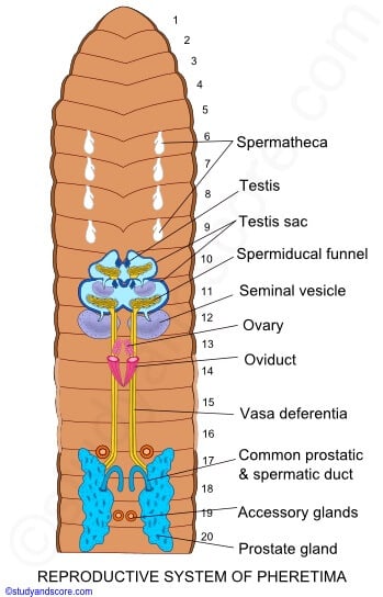
Image Source: Study and Score.
Male reproductive organs of earthworm
It consists of testes, testis sacs, seminal vesicles, vasa deferentia, prostate glands, and accessory glands.
1. Testes
- 2 pairs of very minutes, whitish and lobed structures testes.
- Situated one pair each in segment 10th and 11th segment and attached with the posterior surface of 9/10 and 10/11 intersegmental septa.
- Lie ventrolaterally beneath the alimentary canal, close to the mid-ventral line, on either side of the nerve cord, and attached to the anterior wall of their respective testis sacs.
- Each testis consists of 4 to 8 finger-like lobules projecting from a compact base.
- Each lobule of the testis contains rounded cells in masses called spermatogonia.
- Well-developed only in the young stage of the worm and become degenerated in adult worm.
2. Testis sacs
- Both the testes of each segment are enclosed within a wide bilobed, thin-walled and fluid-filled sacs called testis sac.
- Thus, there are 2 testis sacs situated in 1o and 11 segments on the ventrolateral sides of the ventral cord below the alimentary canal.
- Each testis sacs encloses a pair of testes and a pair of ciliated spermiducal funnels.
- The testis sacs remain in communication with the seminal vesicles.
- Testis sacs of the 11th segment are large enough so as to enclose also the seminal vesicles of that segment.
- The spermatogonia are shed into the testis sacs and pass on into seminal vesicles where they undergo maturation and form spermatozoa.
3. Seminal vesicles
- There are 2 pairs of the large, white, sac-like body called seminal vesicles.
- Lie on segments 11th and 12th into which the testis sacs open by narrow ducts.
- Also referred to as septal pouches since they grow as outgrowths of the septa.
- The anterior seminal vesicles are smaller than the posterior ones.
- The seminal vesicles of the 11th segment are found enclosed in the posterior larger testis sac.
- while those of the 12th segment are exposed in the coelomic cavity.
4. Vasa deferentia
- Each spermatic funnel(posteriorly) leads into a thin, narrow, thread-like sperm duct or vas deferens.
- 2 vasa deferentia of the same side run close together posteriorly along the ventral body wall up to the 18th segment to join the prostatic duct.
5. Prostate glands
- These are pairs of large, white, solid, and irregularly shaped glandular masses
- situated on either side of the gut in the segments from 16th or 17th to 20th or 21st.
- Each gland consists of big glandular parts and small non-glandular parts.
- The glandular part is a racemose gland consisting of several lobes closely fixed together.
- The non-glandular portion consists of several small ductules that unite to form a short, thick, muscular, and curved prostatic duct in the 18th segment.
- The ducts join the 2 vasa deferentia of its side and these 3 ducts are enclosed in a common sheath to for, a common prostatic spermatic duct.
- Both common ducts curve inwards to open to the exterior independently by a pair of male genital pores ventrally on the 18th segment.
- Prostate glands manufacture a fluid, the prostatic fluid, of not definitely known.
6. Accessory glands
- In each of the segments, 17th and 19th are found a pair of rounded, white fluffy masses, called accessory glands.
- Situated on the ventrolateral body wall, one on either side of the nerve cord.
- These glands open to the exterior by a number of small ductules on the 2 pairs of genital papillae, situated externally upon the 17th and 19th segments, one on either side of the mid-ventral line.
- The secretion of these glands helps probably in uniting the 2 worms together during copulation.
- The spermatogonia are shed into testis sacs from testes and pass on to the seminal vesicles where spermatogenesis is completed, and tailed spermatozoa are formed.
- Mature sperm move back into testis sacs, enter spermiducal funnels, travel along vasa deferentia, and discharge through male genital apertures along with the secretion of prostate glands.
Female reproductive organs of earthworm
The female reproductive organs consist of ovaries, oviducts, and spermathecae.
1. Ovaries
- A pair of white, small, lobulated ovaries.
- Lies on the 13th segment attached to the posterior face of septum 12/13in front of it, one on either side of the ventral nerve cord.
- Made of the finger-like process with developing ova in arrow providing it beaded appearance.
- Ova remains in each ovarian lobe in various stages of development being mature in the distal part and immature in the proximal part.
2. Oviducal funnels
- Is a large-saucer-shaped with much folded and ciliated margin.
- Lies immediately below each ovary in the 13th segment.
3. Oviducts
- Each ovarian funnel leads into a short, conical, and ciliated oviduct.
- The oviducts of both sides run backward perforate septum 13/14.
- And converge to meet below the nerve cord, forming a very short common oviduct.
- It opens to the exterior by a single median female genital aperture, situated mid-ventrally on the 14th segment.
4. Spermathecae
- There are 4 pairs of small flask-shaped structures, called spermathecae or receptacula seminales.
- 1 pair each in segments 6th, 7th, 8th, and 9th situated ventrolaterally.
- Each spermathecae has a broad, pear-shaped body, the ampulla.
- The ampulla is continued into a narrow duct-the neck with a small diverticulum or blind caecum on its inner side.
- Also called seminal receptacles because they store the spermatozoa from another worm during copulation.
- The 4 pairs of spermathecae open to outside by 4 pairs of separate spermathecal pores situated inter-segmentally between segments 5/6, 6/7, 7/8, and 8/9.
- In pheretima, unlike other earthworms, the sperms are store in diverticula not in the ampulla.
- Mature ova shed from ovaries are entangled by Oviducal funnels, travel along oviducts.
- And pass out to the exterior through the female genital aperture, to be laid inside the cocoon.
Copulation and fertilization of Earthworm
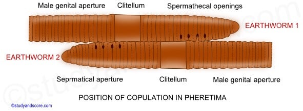
Image Source: Study and Score.
- Copulation has been observed in different species of earthworms.
- Usually occurs in the rainy season during months of July to October at morning hours before sunrise.
- Earthworms are bisexual, still, self-fertilization does not occur because they are protandrous.
- Copulation is a reciprocal cross-fertilization that occurs between two worms where spermatozoa of one earthworm are transferred to another.
- The copulation lasts for 1 hour. There is no penis or vagina for the transfer of sperm.
- During copulation, 2 worms of adjacent burrows half emerge from their burrows and come closer to each other by their ventral surface with head ends pointing in opposite directions.
- In this position the male genital pores of each lie against a pair of spermathecal pores of others.
- The areas surrounding the male genital pores are raised into papillae and inserted successively from behind to forward into the spermathecal pores of other worms.
- During this, the spermatic and prostatic fluid containing the spermatozoa are discharged and stored in spermathecae.
- After this mutual interchange of sperm, the 2 worms separate and later they lay eggs in cocoons.
- Fertilization is thus external, taking place in the cocoons.
Cocoon formation in Earthworm
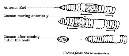
Image Source: ExpertsMind.
- It starts after copulation when ovaries mature.
- The formation of a cocoon in Pheretima has not been studied yet like in other worms, such as Eisenia and Rhynchelmis, etc.
- The epidermis of clitellar segments i.e.,14th, 15th, and 16th segments contain 3 kinds of glands, they are unicellular mucous glands that produce mucus for copulation.
- cocoon-secreting glands secrete the wall of the cocoon and albumen glands produce albumen in which eggs are deposited in the cocoon.
- Cocoon is secreted as a viscid and gelatinous substance by clitellar glands, forming broad membranous band or girdle around clitellum.
- It hardens gradually on exposure to air into a tough but elastic tube which becomes the cocoon or egg capsules.
- Epidermal mucous cells of clitellum also secrete a slime tube over the cocoon.
- As the worm wriggles behind, the slime tube and cocoon are slipped forward over the head.
- In this way, the cocoon receives ova from female genital apertures and sperms of other worms, from spermathecae, so that cross-fertilization is ensured, and zygotes are formed.
- Lastly, the girdle is thrown off from the worm and soon elasticity of its wall closes up its two ends to form a cocoon or ootheca.
- Several cocoons are formed after each copulation because the spermatozoa contained in the spermatheca do not pass out all at one time.
- Cocoons are oval, light yellow in color.
- They are about 2 to 2.4 mm long and 1.5 to 2 mm broad.
- Fertilization occurs after the cocoon has been deposited in a moist place.
- Fertilization takes place inside the cocoon and generally there is only one embryo in a cocoon.
- Cocoons are laid from August to October in damp situations.
Development of Earthworm
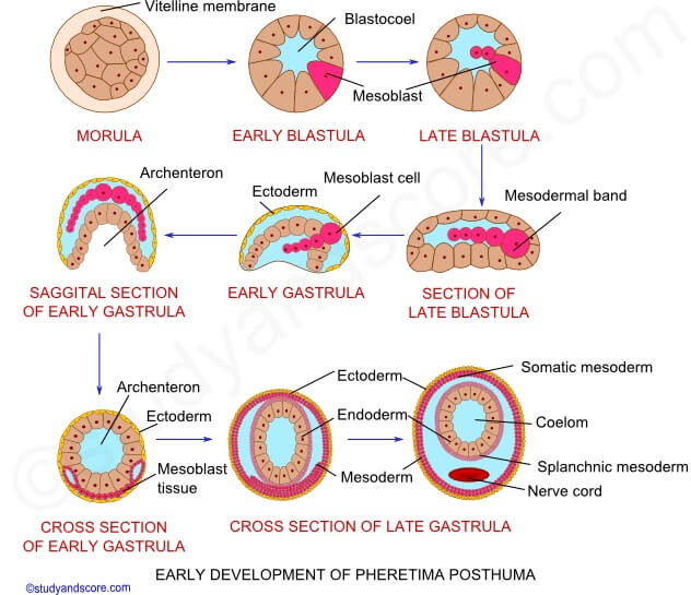
Image Source: Study and Score.
- The cocoon contains fertilized eggs and but only one embryo develops.
- Growing at expense other eggs serving as nurse cells.
- Cocoon also contains albuminous substances secreted by the glands of the clitellum for the nourishment of the embryo.
- Direct development occurs in the cocoon with no larval stages.
- The zygote undergoes holoblastic and a modified spiral cleavage resulting in a hollow ball of cells, the blastula, enclosed in a vitelline membrane.
- The lower cells of the blastula are endodermal and those of the upper cells are ectodermal.
- Gastrulation occurs by the invagination of endodermal cells into ectodermal cells to form a cylindrical gastrula with an archenteron cavity.
- A blastopore narrows to form the mouth.
- Mesoderm develops from 2 large cells of blastula, called mesoblasts.
- Mesoblasts divided to form 2 mesoblastic bands which later give rise to the coelomic epithelial lining.
- Young worm, when fully grown, crawls out of the cocoon in about 2 or 3 weeks to lead an independent life.
References and Sources
- Kotpal RL. 2017. Modern Text Book of Zoology- Invertebrates. 11th Edition. Rastogi Publications.
- Jordan EL and Verma PS. 2018. Invertebrate Zoology. 14th Edition. S Chand Publishing.
- 18% – https://www.biologydiscussion.com/invertebrate-zoology/earthworms/reproductive-system-of-pheretima-earthworm/29368
- 3% – https://www.slideshare.net/YuveenaGokool/phylum-annelidespptx
- 1% – https://www.nepeducation.com/wp-content/uploads/2018/06/Reproductive-System-of-Earthworm.pdf
- 1% – https://www.biologydiscussion.com/invertebrate-zoology/earthworms/earthworm-habitat-nervous-system-and-life-history-zoology/49355
- <1% – https://www.sciencedirect.com/topics/neuroscience/submandibular-gland
- <1% – https://www.quora.com/How-is-cocoon-formed-in-earthworms
- <1% – https://www.merckmanuals.com/home/women-s-health-issues/normal-pregnancy/stages-of-development-of-the-fetus
- <1% – https://www.aplustopper.com/plus-one-zoology-notes-chapter-3/
- <1% – https://www.answers.com/Q/What_are_mature_male_gametes_called
- <1% – https://unclejimswormfarm.com/worm-life-cycle/
- <1% – https://edurev.in/studytube/Earthworm–Chapter-Notes–Class-11–Biology/cbe09ba4-7661-4d5c-8083-b4ac8fcfa960_t
- <1% – https://doku.pub/documents/acarologypdf-z408j4pk4wqx
- <1% – https://courses.lumenlearning.com/suny-biology2xmaster/chapter/features-used-to-classify-animals/
- <1% – https://animals.howstuffworks.com/animal-facts/earthworm3.htm