Interesting Science Videos
Drosophila Development- Stages, Significance
- The fruit fly Drosophila melanogaster has been extensively studied for over a century as a model organism for genetic investigations.
- It also has many characteristics that make it an ideal organism for the study of animal development and behavior, neurobiology, and human genetic diseases and conditions.
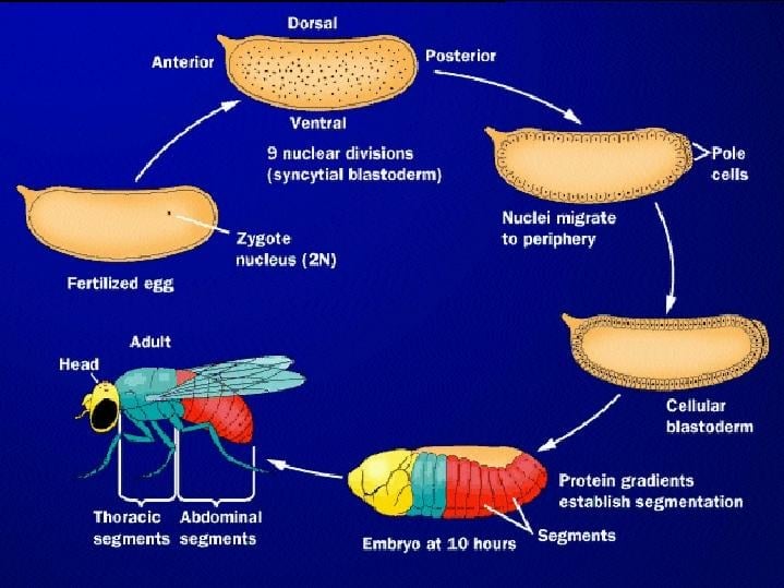
Image Source: biology.kenyon.edu
Developmental Stages of Drosophila
The development of Drosophila can be divided into the following stages:
Embryogenesis
1. Fertilization
- Fertilization of Drosophila can only occur in the region of the oocyte that will become anterior of the embryo. Moreover, the sperm tail appears to stay in this region.
2. Cleavage
- Most insect eggs undergo superficial cleavage, wherein a large mass of centrally located yolk confines cleavage to the cytoplasmic rim of the egg.
- One of the fascinating features of this cleavage type is that cells do not form until after the nuclei have divided.
- The zygote nucleus undergoes several mitotic divisions within the central portion of the egg.
- In Drosophila, 256 nuclei are produced by a series of eight nuclear divisions averaging 8 minutes each.
- The nuclei then migrate to the periphery of the egg, where the mitoses continue, albeit at a progressively slower rate.
- During the ninth division cycle, about five nuclei reach the surface of the posterior pole of the embryo.
- These nuclei become enclosed by cell membranes and generate the pole cells that give rise to the gametes of the adult.
- Most of the other nuclei arrive at the periphery of the embryo at cycle 10 and then undergo four more divisions at progressively slower rates.
- During these stages of nuclear division, the embryo is called a syncytial blastoderm, meaning that all the cleavage nuclei are contained within a common cytoplasm.
- No cell membranes exist other than that of the egg itself.
3. Blastoderm Formation
- Following cycle 13, the oocyte plasma membrane folds inward between the nuclei, eventually partitioning off each somatic nucleus into a single cell.
- This process creates the cellular blastoderm, in which all the cells are arranged in a single-layered jacket around the yolky core of the egg.
- Like any other cell formation, the formation of the cellular blastoderm involves a delicate interplay between microtubules and microfilaments.
- In Drosophila, the cellular blastoderm consists of approximately 6000 cells and is formed within 4 hours of fertilization.
4. The Midblastula Transition
- After the nuclei reach the periphery, the time required to complete each of the next four divisions becomes progressively longer.
- While cycles 1–10 are each 8 minutes long, cycle 13, the last cycle in the syncytial blastoderm, takes 25 minutes to complete.
- Cycle 14, in which the Drosophila embryo forms cells (i.e., after 13 divisions), is asynchronous. Some groups of cells complete this cycle in 75 minutes, whereas other groups of cells take 175 minutes.
- Transcription from the nuclei (which begins around the eleventh cycle) is greatly enhanced at this stage.
- This slowdown of nuclear division and the concomitant increase in RNA transcription is often referred to as the midblastula transition.
5. Gastrulation
- At the time of the midblastula transition, gastrulation begins.
- The first movements of Drosophila gastrulation segregate the presumptive mesoderm, endoderm, and ectoderm.
- The prospective mesoderm—about 1000 cells constituting the ventral midline of the embryo—folds inward to produce the ventral furrow.
- This furrow eventually pinches off from the surface to become a ventral tube within the embryo.
- It then flattens to form a layer of mesodermal tissue beneath the ventral ectoderm.
- The prospective endoderm invaginates as two pockets at the anterior and posterior ends of the ventral furrow.
- The pole cells are internalized along with the endoderm. At this time, the embryo bends to form the cephalic furrow.
- The ectodermal cells on the surface and the mesoderm undergo convergence and extension, migrating toward the ventral midline to form the germ band, a collection of cells along the ventral midline that includes all the cells that will form the trunk of the embryo.
- The germ band extends posteriorly and, perhaps because of the egg case, wraps around the top (dorsal) surface of the embryo.
- Thus, at the end of germ band formation, the cells destined to form the most posterior larval structures are located immediately behind the future head region.
- At this time, the body segments begin to appear, dividing the ectoderm and mesoderm.
- The germ band then retracts, placing the presumptive posterior segments into the posterior tip of the embryo.
- While the germ band is in its extended position, several key morphogenetic processes occur; organogenesis, segmentation, and the segregation of the imaginal discs.
- In addition, the nervous system forms from two regions of ventral ectoderm.
Body Segmentation
- The germband (ventral blastoderm) is the main trunk region.
- The process of germband extension pushes the posterior end over dorsal side.
- The first signs of segmentation grooves appear to outline parasegments which give rise to segments.
- Segments are formed from the posterior of one parasegment and the anterior of the next.
- There are 14 parasegments: 3 mouths, 3 thoraces, 8 abdominal.
Larval Stages
- The larva is a white, segmented, worm-shaped burrower with black mouthparts (jaw hooks) in the narrower head region.
- For tracheal breathing, it has a pair of spiracles (air intakes) at both the anterior and posterior ends.
- Since insect skin will not stretch, the young small larvae must periodically shed their skins (cuticle) in order to reach adult size.
- There are two such molts in Drosophila larval development that are accompanied by shedding of the mouthparts as well as the skins.
- During each period between molts, the larva is called an instar, i.e. the first instar is between hatching and the first molt.
- Both the size of the larva and the number of teeth on the dark-colored jaw hooks are an indication of which instar the larva has reached.
- After the second molt, the larva (now third instar) feed until ready to pupate.
- At this stage, the larva crawls out of the food medium onto a relatively dry place, ceases moving, and everts its anterior breathing spiracles.
Pupal Stage
- Soon after everting its anterior spiracles, the larval body shortens and the cuticle becomes hardened and pigmented.
- A headless and wingless pre-pupa form.
- This stage is followed by the formation of the pupa with everted head, wing pads, and legs.
- The puparium (outer case of the pupa) thus utilizes the cuticle of the third larval instar.
- The adult structures that seem to appear first during the pupal period have actually been present as small areas of dormant tissues as far back as the embryonic stage. These localized pre-adult tissues are called Anlagen (or imaginal discs).
- The main function of the pupa is to permit the development of the Anlagen to adult proportions.
- The breakdown of larval tissues to furnish material and energy for this development is, therefore, a prime feature of pupal metabolism.
Adult Stage
- Adults exhibit typical insect anatomy, including compound eyes, three-part bodies (head, thorax, and abdomen), wings, and six jointed legs.
- The various types of bristles and hairs found on the body are characters that we will use to identify different phenotypes of flies.
Significance of Studies on Drosophila Development
- Drosophila and human development are homologous processes.
They utilize closely related genes working in highly conserved regulatory networks. - Unlike humans, Drosophila is subject to easy genetic manipulation.
- As a result, most of what we know about the molecular basis of animal development has come from studies of model systems such as Drosophila.
References
- http://biology.kenyon.edu/courses/biol114/Chap13/Chapter13A.html
- http://www.mun.ca/biology/desmid/brian/BIOL3530/DB_02/DBNDros.html
- https://www.ncbi.nlm.nih.gov/books/NBK10081/
- http://modencode.sciencemag.org/drosophila/introduction
- https://www.jove.com/science-education/5093/drosophila-development-and-reproduction
- http://researchguides.library.vanderbilt.edu/c.php?g=156859&p=1161911
- http://www.mun.ca/biology/desmid/brian/BIOL3530/DB_02/DBNDros.html
- https://animaldiversity.org/accounts/Drosophila_melanogaster/