The digestive system of earthworms consists of the alimentary canal and the digestive glands.
Earthworm Alimentary Canal
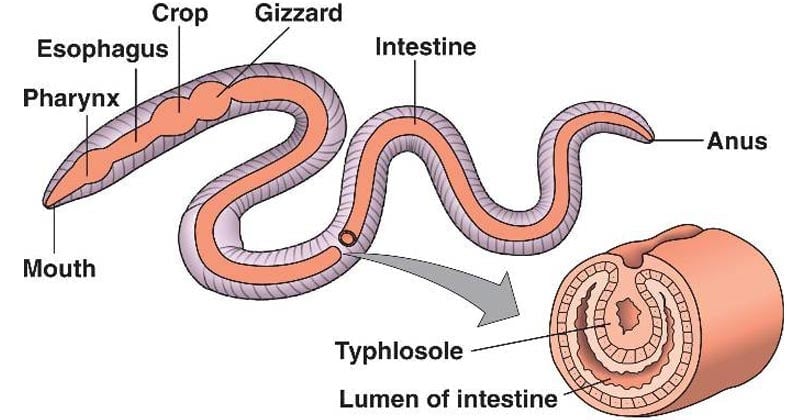
- Complete straight tube throughout the length of the body from mouth to anus.
- Mouth constitutes its anterior and anus its posterior openings receptively.
- Functionally regionated into various parts i.e. mouth and buccal chamber, pharynx, esophagus, gizzard stomach, and intestine.
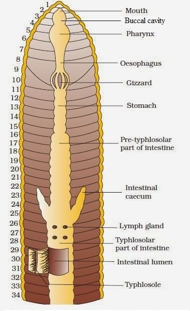
| Parts of the alimentary canal | No. of the segment in the body |
| Mouth | 1st segment |
| Buccal Cavity | 2-3rd segment |
| Pharynx | 3-4th segment |
| Oesophagus | 5-7th segment |
| Gizzard | 8-9th segment |
| Stomach | 9-14th segment |
| Intestine | 15th-last segment except for anus |
| Anus | last segment |
1. Mouth and Buccal Chamber
- Crescentic aperture situated ventral to prostomium.
- The mouth leads into a short, narrow, thin-walled protrusible buccal chamber.
- chamber extends up to the middle of 3 segments.
- Buccal cavity folded and surrounded by muscular strands.
2. Pharynx
- Followed by a buccal chamber.
- Extends up to the 4th segment.
- Pear-shaped broad and muscular separated from the buccal cavity by a groove.
- The pharynx roof is thick and projected into the pharyngeal bulb.
- Pharyngeal bulb lateral walls internally form narrow horizontal shelves.
- Two shelves meet anteriorly and posteriorly and divide the pharyngeal cavity into the dorsal salivary chamber and ventral conducting chamber.
- The roof of pharynx lined by ciliated epithelium.
- Many muscles with connective tissues and blood vessels present above epithelium.
- Outside these present salivary glands
- Glands are small, whitish unicellular glands of chromophil cells.
- Glands open through fine ducts.
- Glands secrets mucin for lubrication of food and
- Proteolytic enzymes for digestion of proteins.
- The ventral conducting system of pharynx serves as a passage for the ingested materials.
- Like the buccal chamber, the pharyngeal wall remains to connect with the body by a thick mass of muscular strands.
- Contraction and relaxation of muscular strands serve to compress or dilate the pharyngeal lumen.
- Acts as a pump during feeding.
- Series of contraction of pharynx resulting in the suckling food into the buccal chamber and pumping the same back into the esophagus.
3. Esophagus or gullet
- Lined behind the pharynx.
- Short, narrow, thin-walled.
- Running up to 8th segments.
- wall folded internally and devoid of any glands.
4. Gizzard
- modification of the esophagus into the prominent, hard, and thick-walled muscular organ.
- Lying in 8th of 8th or 9th segments.
- the wall consists of circular muscles lined by the columnar cells.
- Columnar cells further lined by the tough cuticles.
- Grinds foods into fine states.
5. Stomach
- It is a gizzard followed by a short, narrow, and thin-walled tube.
- Extends up to 14th segments.
- Anterior and posterior opening sphincter.
- Walls highly vascular and glandular but less muscular.
- Internal wall folded transversely.
- The epithelial lining consists of glandular cells and some calciferous glands.
- Glandular Cells secretes a proteolytic enzyme.
- Calciferous glands secrete calcium and CO2.
- Calcium neutralizes the contents of the alimentary canal.
- Calciferous glands are excretory removes ions of calcium and carbonates from the blood.
- Calcite excreted into the stomach when the level of ion becomes excessive and passed out with mud through the anus.
6. Intestine
- The region next to the stomach.
- Long, wide, and thin-walled tube.
- Extends from 15th segments to the anus.
- Beaded appearance due to circular constriction corresponding to septa.
- The internal lining has ciliated and glandular cells.
- Internal lining folded to form villi.
- one of those villi become larger and well developed to form typhlosole.
- Typhlosole runs mis-dorsally from 26th to last segments except 24, 25 segments.
- Divisible into 3 parts
1. Pre-typhlosolar region
- First or anterior parts lying between 15th to 26th segments.
- Walls folded internally to forms minutes process, the villi.
- Villi are highly vascular.
- No typhlosole is found in this region.
- 26th segment gives outs externally a pair of forwardly- directed conical outgrowth, intestinal caeca.
- Intestinal caeca extended up to 22nd or 23rd segment
- Internally have any folds to form a villi-like process.
- Highly vascular and filled with secretory cells.
- Are digestive glands and secrete amylolytic enzymes for digestion of starch (Chen and Push (1941).
- In this region active digestion occurs.
2. Typhlosolar region
- Second or middle part of the intestine.
- Lies between 26th to last segments except for 24, 25th segments.
- Characterized by the presence of highly glandular and vascular longitudinal ridge.
- Provided with internal median folds of the dorsal wall of intestine i.e. Typhlosole.
- It increases the absorptive surface of the intestine.
- Process of digestion complete in this region.
3. Post- typhlosolar region
- Third or last parts i.e. 24, 25 segments.
- Has no typhlosole.
- Called rectum.
- Thin-walled, vascularized without villi and glandular cells.
- Casting occurs here.
7. Anus
- The small circular opening at the terminal end.
- Last or anal segments of the body.
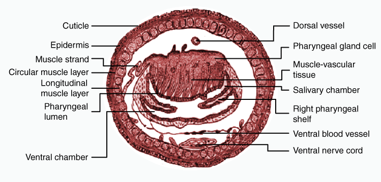
Histology of Alimentary Canal
Peritoneum
- Outermost layer.
- Consists of tall and narrow cells.
- Cells modified around the stomach and intestine called chloragogen cells or chloragocytes.
- Cells contain yellow refractile granules called chloragosomes.
- The exact function is controversial.
- But said to serve for the storage of food, deamination of proteins, deamination of proteins, the formation of urea from ammonia, excretion, etc.
Muscles
- Lying below peritoneum.
- Include the outer layer of the longitudinal and inner layer of circular muscle fibers.
- They are well developed around pharynx and esophagus.
- But poorly developed in intestines.
- Muscles arrangements in the gut wall are just reverse to the body wall.
- In gizzard, longitudinal muscles are absent.
- But circular muscles are much developed.
- All Gut wall muscles are involuntary and unstriped.
Enteric epithelium
- Consists single layer of columnar cells
- They become ciliated in the roof of the pharynx.
- Mostly Glandular and absorptive in the stomach.
- Glandular in intestine.
- Internally thrown into folds in the esophagus, stomach, and intestine.
Cuticle
- Present in the buccal cavity as thin lining
- Present in the gizzard as thick lining.
Food and Feeding Mechanism
- The earthworm is omnivorous.
- Feeds directly upon organic humus, decaying matters.
- Also feeds directly upon leaves, grasses, seeds, small protozoans, nematodes, insects, algae, and other microorganisms found in soil.
- Ingest soil in larger quantities so, the gut is always full of soil.
- Ingest food by pumping action of its pharynx.
- The contractile sucking action of pharyngeal walls draws fragments of soil into the buccal chamber.
- Action is accelerated by the action of strands of muscle fibers, extending from pharynx to body wall.
Earthworm Digestive System Video
Physiology of Digestion
- Various types of enzymes are said to be secreted by digestive glands due to the omnivorous mode of feeding habit.
- Ingested food is pressed to move posteriorly.
- No digestion occurs in the buccal chamber.
- In the ventral conducting chamber of the pharynx, it receives the salivary secretion from salivary gland cells.
- Salivary secretion contains mucin and proteolytic enzymes.
- Mucin lubricates food and food passages.
- Proteolytic enzymes hydrolyze proteins into peptones and proteases.
- Then, food comes into gizzard through the esophagus.
- Gizzard acts as a grinding machine that further grind food and soil.
- This is facilitated by contractile movements of its muscular wall which causes the food to roll about, internal cuticle lining, striking against food particles are ground up fully.
- Then, Food reaches in the stomach in fine states.
- chalky secretion of calciferous glands located in the stomach wall neutralizes the humic acid present in the soil.
- Then food reaches the intestine.
- The intestine is the principal site of digestion.
- Enzymes are secreted by glandular cells of the intestine and intestinal caeca.
- Enzymes like pepsin, trypsin, amylase, lipase cellulase and chitinase are secreted.
- Pepsin hydrolyzes proteins into proteases and peptones
- Trypsin hydrolyzes the product into amino acids.
- Amylases acting upon carbohydrates and converting them into monosaccharides.
- Lipase brings hydrolysis of fats into glycerol and fatty acids.
- Cellulase hydrolyses the cellulose into cellobiose.
- Chitinase hydrolyzes chitin present in food.
- Digestion is extracellular in the earthworm, as in higher animals such as frog and rabbits.
- Digestion occurs in the stomach and fully completed in the stomach.
- Intestine function for absorbing the digestive nutrients.
- Digested food is absorbed by intestinal villi, more particularly by typhlosole.
- Absorbed food are passed to blood capillaries in the intestinal wall.
- Coelomic fluid also serves to transport digested food to tissues.
- Undigested food and soils are passed out through anus as earthworm casting at the opening of burrows.
- The casting of earthworm consists of small and round pellets of balls.
References
- Kotpal RL. 2017. Modern Text Book of Zoology- Invertebrates. 11th Edition. Rastogi Publications.
- Jordan EL and Verma PS. 2018. Invertebrate Zoology. 14th Edition. S Chand Publishing.