Circulatory system of earthworm
- The circulatory or blood vascular system of an earthworm is a closed type.
- It consists of the blood vessel, heart, capillaries, and blood glands.
- Blood is composed of fluid plasma and colorless corpuscles.
- Blood is red in color due to the presence of respiratory pigment hemoglobin (erythrocruorin) in it.
- Hemoglobin is not contained in the corpuscles like vertebrates but occurs dissolved in plasma.
- Hemoglobin aids in the transportation of oxygen for respiration.
Blood vessels
They are of 2 types: collecting blood vessels and distributing blood vessels.
- They are closed tubes with a definite wall, and they break into capillaries to ramify in the different parts of the body.
- The arrangement of blood vessels in the anterior 13 segments is different from that behind the 13th segments i.e., in the region of the intestine.
- So, the blood vessels can be described under 2 heads: A. Blood vessels and their arrangements behind the 13th, i.e, intestinal region. B. Blood vessels and their arrangement in the anterior 13th segments.
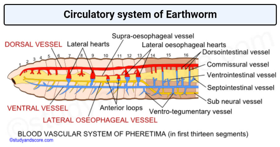
Image Source: Study and Score.
A. Blood vessels and their arrangements behind 13th, i.e. intestinal region
The blood vessels of this region include:
- Median longitudinal blood vessels
- The intestinal blood plexus
- The commissural vessel
- The integumentary vessel
- The nephridial vessel
1. Median longitudinal blood vessels
a. Dorsal vessel
- The largest blood vessel of the body.
- Runs mid-dorsally above the alimentary canal, from one end of the body to another.
- Thickest vessel with contractile muscular walls visible from outside as a dark line through the thin and semitransparent body wall.
- Provided with pair of valves in the front of the septum in each segment.
- The direction of blood flow in this vessel is from backward to forward (from posterior to anterior).
- Contractile and pulsates rhythmically to force blood from posterior to anterior side.
- Behind the 13th segment, it is collecting vessels, receiving blood through two pairs of dorso-intestinal vessels from the intestine, and a pair of commissural vessels from a sub-neural vessel in each segment.
- The commissural vessels from the loop behind each septum and they receive blood from the body wall, nephridia, and prostate glands.
- The commissural vessels also give out blood in each segment through a septointestinal branch to the intestine.
b. Ventral vessel
- It is also a large vessel that runs mid-ventrally below the alimentary canal and above the nerve cord from the 2nd segment to the last segment of the body.
- Its walls are thin and without muscles and valves.
- The direction of blood flow in this vessel remains from anterior to the posterior side or from in front to backward.
- It is a principally distributing vessel. It supplies blood, each segment, through a pair of ventro-tegumentary vessels to integumentary nephridia, body wall, septa, and reproductive organs.
- This vessel also gives out a ventro-intestinal vessel to lower parts of the intestine in each segment behind the 13th segment.
- Behind the 13th segment, each ventro-tegumentary vessel sends a small branch, a septo-nephridial branch, supplying the septal nephridia.
c. Sub-neural vessel
- It is also a long and slender vessel that runs immediately beneath the nerve cord in the mid-ventral position.
- It extends from the 14th segment to the last segment and is formed by the union of two lateral oesophageal vessels.
- It is without muscular walls and internal valves.
- The direction of blood flow is from in front backward.
- It is mainly a collecting vessel.
- It receives blood from the ventral nerve cord and ventral wall in each segment through a pair of small branches.
- It gives a pair of commissural vessels in each segment that joins the dorsal vessel.
2. Intestinal blood plexus
- The intestine is richly supplied with blood capillaries that form a close network.
- Consists of a close network of capillaries in the wall of the intestine.
- There are 2 capillaries networks in the intestine i) external plexus ii) internal plexus.
- External plexus lies on the surface of the gut and receives blood from the ventral vessel through ventro-intestinal and septo-intestinal and passes it on to internal plexus.
- The internal plexus is present in between the circular muscle layer of the intestine and the internal epithelial lining.
- Internal plexus passes on blood, along with absorbing nutrients, to the dorsal vessel through dorso-intestinal.
3. Commissural vessels
- It connects the dorsal and sub-neural vessels.
- They receive blood from nephridia, body wall, and the reproductive organs through capillaries and send them to the dorsal blood vessel.
4. Integumentary vessels
- These vessels coming from ventral vessels supply blood to integument for aeration.
- The aerated blood is collected by numerous capillaries of the commissural vessels in each segment.
- Thus, there is a close parallelism between venous and arterial capillaries throughout the body wall.
5. Nephridial vessels
- Originate from the ventro-tegumentary vessels of the ventral vessel.
- They supply blood to nephridia.
B. Blood vessels and their Arrangement in Anterior 13 segments
It consists of the following:
- Median longitudinal vessels
- Herat and anterior loops
- Blood vessels of the gut
The function of collecting blood from the anterior region of the gut is taken over by a new vessel supra-oesophageal, while the blood from the peripheral structures is collected by the right and left lateral oesophageal.
1. Median longitudinal blood vessels
a. Dorsal vessel
- The blood vessel becomes the distributing vessel in these segments instead of the collecting vessel.
- Structurally it is like that of anterior segments.
- But it has neither dorso-intestinal nor commissural vessels opening into it.
- It sends out all the collected blood from the posterior region of the body into the hearts and the anterior region of the gut.
- In the gut it divides into 3 branches distributes over the pharyngeal bulb and the roof of the buccal chamber.
- However, it supplies to the stomach, gizzard, esophagus, pharynx, and other related parts.
b. Ventral vessel
- The blood vessel remains to distribute in these segments also but extends only up to the second segment.
- No ventro-intestinal, hence it does not supply to the alimentary canal in this region.
- Ventral vessels give off a pair of ventro-tegumentary vessels in each segment supply blood to the body wall, septa, nephridia, and reproductive organs.
c. Supra- oesophageal vessel
- It is the shortest, thin-walled collecting vessel lying mid-dorsally above the stomach and confined to segments 9 to 13.
- Connected to the lateral oesophageal vessel through 2 pairs of anterior loops.
- Connected to the ventral vessel through 2 pairs of latero-oesophageal hearts.
- At places, it divides into separate vessels that reunite to form a single vessel.
- It collects blood from the stomach, gizzard, and (through anterior loops) from lateral oesophageal.
- And pumps it through lateral oesophageal hearts into the ventral vessel.
d. Lateral oesophageal
- In fact, the subneural vessels bifurcate in the 14th segments to form 2 lateral oesophageal.
- These vessels are thick and lie one on the ventrolateral side of the gut, running from the anterior end of the body up to the 13th segment.
- Closely attached to the wall of the stomach from 10th to 13th segments and communicate with the ring vessels.
- These receive a pair of ventro-tegumentary vessels in each segment, collecting blood from the body wall, septa, nephridia, and reproductive organs.
- Some of the blood passes to supra-oesophageal vessels through anterior loops in each of the segments 10 and 11 and through several ring vessels running through the wall of the stomach.
- The rest of the blood flows back into the sub-neural vessels.
- They function likes subneural and commissural vessels of the posterior region e. these are collecting vessels.
2. Hearts and anterior loop
- In each segment 7, 9, 12, and 13 is found a pair of large, thick, muscular, and rhythmically contractile vertical vessels, called hearts.
- They are neurogenic i.e., the heart originates in the nerve cells of the heart.
- They pump blood from dorsal to the ventral vessels, while flow in opposite direction is prevented by internal valves.
- The hearts of the 12th and 13th segments are joined above to both the dorsal and the oesophageal vessels, called latero-oesophageal hearts.
- These hearts have thick muscular walls and a pair of valves at each junction with dorsal vessels and supra-oesophageal vessel, and another pair of valves at the ventral end.
- These allow flowing blood downwards only.
- Another heart of 7th and 9th segments connect dorsal and ventral vessels only and are called lateral hearts.
- They have 4 pairs of valves that allow blood to flow only downwards.
- Besides 4 pairs of heart, there are 2 pairs of loop-like vessels called anterior loop.
- The anterior loop is a pair of thin-walled, non-pulsatile, non-muscular, and loop-like broad vessels, without valves, in each of the 10th and 11th segments.
- Anterior loop covey blood from lateral-oesophageal into the supra-oesophageal vessel.
3. Blood vessels of the gut
- On another side of the stomach situated ring-like vessels.
- Ring vessels are characteristic circular vessels of the stomach situated within the muscular coat, about 12 vessels per segment.
- These vessels connect the supra-oesophageal and lateral-oesophageal vessels.
- Through these vessels, blood flows upwards from the lateral-oesophageal into the supra-oesophageal.
- The buccal cavity, pharynx, and gizzard receive their blood supply from dorsal blood vessels directly.
Video: Circulatory system in Earthworm by Studio Biology.

Circulation of blood
- Blood flows from behind to forward in the dorsal vessel.
- And from front to backward in ventral, latero-oesophageal, supra-oesophageal, and sub-neural vessels.
- The ventral vessel is the main distributing vessel, supplying blood to all parts of the body.
- In the first 13 segments, it supplies blood to the body wall, septa, nephridia, and reproductive organs through ventro-tegumentary.
- Behind the 13th segment, it supplies blood to the body wall and nephridia through ventro-tegumentary.
- Supplies blood to gut wall through ventro-intestinal.
- Sub-neural, lateral oesophageal, and supra-oesophageal are the main collecting vessels.
- Lateral oesophageal collects blood in the first 13 segments from the alimentary canal, body wall, nephridia, septa, and reproductive organs.
- And they discharge into supra-oesophageal through anterior loops and ring vessels.
- Supra-oesophageal also collects blood from the gizzard and stomach and pours into the ventral vessel through latero-oesophageal hearts.
- Sub-neural collects blood in the intestinal region from the ventral body wall and nerve cord.
- And send into dorsal vessels through the commissural which also receives blood from the body wall, septa and nephridia.
- Commissural also pours some blood into the gut wall through septo-intestinal.
- Dorsal vessel functions both collecting and distributing vessels.
- It collects blood through dorso-intestinal in the intestinal region from the gut wall.
- And through commissural from sub- neural vessel, septa, and nephridia.
- In the first 13 segments, it distributes some blood through branches to the alimentary canal and pours the remaining blood through hearts into the ventral vessel.
- The blood distributes digested food to various body regions.
- And it collects waste substances like nitrogenous waste and CO2 which are eliminate through nephridia, skin, and the coelomic fluid.
Read Also: Nervous System of Earthworm
Blood glands
- In the segments, 4, 5, and 6 segments above pharyngeal mass and connected with pharyngeal or salivary glands are found small, red-colored, follicular bodies, the blood glands.
- Each gland consists of a mass of loose cells surrounded by a capsule with a syncytial wall.
- These glands are connected with pharyngeal nephridia and with salivary glands.
- These glands manufacture blood corpuscles and hemoglobin.
- They are also regarded to be excretory by some workers.
References and Sources
- Kotpal RL. 2017. Modern Text Book of Zoology- Invertebrates. 11th Edition. Rastogi Publications.
- Jordan EL and Verma PS. 2018. Invertebrate Zoology. 14th Edition. S Chand Publishing.
- 16% – https://www.slideshare.net/SSMV2016/circulatory-system-of-earthworm-130564375
- 7% – https://www.notesonzoology.com/earthworm/earthworm-digestive-and-reproductive-system-zoology/13337
- 7% – https://www.biologydiscussion.com/invertebrate-zoology/earthworms/circulatory-system-of-earthworm/29388
- 2% – https://www.biology-today.com/general-zoology/invertebrate-zoology/circulation-in-pheretima/
- 1% – https://www.slideshare.net/SoniaBajaj10/circulatory-system-of-earthworm
- 1% – https://www.shareyouressays.com/knowledge/biology-question-bank-144-mcqs-on-animal-kingdom-answered/114611
- <1% – https://zoologyforeamcet.blogspot.com/2011/
- <1% – https://www.slideshare.net/prof_aarif/earthworm-79257978
- <1% – https://www.rbsesolutions.com/class-11-biology-chapter-35-english-medium/
- <1% – https://answers.yahoo.com/question/index?qid=20070320085114AAPQE9e
- <1% – http://droualb.faculty.mjc.edu/Lecture%20Notes/Unit%204/link%20blood_vessel_distribution%20with%20figures.htm