Strasburger discovered the chromosome in 1815; W. Waldeyer introduced the term chromosome in 1888.
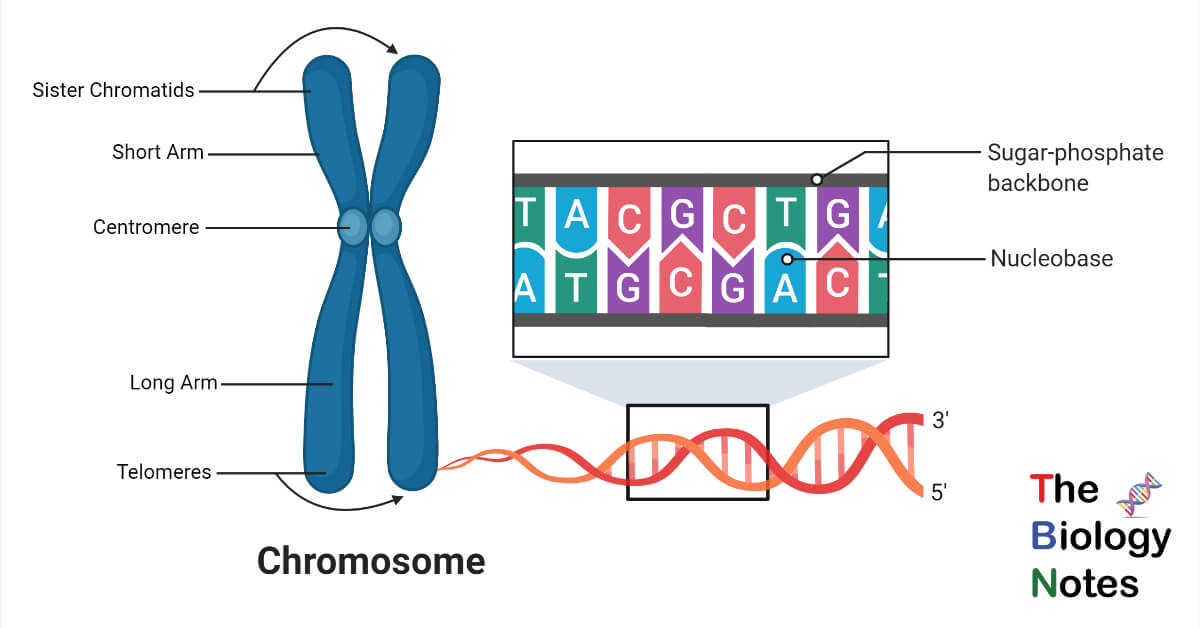
A chromosome is a thread-like structure in which DNA is packed and coiled around a protein called histone. They are present in the center of the nucleus.
Chromosomes hold genes responsible for different characteristics; so, 23 pairs (total 46) of chromosomes are present in the human body. There are 22 pairs of autosomes and one last pair of sex chromosomes (XX for female, XY for male).
Interesting Science Videos
Structure of Chromosome
a. Chromatids- During cell division, two identical strands are formed and joined by a centromere; both are called sister chromatids.
b. Centromere- These are also called kinetochores. It is present at the center, where the chromatid and spindle fibers are attached. It helps in movement at the anaphase stage of cell division.
c. Telomere- This is the terminal region of the chromosome.
d. Chromatin- Chromatin is condensed, enveloped around nuclear proteins, and contains DNA, RNA, and proteins that form chromosomes in the nucleus.
The chromosomes have a p and a q arm; p (petit) is a short arm and, q is a long arm. Chromosomes like 13,14, 15 have small p-arms.
History of Chromosomes
Theodor Boveri- represented two significant properties of the chromosome that are the identity of each chromosome; the other is passing the genetic information to further generations.
1860- Mendel gave the law of inheritance.
1875- Cell division in somatic cells or mitosis discovered.
1890- Reduction of cell division in gamete formation or meiosis discovered.
1902- Walter Sutton and others found inheritance and chromosomal behavior. They observed that in somatic cells, chromosomes paired together in which meiosis occurs in gametes.
Types of Chromosomes
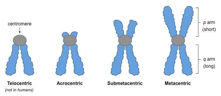
- Metacentric- they have a centromere in the center. All arms are of equal length. Example chromosome 1, 3, 16, 19, 20.
- Submetacentric- they have an off-center centromere p arm slightly shorter than q. Example chromosome 2, 4-12, 17,18, X.
- Acrocentric- they have an off-center centromere having a much shorter p-arm. Example 13-15, 21, 22, Y.
- Telocentric- centromere is present at the end of the chromosome; no p-arm exists. Humans don’t have telocentric chromosomes.
Chromosome and Cell division
Mitosis is a process in which all content in a cell duplicates like chromosomes and forms two identical daughter cells. Another division is meiosis, in which humans contain the same number of chromosomes in each generation. In this, chromosomes are reduced from 46 to 23 for the formation of sperm and egg cells.
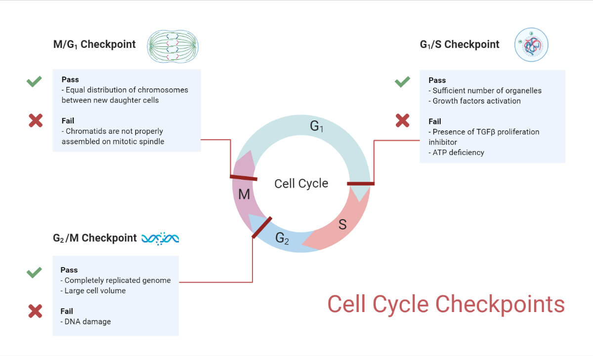
Cell division cycle has 4 phases
G1 phase- it is the phase after cell division and before DNA replication. This cell decides and observes the environment to start another phase of cell division.
S phase- where a cell starts replication of chromosomes and DNA synthesis.
G2 phase- it is the phase where replication ends and cell division starts. The cell makes sure DNA replication is completed and does repair if needed.
M-phase- cell division starts having prophase, metaphase, anaphase, telophase, and cytokinesis.
Mitosis Cell Division Cycle
It is a process of two daughter cells production that are genetically similar to each other and, to their parental cells. During the process, a diploid cell has 2N chromosomes and 2X DNA.
When replication starts, the cell remains genetically identical to 2N, but the number of the chromosome gets doubled to 4X DNA; this happens because each of the chromosomes replicates its DNA. Now there are pairs of exact sister chromatids that get separated and move to the opposite end of the dividing cell; mitosis results in 2N chromosomes and 2X DNA.

Mitosis cell cycle steps
Interphase– This is the phase where the cell spends most of its time starting the cell cycle, ready to divide and assemble nutrients and energy.
Prophase– Chromatin condenses and the formation of mitotic spindles makes chromosomes visible.
Metaphase– In the middle of the cell chromosome lined up and pulled by the microtubule of the spindle.
Anaphase– The separation of chromatid towards the opposite ends of the cell.
Telophase– Daughter chromosomes move at the pole and two new nuclei are formed.
- Prophase- it is the longest phase in mitosis; chromatin gets condensed to form chromosomes and breaking of the nuclear envelope. Centrioles in animal cells present close to the nucleus, get split, and move to opposite poles; centrioles are small organelles present in eukaryotic cells; it helps to confirm each cell has a complete set of chromosomes after cell division. When centrioles move separated, the formation of spindles starts having fiber made up of microtubules.
- Metaphase- metaphase having spindle fibers linked to centromeres of each sister chromatid. The sister chromatids form a line in the center of the cell, during cell division spindle fibers ensure sister chromatids are detached and move to different daughter cells. Some spindles are not attached to the kinetochore protein of the centromeres; they are known as non-kinetochore spindles that aid in cell elongation.
- Anaphase- the division of the centromere occurs and sister chromatids are detached. Spindle fibers shorten and sister chromatid pulls apart. Each sister chromatid moves to the opposite poles of the cell; now each pole gets a complete set of chromosomes.
- Telophase- in this chromosomes condensed and move to the opposite ends where they are stretched into chromatin arrangement. Mitotic spindles get depolymerized in the form of tubulin monomers and they are used for creating cytoskeleton components for each daughter cell, now a nuclear envelope is formed, and in the nuclear area, nucleosomes occur.
- Cytokinesis– this is the final stage in both eukaryotic and prokaryotic cell division, here splitting of cytoplasm into two and cell divides.
Chromosome Mutation
A chromosome mutation occurs when there are changes in the segment of DNA/ sequence, an increase or decrease in the genome, and mutations caused by radiation, chemicals. Some mutations are hereditary as they pass from the parent germline to the offspring; non-hereditary mutations occur outside the germline in cells, it is called a somatic mutation. There are types of mutational changes in chromosomes. For example- deletion, duplication, inversion, translocation.
Cause of Mutation in Chromosome
The causes of mutations that lead to a genetic disorder are changes in the structure of chromosomes, mutations in DNA sequence, rearrangement, missing chromosome numbers, etc.
These events happened during meiosis:-
- During cell division, a chromosome segment accidentally gets duplicated and forms different mutations.
- At crossing over chromosomes breaks and get detached abnormally causing genetic disorder due to chromosomal mutation.
- Numerical mutation occurs when nuclear cell division is deformed.
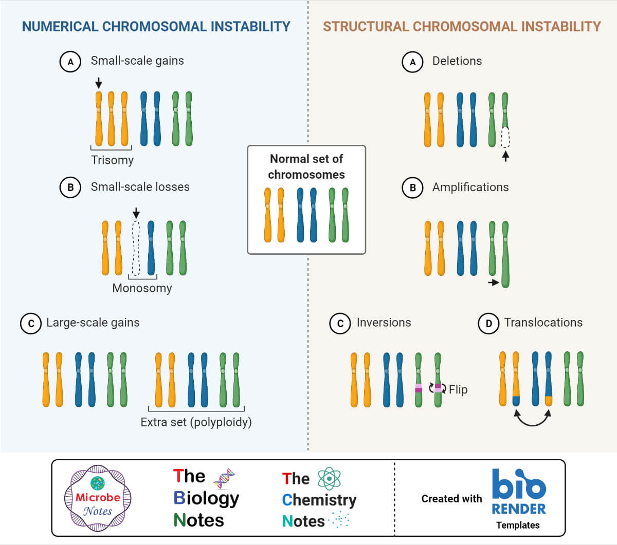
Types of mutation- Mutation I and Mutation II
Mutation I include- deletion, duplication, inversion, and translocation.
- Deletion- In this, a part of the chromosome is deleted during cell division. Deletion can be a terminal where deletion occurs at the end; another is interstitial where deletion happened within the chromosome and at the end. For example, a part that is deleted at chromosome 5 causes Cri-du-chat syndrome, in which head size is reduced and high crying-pitch like a cat in infants.
- Duplication- A part of a chromosome gets duplicated and results in Charcot-Marie- tooth disease I which leads to the duplication of chromosome number 17 and causes muscle weakness. MECP2 duplication syndrome where the MECP2 gene is duplicated and causes weak muscle seen in males.
- Inversion- This affects chromosome 2; an inversion is a segment of a chromosome that is reversed from end to end and reattached in the opposite direction. There are two types- paracentric, in which centromere is not involved; pericentric in which centromere is involved. For example, an inversion of chromosome 12 creates the tallest teenager.
- Translocation – Here a chromosome breaks and relocates to different chromosomes; for example, Robertsonian in this elongation of a chromosome happens when one segment of a chromosome is attached to another. Another example is when the 21 chromosomes are translocated to chromosome 14 and cause Down syndrome with symptoms- pale skin, cardiovascular problems, small head, brush field spots, dysmorphic features, etc.
Mutation II include- aneuploidy and polyploidy.
- Aneuploidy- This happens when an error occurs during meiosis; chromosome sets don’t get separated and leave one extra chromosome or less chromosome with two or one sex cells. Down syndrome is an example of aneuploidy causing trisomy.
- Polyploidy- This causes two sets of genomes in organisms and causes gigantism and reduce fertility. Mostly happen in plants.
Common chromosome disorder
Trisomy 18 causes Edward syndrome, and trisomy 13 causes Patua syndrome. Edward syndrome symptoms include clenched hands, severe mental disability, small-mouth, low set ear, large back head, rocker feet. Patua syndrome includes a small head, small eyes, intellectual disability, more than five fingers, cleft lips, and malformations of the forebrain. The sex chromosome disorder is Klinefelter syndrome, in which a person with an XY chromosome has an X-added chromosome. Its genotype is XXY which is male; another trisomy is XYY syndrome and XXX syndrome.
Turner syndrome is a monosomy sex chromosome in which a person has only X and births as female.
Identification of genetic disorders
DNA testing is done to identify these disorders; these are-
Carrier testing- In this blood test is done to check if there is a mutation related to a genetic disorder.
Prenatal screening- A blood test is done on a pregnant woman that shows whether the baby has a genetic disorder or has a common chromosome condition.
Diagnosis of chromosomal/ genetic disorder
It needs analysis of chromosomes, clinical diagnosis is done and conformation cytogenetic study is performed. Standard cytogenetics of chromosomes is done at the metaphase stage where all chromosomes are visible and identified by their size, centromere position, and dark-light band pattern by staining.
Physical examination is done by measuring the head circumference, face features, eye distance, arms, and legs length. Family history is important for the diagnosis of a genetic disorder; family history gives the idea about the genetic error, if it happened, and the inheritance pattern.
Chromosome functions
The production of protein depends on chromosomes and DNA; for all cells to function, protein is required. The chromosomal DNA has genes that code for proteins called gene expression, during the production of protein DNA unfolds and coding is transcribed into an RNA transcript. DNA gets exported from the nucleus and gets translated into protein formation.
- Determine gender of the human
- Have instructions to form new cells
- Determine traits like eye color, hair, height, etc.
- Holds the genes which are heredity units.
- It controls the activity of cell
References
- https://bio.libretexts.org/Bookshelves/Human_Biology/Book%3A_Human_Biology_(Wakim_and_Grewal)/07%3A_Cell_Reproduction/7.3%3A_Mitotic_Phase_-_Mitosis_and_Cytokinesis
- https://bioprinciples.biosci.gatech.edu
- https://askabiologist.asu.edu/cell-division
- https://ib.bioninja.com.au/standard-level/topic-3-genetics/32-chromosomes/chromosome-types.html
- https://sciencing.com/four-major-types-chromosomes-14674.html
- https://www.news-medical.net/health/Chromosome-History.aspx
- https://www.savemyexams.co.uk/a-level/biology/cie/22/revision-notes/5-the-mitotic-cell-cycle/5-1-replication–division-of-nuclei–cells/5-1-1-chromosome-structure/
- https://www.healio.com/hematology-oncology/learn-genomics/genomics-primer/what-are-chromosomes
- https://knowgenetics.org/introduction-to-chromosomes
- https://www.osmosis.org/answers/chromosomal-aberrations
- https://www.thoughtco.com/chromosome-373462
- https://medlineplus.gov/genetics/understanding/consult/diagnosis/
- https://my.clevelandclinic.org/health/diseases/21751-genetic-disorders
- https://chromodisorder.org/introduction-to-chromosomes/
- https://www.thoughtco.com/chromosome-mutation-373448
- https://www.biologyonline.com/dictionary/chromosomal-mutation
- Images created with biorender.com