Chiasmata (singular: chiasma, from the Greek, meaning “X-shaped cross”) is the point of contact that occurs in the Prophase I pachytene phase and links two non-sister chromatids belonging to homologous chromosomes together until Anaphase I of meiosis I cell division.
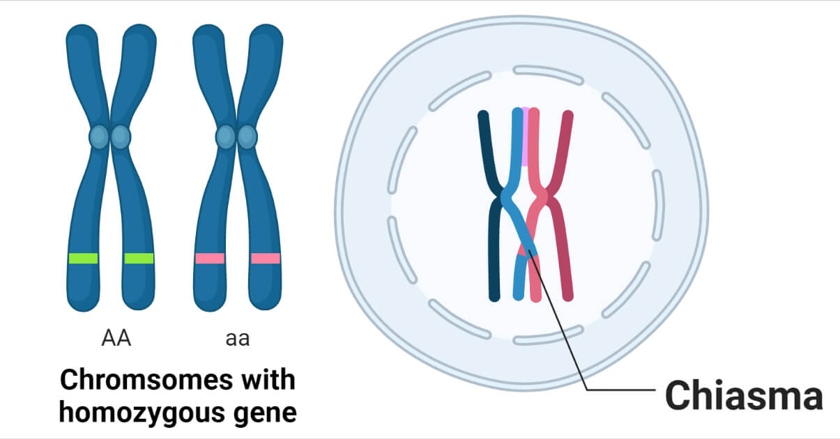
The phenomenon of chiasmata was discovered and described by Frans Alfons Janssens in 1909 which he called ‘chiasmatypie.’
Formation of Chiasmata
Pairing and recombination of homologous chromosomes take place during the prophase of meiosis I.
In this phase, exchanges between homologous chromosomes occur regularly by crossing over.
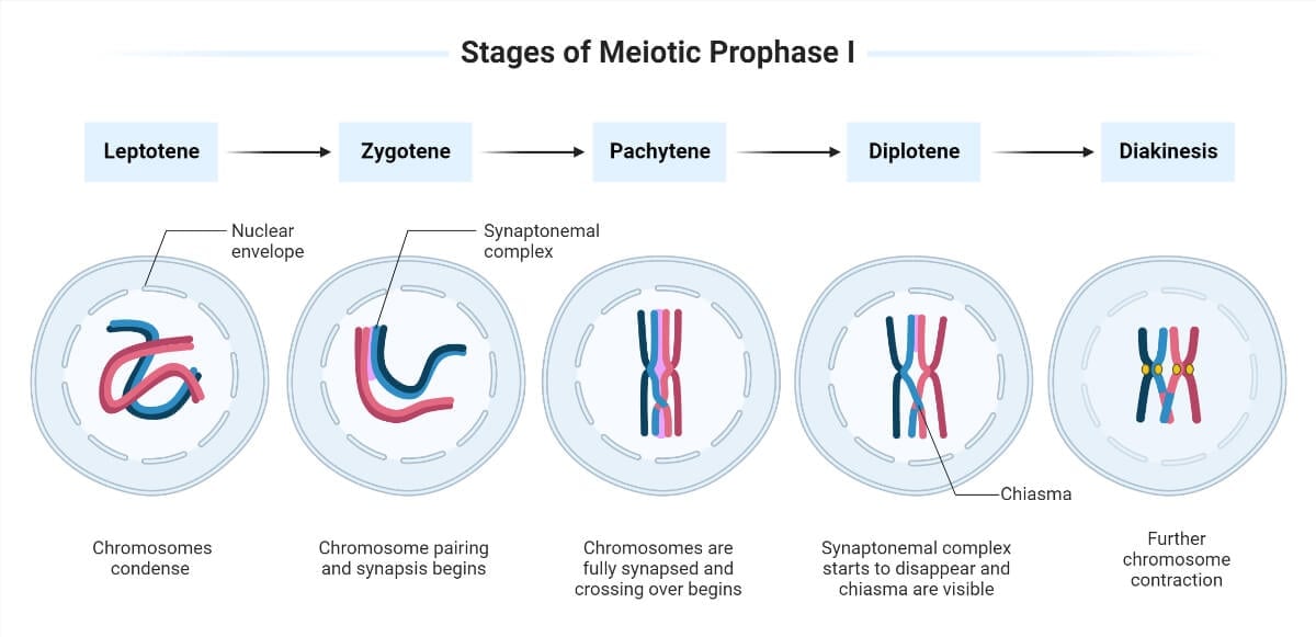
Prophase I involves five different phases. They are:
- Leptotene
- Zygotene
- Pachytene
- Diplotene
- Diakinesis
- Chiasmata form at sites where programmed DNA breaks, generated by specialized enzyme Spo11, undergo the full recombination pathway to generate crossovers.
- It is unknown how crossing over occurs, representing exchanges of DNA sequence data into chiasmata.
- The centromeres of the two sister chromatids join to produce a single kinetochore that binds microtubules when a homologous chromosome orients at the metaphase plate of meiosis I.
- Homologous chromosomes move closely together and form a synaptonemal complex just before the pachytene stage begins. This initiates chiasma formation and is the prerequisite for crossing-over and subsequent recombination.
- The synaptonemal complex is a protein lattice between homologous chromosomes that first forms at specific sites before spreading outward to cover the whole length of the chromosomes.
- This close pairing of the homologous chromosomes is called synapsis.
- The exchange of chromosomal segments between non-sister homologous chromatids (crossing over) takes place, which is facilitated by the synaptonemal complex.
- Homologous chromosomes remain paired until anaphase of meiosis I due to cohesion between chromosome arms distal to chiasmata, which counters the bipolar pulling force of the spindle on the homologs.
- At anaphase I, the distal cohesion is released from chromosomes allowing the chiasmata to separate, and the sister chromatids from the other parent move to the other daughter cell, the two sister chromatids (at least one of which has undergone a crossover exchange) move together toward the same spindle pole.
- As a result, the two daughter cells produced during meiosis I have a haploid number of chromosomes drawn randomly from the two parents, each with two sister chromatids.
- The number of chiasmata varies according to the species and the length of the chromosome.
- For effective homologous chromosomal separation during meiosis I, there must be at least one chiasma per chromosome, but there may be as many as 25.
Structure of Chiasmata
- The ultrastructure of chiasmata is unknown, although it is likely that each chiasma is made up of two sister chromatid arms that have not been altered. Two recombinant arms that have DNA molecules and the corresponding protein structures spliced into them.
- The distal sister chromatid arms that are cohesive between the chiasma and the telomeres stabilize this DNA complex on the chromosome.
- A single chiasma can link homologous chromosomes together during meiosis I.
- In the majority of species, the total number of chiasmata (in males and females) is far more than the number of chromosomes.
- Except for the five acrocentric short arms, which do not typically undergo crossovers, humans have 39 such arms on the 23 pairs of homologous chromosomes.
- There is typically only one chiasma produced for most arms.
- Human males typically have 46 to 53 chiasmata.
- In female humans, a single chiasma can maintain homologous stable pairing for more than 40 years while being released on schedule as the oocyte develops into an egg.
Measurement of Chiasmata
The distances between neighboring chiasmata can be quantified in cytological units (such as microns) in animals with cytologically advantageous meiotic chromosomes, and a frequency distribution of inter-chiasmata distances can be established.
Instead of being exponential, as is typical of linkage distance distributions, these distributions are frequently modal.
Significance of Chiasmata
- During meiosis I, chiasmata are necessary for the homologous chromosomes to adhere to the opposing spindle poles and then segregate to the opposing poles.
- A chromosomal crossover, an exchange of genetic material between both chromatids take place at a specific chiasma.
- In meiosis, absence of a chiasma generally results in improper chromosomal segregation and aneuploidy.
- Formation of chiasmata indicates the locations of completed exchanges.
- People who have mutations in the cohesion subunit gene are infertile because they lack chiasmata, which prevents meiosis from being completed.
References
- Andersen, S. L., & Sekelsky, J. (2010). Meiotic versus mitotic recombination: two different routes for double-strand break repair: the different functions of meiotic versus mitotic DSB repair are reflected in different pathway usage and different outcomes. BioEssays. 32(12), 1058–1066. https://doi.org/10.1002/bies.201000087
- Fledel-Alon, A., Wilson, D. J., Broman, K., Wen, X., Ober, C., Coop, G., & Przeworski, M. (2009). Broad-scale recombination patterns underlying proper disjunction in humans. PLoS genetics, 5(9), e1000658. https://doi.org/10.1371/journal.pgen.1000658
- Hirose, Y., Suzuki, R., Ohba, T., Hinohara, Y., Matsuhara, H., Yoshida, M., Itabashi, Y., Murakami, H., & Yamamoto, A. (2011). Chiasmata promote monopolar attachment of sister chromatids and their co-segregation toward the proper pole during meiosis I. PLoS genetics, 7(3), e1001329. https://doi.org/10.1371/journal.pgen.1001329
- Passarge E. (2001). Crossing-Over in Prophase I. In Color Atlas of Genetics. Thieme
Stuttgart. New York, pg. 118 - Pollard T. D., Earnshaw W. C., Lippincott-Schwartz J. & Johnson G. T. (2017). Chapter 45 – Meiosis. In Cell Biology (Third Edition). Elsevier, pg. 779-795, ISBN 9780323341264. https://doi.org/10.1016/B978-0-323-34126-4.00045-1.
- Roger Williams University. (2022). Meiosis & Sexual Reproduction. Accessed from: https://rwu.pressbooks.pub/bio103/chapter/meiosis-and-sexual-reproduction/
- Stahl F.W. (2013). Crossover Interference. In Brenner’s Encyclopedia of Genetics (Second Edition), Academic Press, Pg. 226-228. ISBN 9780080961569. https://doi.org/10.1016/B978-0-12-374984-0.00357-0.