The centromere is derived from the Greek words “Centro” and “mere” which mean “central” and “part” respectively. When a cell divides, the centromere, which resembles a constrictive area of a chromosome, is fundamental in assisting in the division of the DNA within the cell (mitosis and meiosis) during the metaphase stage.
Centromeres can be identified morphologically by their heterochromatin staining pattern and appearance at metaphase and anaphase in the chromatin. The area where the cell’s spindle fibers are tethered is the centromere. The two identical sister chromatids that make up the replicated chromosome are dragged to opposite sides of the dividing cell once the spindle fibers have been anchored to the centromere, resulting in identical DNA in the two daughter cells.
The centromere serves as the foundation for a protein called the kinetochore. The kinetochore is important as it attaches the sister chromatids to the mitotic spindle, thereby facilitating chromosome division. Before the onset of mitosis, the centromere is present. The centromeric region is thought to contain constitutive heterochromatin. The lengthy repeating base sequences that make up centromeres in higher eukaryotes are typical. These repetitive sequences in humans are termed ‘Alpha satellites’. The centromere in eukaryotes like Saccharomyces cerevisiae is much shorter (126bp) and does not repeat. Despite being a chromosome component, the centromere is a piece of non-coding DNA devoid of genetic material.
Interesting Science Videos
Structure of Centromere
There is a huge variation in centromere architecture among various creatures. The kinetochore and DNA-associated proteins make up the majority of the centromere’s components.
Kinetochore: For precise chromosomal segregation, the vertebrate kinetochore, a complex megadalton protein structure, determines the attachments between the chromosomes and microtubules of the mitotic and meiotic spindles to transfer the mother cell’s duplicated genome to its daughters. Through intricate mechanisms synchronizing with the cell cycle, kinetochores are assembled on centromeric chromatin. Additionally, kinetochores regulate the feedback systems coordinating the attachment of chromosomes to the cell cycle and correcting erroneous microtubule attachments. These help to ensure their preservation across successive generations.
DNA-associated proteins: The centromere’s DNA is typically in a heterochromatin form, which is necessary for separation during anaphase and the attachment of sister chromatids, which is mediated by the cohesin complex.
Position of Centromere
Acentric describes a chromosome that lacks a centromere. During cell division, it is improperly segregated, and it quickly disappears in succeeding cell cycles. Monocentric chromosomes have only one centromere. Centromeres are almost invariably located within heterochromatin segments on the monocentric chromosomes that are present in the majority of metazoan plants and animals.
These are categorized based on the position of the centromere into the following types:
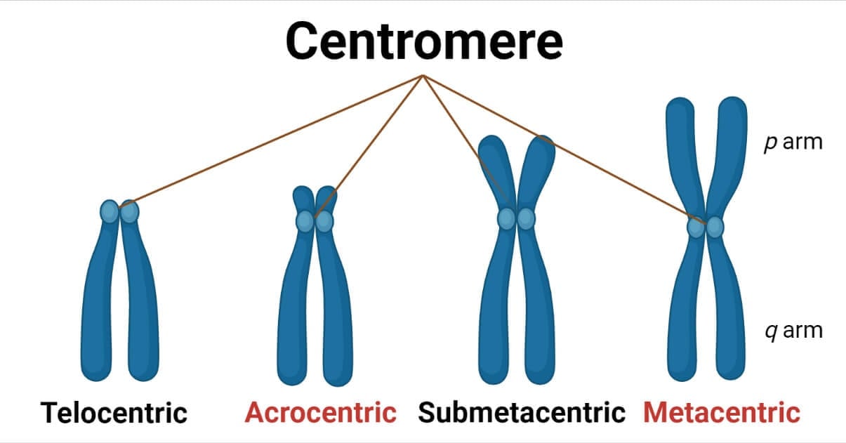
Metacentric chromosome
The centromere is situated in the center of metacentric chromosomes, ensuring that both parts are of identical length. The metacentric human chromosomes are 1, 3, 16, 19, and 20.
Submetacentric chromosome
There is a little imbalance in the length of the two parts of submetacentric chromosomes because the centromere is slightly off-center. Submetacentricity is present in human chromosomes 2, 4 – 12, 17, 18, and X.
Acrocentric chromosome
A substantially off-center centromere on an acrocentric chromosome causes one very long and one very short segment. Acrocentric human chromosomes include 13-15, 21, 22, and Y.
Telocentric chromosome
The centromere is located at the end of a telocentric chromosome. Telocentric chromosomes are not present in humans but in some other animals, such as mice.
Some organisms contain holocentric chromosomes with dispersed centromeres, such as plants in the genus Luzula. These chromosomes have centromeric heterochromatin dispersed along their whole length, to which the spindle attaches.
Chromosomes with numerous centromeres can infrequently form in mammalian tissue culture cells.
Diacentric describes a chromosome that has two centromeres. Chromosome breakdown followed by fusion frequently results in dicentric chromosomes. At anaphase, when the centromeres are tugged in opposite directions, a dicentric chromosome typically breaks.
Certain plants have holocentric chromosomes that have evolved yet-unknown defenses against this issue.
Hence, the centromere’s location offers a helpful marker for categorizing chromosomes into karyotype groups and creating a uniform nomenclature for mapping the locations of genes on chromosomes.
Types of Centromere
Point Centromeres
These are centromeres where mitotic spindle fibers draw to DNA sequences. These DNA sequences of the point centromere will often be found anywhere, resulting in the appearance of mitotic spindle fibers. The protein that initiates the formation of the mitotic spindle fiber complex will bind to that DNA sequence regardless of its position or other variables. Saccharomyces cerevisiae is shown to possess a point centromere.
Regional Centromeres
Most eukaryotic cells, including humans, use regional centromeres. These are centromeres, where a mixture of traits acting together to signal the position of a centromere determines mitotic spindle binding rather than a precise DNA sequence. Thus, the centromere’s placement is not determined by the DNA sequence.
The epigenetic markers provide information to the proteins regarding the position of these centromeres. To connect with the mitotic spindle complex, this is necessary. Enzymes are used to alter the DNA to create these epigenetic markers chemically. Without changing any of the information already existing in DNA, they are simply added to or removed from the DNA.
Functions of Centromere
- Centromeres play a role in the adhesion and separation of sister chromatids, chromosomal movement, attachment of microtubules, regulation of mitotic checkpoint, and heterochromatin formation. Cohesin protein molecules aid in adhering sister chromatids together.
- Centromeres serve as complex signaling hubs that control how quickly the cell cycle progresses.
- When eukaryotic cells divide, centromeres aid in the appropriate chromosome alignment and segregation.
- The centromeres serve as the kinetochore’s point of attachment. Sister chromatid attachment, as well as spindle fiber attachment are two of its primary activities.
- Centromeres are crucial in the development of a new cell. The centromere acts as a binding location for the two replicated chromosomes, known as sister chromatids, when the chromosomes are copied.
- Centromere dysfunction results in cancer and miscarriage in some cases.
References
- https://www.cell.com/trends/cell-biology/fulltext/S0962-8924(05)00234-5
- https://www.ncbi.nlm.nih.gov/pmc/articles/PMC5371998/
- https://www.genome.gov/genetics-glossary/Centromere
- https://www.nature.com/scitable/topicpage/chromosome-segregation-in-mitosis-the-role-of-242/
- https://byjus.com/biology/centromere/
- https://www.vedantu.com/biology/centromere
- https://biologydictionary.net/centromere/
- https://mail.almerja.com/reading.php?idm=29420