
Cell Organelles Definition
The cell is the basic unit of organization or structure of living matter bound by the semipermeable membrane and is capable of self-replicating in a medium free of other living systems. Cell organelle is the cellular component that can be both membranous and non-membranous present within a cell having distinct structures and functions.
Cell Membrane
Cell membrane or plasma membrane or plasmalemma is the thin and delicate layer that encloses the cell by defining the boundaries and maintaining the differences between the cytosol and the outer environment.
Structure of Cell Membrane
- The cell membrane is the tri-laminar structure containing two-layer of lipid and a protein.
- The major phospholipids found in the cell are phosphoglycerides, sphingolipids, and sterols.
- Phospholipids are the most abundant membrane lipid having a polar head group containing a hydrophilic head and two hydrophobic hydrocarbon tails.
- Some of the carbohydrate molecules get attached to the external surface of the cell membrane, either to the protein to form glycoprotein or to lipids to form glycolipids.
Functions of Cell Membrane
- The cell membrane provides mechanical support to the cell which helps in maintaining the shape of the cell and serves as a protection layer for its components.
- It is selectively permeable which controls the entry and exit of the different molecules.
- Transmembrane protein plays a role to connect the cytoskeleton through the lipid bilayer extracellular matrix or the adjacent cells.
Cell Wall
The cell wall is an integral part of the cell which is a rigid structure.
Structure of Cell Wall
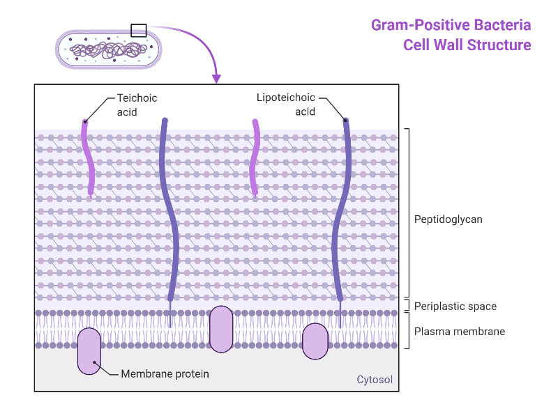
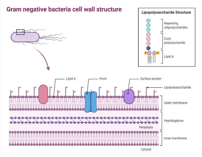
- The structure of the cell wall has divided bacteria into two groups i.e. Gram-positive and Gram-negative where Gram-positive bacteria have a thicker cell wall and Gram-negative have a thinner cell wall.
- The bacterial cell wall is composed of peptidoglycan where linear polysaccharide chains are crosslinked by the peptides.
- The cell wall of most algae and higher plants is composed of cellulose, hemicellulose, lignin, and pectin, and the fungi are chitin.
- A cell wall is a three-layered structure having a middle lamella, primary cell wall, and secondary cell wall.
- Middle lamella is the pectin-rich layer found between the adjacent cell walls.
- The primary cell wall is found just beneath the middle lamella which is formed immediately after the cell division, mainly composed of pectin and hemicellulose.
- In certain types of cells, an additional layer is found to the inner surface of the primary cell wall called the secondary cell wall, it is composed of cellulose, hemicellulose, and lignin.
Functions of Cell Wall
- The cell wall in bacteria determines the shape and also prevents the cell from bursting as the result of osmosis.
- The cell wall exhibits as the exoskeleton of the plant by providing the protecting layer and the mechanical support.
- It also determines the shape of the plant and prevents drought.
- It acts as the barrier between the interior cellular components and the external compartments.
Centrioles
The centriole is the hollow cylindrical structure formed by the arrangement of the tubulins.
Structure of Centrioles
- Centrioles are the tubular structure that measures about 0.2µm × 0.5µm which is open on both sides until it carries a flagellum or cilium.
- The wall of the centriole is made up of nine groups of microfilament which is arranged in a circle.
- Each group is a triplet formed by the A, B, and C tubules. Tubule A has 13 protofilaments while B and C have 10 protofilaments.
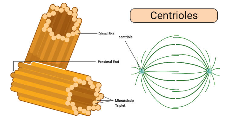
Functions of Centrioles
- It acts as the nucleating center from where the microtubules grow.
- During cell division, it forms the microtubule of spindle fiber which helps in the movement and separation of chromosomes.
- It is involved in the formation of the cilia and flagella.
Chloroplasts
Chloroplasts are one of the types of plastids that are colored due to the presence of pigment molecules.
Structure of Chloroplasts
- Chloroplast is oval or discoidal shaped in the higher green plants and are of different shapes like a cup or horse-shoe-shaped, girdle-shaped, and ribbon-shaped in case of algae.
- It is a double membranous structure with the outer membrane having less protein and is permeable, and the inner membrane having higher protein content and is selectively permeable.
- The inner space of the chloroplast is filled with the matrix namely stroma.
- The inner membrane of the chloroplast have invaginations carrying several membranous sacs like structure called thylakoids, each stack of the thylakoid is called a granum.
- The granum is connected with a pipe lie structure called stroma lamella.
- It is a dimorphic organelle i.e. it exists in two forms; granal and agranal.
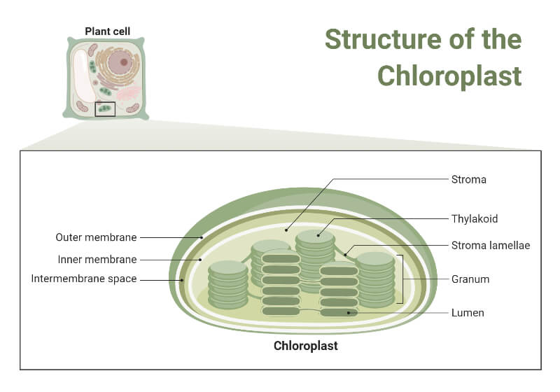
Functions of Chloroplasts
- It consists of different enzymes and molecules of green pigment which traps the light energy for the photosynthesis process.
- It consists of DNA, ribosome, and a complete protein synthesis mechanism that aids in the inheritance.
Cilia and Flagella
Cilia and flagella are the locomotory organelle having a similar type of structure but differ in number and size.
Structure of Cilia and Flagella
- Cilia and flagella arises from the centriole.
- These are the minute hair-like structure which is present extracellularly but originated intracellularly from the basal bodies.
- These are made up of a bundle of microtubules called the axoneme which is the part of the plasma member.
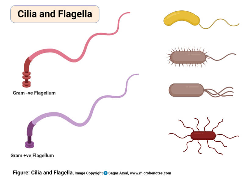
Functions of Cilia and Flagella
- They help in the locomotion of the organism.
- In aquatic organisms, cilia create water current for obtaining food, renewal of oxygen supply, and quick diffusion of carbon dioxide.
Cytoplasm
The cytoplasm is the semisolid substance that lies external to the nuclear member and internal to the plasma membrane.
Structure of Cytoplasm
- Cytoplasm can be distinguished into the cytosol and cytoplasmic structure.
- The cytosol is the liquid portion of the cytosol which contains 20-25% of protein and enzymes of the cell.
- In cytoplasmic structure living and non-living elements are suspended, the non-living structure is called periplasm and the living structure are called organelles.
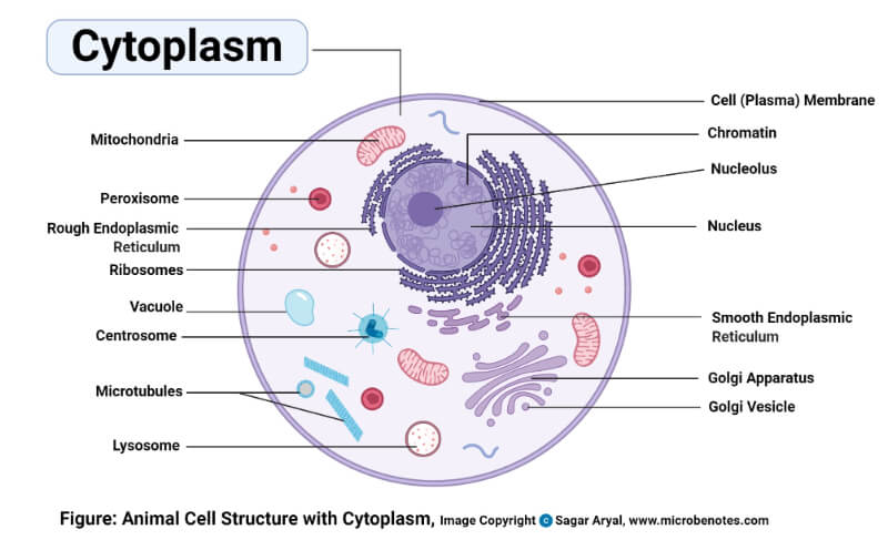
Functions of Cytoplasm
- Its fibers help to maintain the cell shape and movement of the other cell organelles by cyclosis.
- It stores food and secretory substances in the form of oil drops, triacylglycerol, secretory granules, glycogen granules, etc.
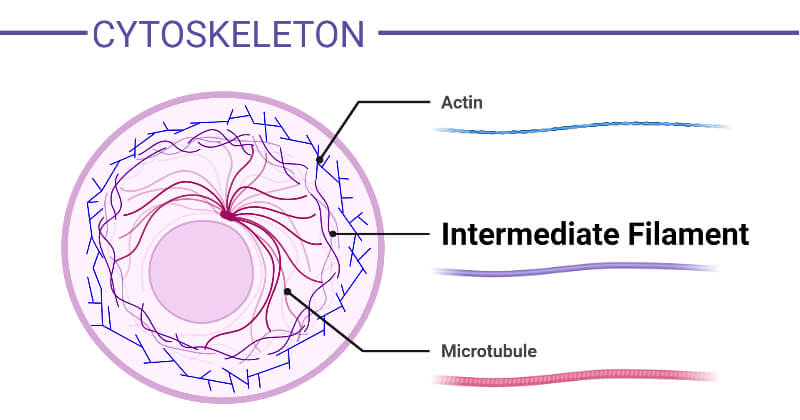
Cytoskeleton
The fibers present in the cytosol are the cytoskeleton.
Structure of Cytoskeleton
- It extends throughout the cytoplasm.
- It is a complex network of three types of protein filaments namely; microtubules, microfilaments, and intermediate filaments.
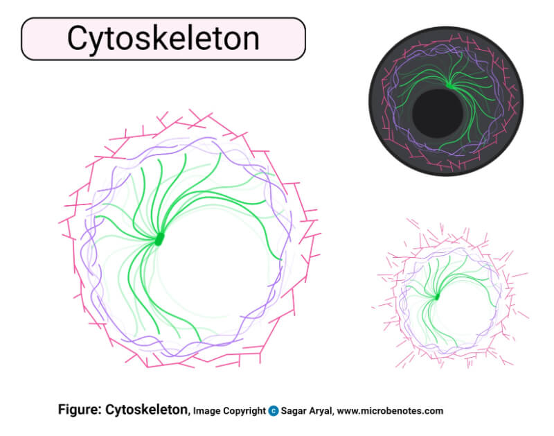
Functions of Cytoskeleton
- It is responsible for maintaining the cell shape.
- It functions in the intracellular and extracellular transport of water, ions, and small molecules.
- It helps in movements such as the crawling of cells on the substratum, muscle contractions, and changes in the shapes of a developing vertebrate embryo.
Cytosol
The cytosol is the fluid and soluble portion found outside the organelles which is the part of the cytoplasm.
Structure of Cytosol
- The cytosol consists of water, proteins, lipids, carbohydrates, various types of RNA molecules, and different types of smaller molecules.
- The peripheral layer of the cytosol is non-granular, viscous, clear, and rigid whereas the inner portion is granular and clear.
Functions of Cytosol
- It acts as a substrate to various cell organelles for their functioning.
- It is the site for the synthesis of organic materials like proteins, lipids, nucleotides, etc.
- It is the site for the catabolic pathways like glycolysis, pentose phosphate pathway, and fatty acid pathway.
- It helps in the exchange and distribution of various materials in the cell.
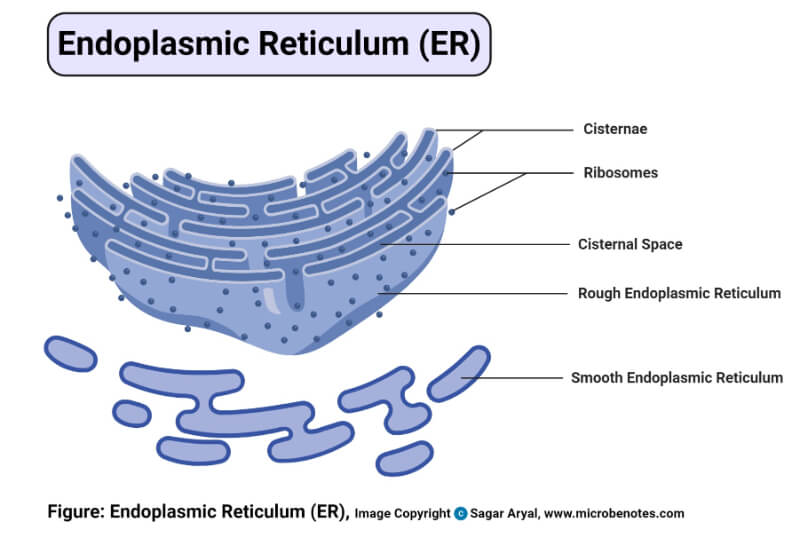
Endoplasmic Reticulum (ER)
The endoplasmic reticulum is the vacuoles or cavities which are concentrated in the endoplasmic portion of the cytoplasm. It is present in all eukaryotic cells except mature RBCs, eggs, embryonic cells and absent in prokaryotes.
Structure of Endoplasmic Reticulum (ER)
- The endoplasmic reticulum forms a network of the membranous system extending from the nuclear membrane to the cell membrane.
- It occurs in three different forms: Cisternae, Vesicle, and Tubules.
- Cisternae are long, flattened, unbranched sac-like structures having a diameter of 40-50 µm.
- Vesicles are oval, membranous structures having a diameter of 25-500 µm.
- Tubules are branched structures having a diameter of 50-190 µm.
- It is of two types according to the presence and absence of ribosomes: Smooth Endoplasmic Reticulum (SER) and Rough Endoplasmic Reticulum (RER).
- SER is found in the cells which are involved in the metabolism of lipids and glycogen such as adipose cells, interstitial cells, conduction fibers of the heart, spermatocytes, and leucocytes.
- RER is found in those cells which are active in protein synthesis such as pancreatic cells, plasma cells, goblet cells, and liver cells.
Functions of Endoplasmic Reticulum (ER)
- It provides an ultrastructural skeletal framework to the cell.
- Intracellular and intercellular exchange of materials by the process of osmosis, diffusion, and active transport.
- It consists of many enzymes which perform various synthetic and metabolic activities.
- These are found to conduct intracellular impulses which help in muscle contraction.
- It helps in the detoxification of harmful chemicals.
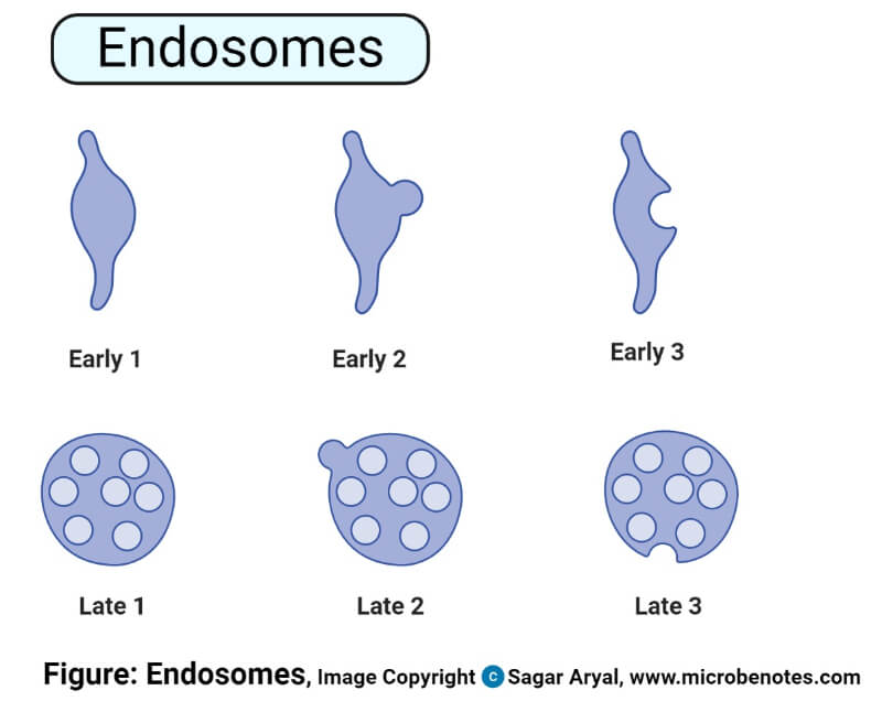
Endosomes
Endosomes are the membrane-bound structure found in the cytoplasm formed by the endocytosis process.
Structure of Endosomes
- These are the simply membrane-bounded vesicles and is similar to the structure of vesicle.
- These are mainly three types; Early endosomes, Late endosomes, and Recycling endosomes.
- Early endosomes contain tubular and vacuolar domains, containing the RAB5A markers.
- Late endosomes are generated from mature early endosomes, containing the RAB7A marker.
- Recycling endosome contains a large tubular network with RAB11 marker.
Functions of Endosomes
- It plays a major role in the synthesis, sorting, and delivery of the macromolecules within the cell.
- It acts as a temporary vesicle for the transport of molecules for degradation in lysosomes.
- Regulation of transmembrane receptor.
- In plants, endosomes are crucial for the maintenance of vacuoles and cell growth.
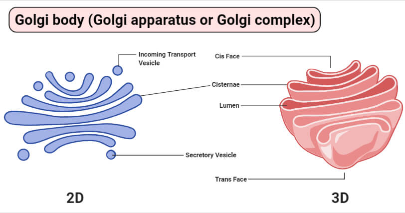
Golgi Apparatus (Golgi Bodies)
Golgi apparatus, also known as Golgi bodies are the cell organelle that helps in transporting, modifying, and packaging protein and lipid molecules. It is present in all cells except prokaryotic cells and certain eukaryotic cells namely; fungi, antherozoids of bryophytes and pteridophytes, mammalian RBCs, sieve tubes of plant, and mature sperms.
Structure of Golgi Apparatus
- The shape and form of the Golgi apparatus vary according to the types of cells.
- The Golgi apparatus appears as the interconnecting array of cisternae, tubules, and vesicles.
- It is formed by vesicles of the Endoplasmic reticulum. They have convex, forming, or cis-face (located next to either nucleus or rough ER) and concave, maturing, or trans-face (located near the plasma membrane).
Functions of Golgi Apparatus
- It plays a key role in packing and storage of cell’s protein and membrane constituents, and in directing them to their proper destinations.
- It helps in cell plate formation during the cytokinesis of mitosis or meiosis in the plant cell.
- Formation of lysosomes, plasma membrane, acrosome in sperms during spermatogenesis, and cortical granules of a variety of oocytes in the animal cell.
- Intracellular transport of different materials.
Intermediate filaments
Intermediate filaments are tough and durable protein fibers found in the cytoplasm of higher eukaryotic cells such as fingernails, claws, horns, feathers, hairs, beaks, turtle shells, etc.
Structure of Intermediate filaments
- It is fibrous, constructed like woven ropes having a diameter of 8-10nm which is the intermediate between microfilament and microtubules.
- In cross-section, it has a tubular appearance; each tubule appears to be made up of 4 or 5 protofilaments.
- It is composed of subunits which are called dimers, each dimer is composed of polypeptides.
- It is of four types of intermediate filament protein namely; Type I, Type II, Type III, and Type IV.
Functions of Intermediate filaments
- It provides support for cells that comes under physical stress such as muscle cells, neurons, and some epithelial cells.
- Supports cellular movement by providing mechanical strength to cells and tissues involved.
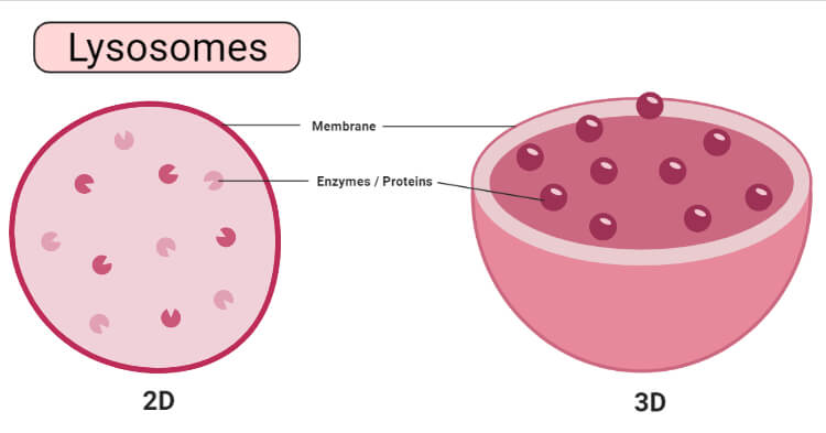
Lysosomes
Lysosomes also called “suicidal bags” of cells are tiny membrane-bound vesicles involved in intracellular digestion mainly found in animal cells.
Structure of Lysosomes
- The lysosomes are the circular vacuolar structure that is filled with solid material and are bounded with a unit membrane.
- Lysosomes are formed by the vesicles of Golgi bodies.
- It contains hydrolytic enzymes which help in the digestion of food.
- Lysosomes may contain up to 40 types of hydrolytic enzymes some of them are; nucleases, glycosidases, proteases, phosphatases, sulphatases, etc.
- Lysosomes exhibits polymorphism, four different types of lysosomes have been recognized they are; Primary lysosomes, Heterophagosomes, Autophagosomes, and Residual Bodies.
Functions of Lysosomes
- Digestion of the large food particles.
- Digestion of the intracellular substances i.e., proteins, lipids, and carbohydrates, and supply to the cell during starvation.
- The dead cell of the membrane of the lysosome ruptures and releases the enzymes which digest the various cell organelle of the cell, this process is called autolysis or self-destruction. It happens during starvation.
Microfilaments
Microfilaments are thin, solid, and smallest cytoskeleton. These are generally distributed in the cortical region of the cell just beneath the plasma membrane.
Structure of Microfilaments
- Microfilament is 7nm in diameter.
- Actin is the main structural protein of the microfilament. These individual actins are called G-actin and this G-actin polymerizes under favorable conditions to form F-actin.
- There are three types of actin – α, β, and γ. Α-form of actin is found in muscle tissues and β and γ are found in non-muscle tissues.
Functions of Microfilaments
- It helps in cell division in animal cells by forming constrictions.
- It helps in the cytoplasmic streaming in plant cells e.g., Nitella and Chara.
- It plays a role in cell migration via lamellipodia and filopodia.
Microtubules
Microtubules are cylindrical, straight, and hollow found in all eukaryotic cells except the erythrocytes of humans.
Structure of Microtubules
- A microtubule consists of long, unbranched, hollow tubules of 24-25 nm in diameter, 200 nm to a few micrometers in length with a 6nm thick wall having protofilaments.
- Protofilaments are formed by the longitudinal array of linear protein polymers called tubulin. Usually, 13 in number but varies according to cell.
- Tubulin occurs in two different forms, namely α-tubulin and β-tubulin each containing 450 amino acids.
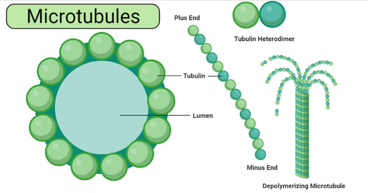
Functions of Microtubules
- It acts as a structural supporter and organizer as it determines the shape of the cell and maintains the internal organization of the cell.
- It helps to determine the shape of developing cells during cell differentiation.
- It plays a role in the contraction of the spindle and movement of chromosomes and centrioles as well as ciliary and flagellar motion.
- It is involved in the transport of vesicles, proteins, granules, and organelles within the cell.
Mitochondria
Mitochondria are the powerhouse of the cell as it converts glucose into Adenosine Tri-Phosphate (ATP) during the cellular respiration. It is a semi-autonomous organelle as it contains specific DNA for cytoplasmic inheritance and ribosomes for protein synthesis. It is found in all eukaryotic cells except mature mammalian RBCs and sieve tubes of phloem.
Structure of Mitochondria
- Mitochondria is a double membrane-bounded cell organelle having a varying size from 0.5 to 2.0 µm.
- The number of mitochondria varies from cell to cell some consist of a large number and even single e.g. the giant Amoeba, Chaos chaos contains 50,000; the egg of sea urchin contains 140,000 to 150,000. Also, some algal cells contain only one mitochondrion.
- Depending upon the physiological structure it may be of club, racket, vesicular, or round shape.
- The outer membrane is smooth whereas the inner membrane is convoluted forming infoldings known as cristae. Cristae contain stalked particles called oxysomes.
- Oxysomes are differentiated into stalk and base. The stalk region contains ATPase which helps in ATP synthesis and the base region helps in the electron transport system.
- It consists of 65 to 70% proteins, 25 to 30% lipids, 0.5% RNA, and a trace amount of DNA.
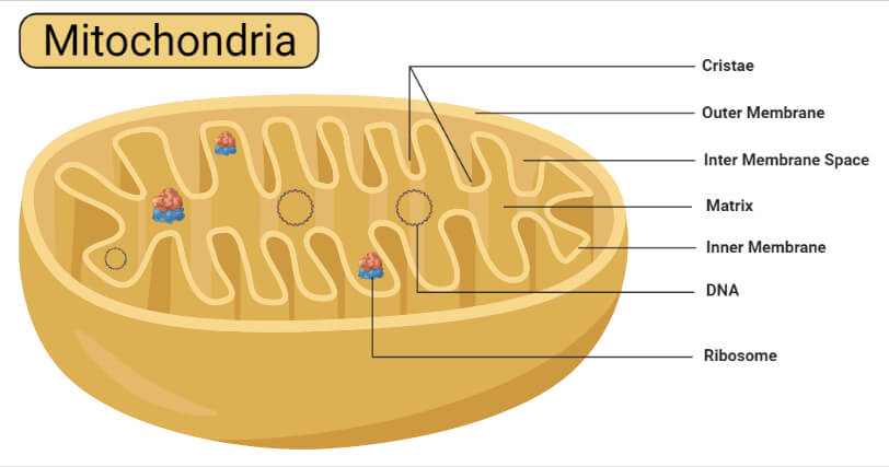
Functions of Mitochondria
- It performs the most important functions such as oxidation, dehydrogenation, oxidative phosphorylation, and respiratory chain in the cell.
- It is the respiratory organ of the cell where carbohydrates and fats are oxidized to form carbon dioxide and water.
- It synthesizes the energy-rich compound adenosine triphosphate.
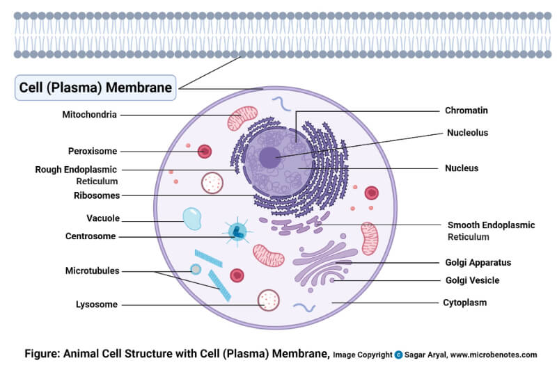
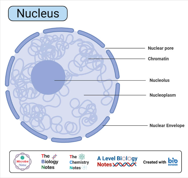
Nucleus
The nucleus is the controlling unit of the cell where almost all of the cell’s DNA is confined, replicated, and transcribed. It is found in all the eukaryotic cells the mature sieve tubes of higher plants and mammalian erythrocytes. The prokaryotic cell has an incipient nucleus.
Structure of Nucleus
- Generally, the cell contains a single nucleus but may vary from cell to cell. According to the number of nuclei it can be mononucleated, binucleated, and polynucleated.
- Nuclei vary in size from 3-25µm in diameter depending upon the type of cell.
- The nucleus is composed of a Nuclear membrane, Nucleoplasm, Chromatin fibers, and Nucleolus.
- A nuclear membrane or nuclear envelope is composed of lipid and protein which is a bilayered structure consisting of pores called nuclear pores.
- Nucleoplasm or nuclear sap is the space between nuclear membrane and nucleolus which is filled with transparent, semi-soli, and granular matrix. It consists of nucleic acids, proteins, enzymes, and minerals.
- Chromatin fiber is a thread-like, coiled, and elongated structure that consists of DNA and proteins. These chromatin fibers become thick ribbon-like structure chromosomes during cell division.
- The nucleolus is the spherical colloidal acidophilic body which is the site for the synthesis of ribosomal units.
Functions of Nucleus
- It consists of the hereditary material; chromatin of an organelle.
- It controls cell metabolism and other activities through the formation of RNAs.
- It helps in the protein synthesis by the formation of ribosomes.
- It directs cell differentiation and replication.
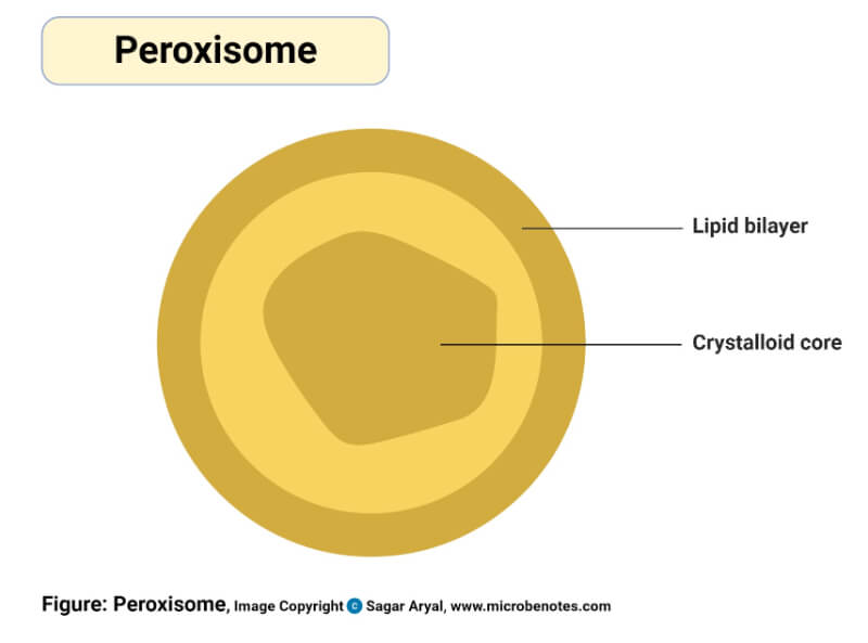
Peroxisomes
Peroxisomes are the microbodies that are common in both plant and animal cells.
Structure of Peroxisomes
- It usually appears circular having a diameter of 0.2-1.5µm.
- It is enclosed with a unit membrane of lipid and protein molecules.
- It consists of catalases and oxidases which participate in the oxidation of the substrate.
Functions of Peroxisomes
- It helps in the β-oxidation of fatty acids and is also associated with the oxidation of amino acids and uric acids.
- In plants, these are involved in photorespiration and catalyzes instruction of hydrogen peroxides into water and oxygen.
Plasmodesmata
Plasmodesmata are the bridge between the cytoplasm of the neighboring plant cell.
Structure of Plasmodesmata
- Plasmodesmata are lined with the plasma membrane.
- These are formed during cell division when a part of the parental endoplasmic reticulum is trapped to form a daughter cell.
Functions of Plasmodesmata
- It helps in cellular communication.
- It plays a crucial role in the transport of macromolecules and micromolecules from one cell to another.
- These have significance in vesicular transport due to their connection with surrounding tissues.
- These convey nutrient molecules towards and away from the veins.
Plastids
Plastids are colored or colorless double membranous organelle found only in plant cells and Euglena (protozoan). In animal cells, chromatophores are present instead of plastids.
Structure of Plastids
- Plastids are developed from protoplastids found in meristematic regions.
- It is of three types based on structure and function; Leucoplasts, Chloroplasts, and Chromoplasts.
- Leucoplast is the largest, colorless plastid responsible for the storage of starch, proteins, and lipids.
- Chloroplasts are green plastids which are the photosynthetic apparatus of the organelle.
- Chromoplast is a colored plastid other than green which provides color to fruits, seeds, roots, and flowers.
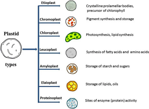
Figure: Types of Plastids. Image Source: Sitanshu Sekhar Sahu (https://doi.org/10.1186/1471-2105-14-S14-S7)
Functions of Plastids
- Storage and synthesis of different components like carbohydrates, lipids, proteins, and several anabolic enzymes.
- It makes plants visually attractive to insects and other animals which helps in the pollination and dispersal of seeds.
- Photosynthesis and photorespiration.
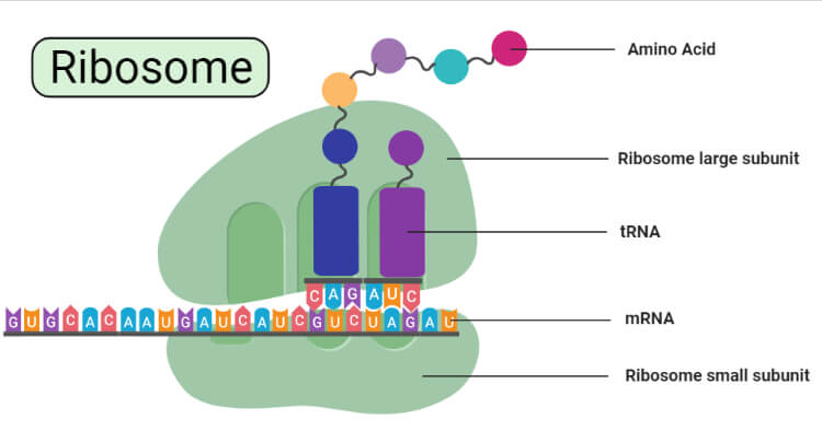
Ribosomes
Ribosomes are small, dense, round, and granular particles found freely in the matrix of mitochondria, chloroplast, and cytoplasm or remain attached to the endoplasmic reticulum and nucleus.
Structure of Ribosomes
- The ribosomes are hydrated, porous, and spheroids structural organelle composed of two subunits.
- It is composed of chiefly two-component; 40-60%RNA and 36-37% protein where these two are intertwined forming a complex arrangement.
- According to the size and sedimentation coefficient, it is of two types; 70s and 80s.
- 70s ribosome is found in prokaryotes and has two subunits, i.e. 50S and 30S.
- 80S ribosome is found in eukaryotes and has two subunits, i.e. 60S and 40S.
- The association and dissociation of these subunits depend upon the concentration of Mg++.
Functions of Ribosomes
- It is the site for protein synthesis.
- These protein acts as enzymes and controls cellular functions.
- Translation of genetic material from mRNA to protein during DNA translation.
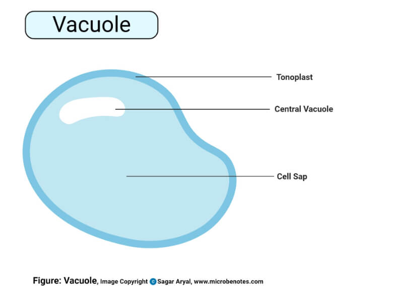
Vacuoles
Vacuoles are fluid-filled vesicles bounded by a single unit membrane called tonoplast.
Structure of Vacuoles
- It has a lipo-proteinous selectively permeable membrane.
- It consists of water, sugars, proteins, acids, and nitrogenous compounds such as alkaloids and anthocyanin pigments.
- In an organelle vacuole can be found as food vacuole, gas vacuole, contractile vacuole, and sap vacuole.
Functions of Vacuoles
- Gas vacuoles are associated with gaseous exchange and buoyancy.
- Food vacuoles help stores water, minerals, sugars, amino acids, pigment compounds, etc.
- The sap vacuoles help in maintaining the shape of the cell acting against turgor pressure.
- Contractile vacuoles are osmoregulatory in function and also help in the cytoplasmic moment in protozoans.
Vesicles
Vesicles are membrane-bound sacs formed during exocytosis and phagocytosis, which can be present inside or outside the cytoplasm.
Structure of Vesicles
- It can also be prepared with artificial techniques and called liposomes.
- Fluids are enclosed by the lipid bilayer.
- Vesicles with a diameter greater than 100nm are considered vacuoles.
- SNARE proteins on its surface help to recognize the specific organelles to which they need to transport materials.
- The main types of vesicles are; Lysosomes, Peroxisomes, Transport vesicles, Secretory vesicles, and Extracellular vesicles.
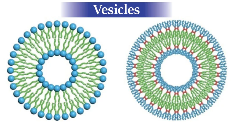
Image Source: University of Pennsylvania.
Functions of Vesicles
- Help in the transport of different biomolecules inside and outside by fusing with the plasma membrane.
- Involved in the transport of neurotransmitters in addition to calcium channels in the synapse of neurons.
- It can fuse with cell organelles such as Golgi bodies and lysosomes and helps in transport between them.
- They are also involved in buoyancy control.
- They also work as a storage tank for proteins, enzymes and, hormones.
- Different chemical reactions can also take place within them.
- They can absorb and involve in the destruction of toxic substances.
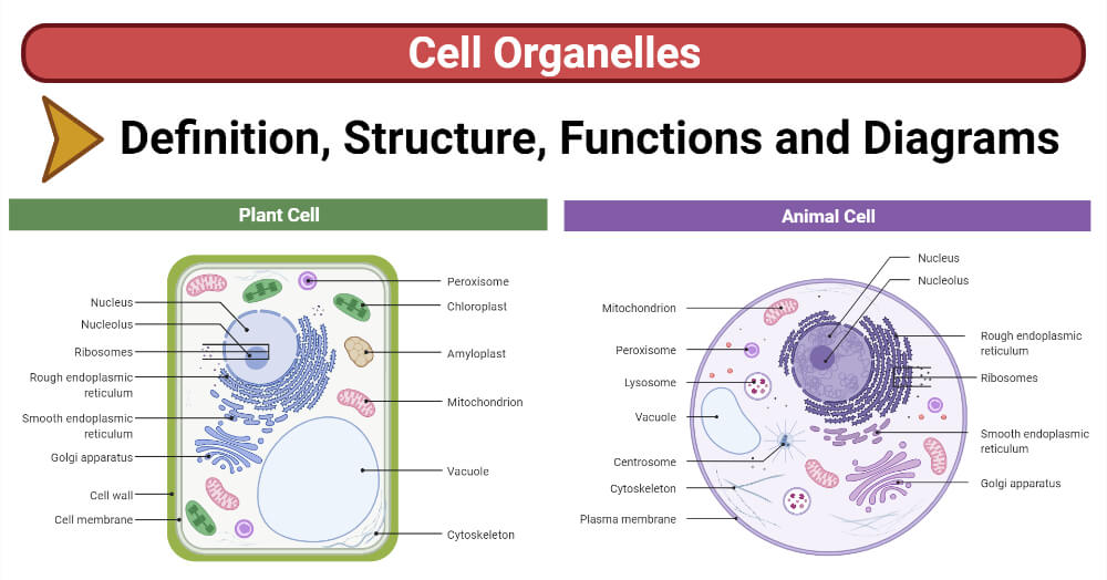
Plant Cell vs Animal Cell (13 Major Differences)
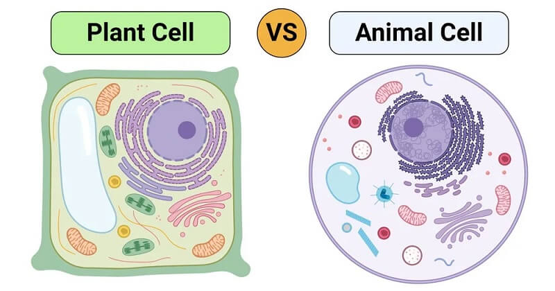
| Characters | Plant Cell | Animal Cell |
| Size | 10-100 µm in length | 10-30 µm in length |
| Shape | Square or rectangular shape | Irregular or circular shape |
| Cell wall | Present, made up of cellulose or peptidoglycan | Absent but the cell is enclosed by the cell membrane. |
| Centrioles and Centrosomes | Absent | Present |
| Cytokinesis | By cell plate formation | By cleavage furrow formation |
| Lysosomes | Rarely present | Present; digests cellular macromolecules |
| Mitochondria | Fewer | Numerous |
| Mode of Nutrition | Autotrophic | Heterotrophic |
| Nucleus | Pushed to one side of the cell by vacuole | Nearly centrally located |
| Plastids | Present | Absent |
| Vacuoles | Large and centrally located | Small numerous and scattered |
| Energy | Stored in form of carbohydrate | Stored in form of starch |
| Amino acids | Synthesizes all 20 amino acids | Capable of synthesizing only 10 amino acids |
References
- Verma, P. S., & Agarwal, V. K. (2016). CELL BIOLOGY, GENETICS, MOLECULAR BIOLOGY, EVOLUTION, AND ECOLOGY.
- Bruce Alberts, Alexander Johnson, Julian Lewis, David Morgan, Martin Raff, Keith Roberts, P., & Walter. (2008). Molecular Biology of the Cell,5th Edition. In Medicine & Science in Sports & Exercise (Vol. 40, Issue 9).
- Cell Membranes – The Cell – NCBI Bookshelf. (n.d.). Retrieved August 10, 2021, from https://www.ncbi.nlm.nih.gov/books/NBK9928/
- Cell Walls and the Extracellular Matrix – The Cell – NCBI Bookshelf. (n.d.). Retrieved August 12, 2021, from https://www.ncbi.nlm.nih.gov/books/NBK9874/
- Plasma membrane and cytoplasm (article) | Khan Academy. (n.d.). Retrieved August 10, 2021, from https://www.khanacademy.org/science/biology/structure-of-a-cell/prokaryotic-and-eukaryotic-cells/a/plasma-membrane-and-cytoplasm
- Britannica, T. Editors of Encyclopaedia (2019, September 11). Cytoplasm. Encyclopedia Britannica. https://www.britannica.com/science/cytoplasm
- Ribosomes – Structure And Functions | A-Level Biology Revision Notes. (n.d.). Retrieved September 10, 2021, from https://alevelbiology.co.uk/notes/ribosomes-structure-and-functions/
- Contento, A. L., & Bassham, D. C. (2012). Structure and function of endosomes in plant cells. Journal of Cell Science, 125(15), 3511–3518. https://doi.org/10.1242/jcs.093559
- 4.4A: Vesicles and Vacuoles – Biology LibreTexts. (n.d.). Retrieved September 10, 2021, from https://bio.libretexts.org/Bookshelves/Introductory_and_General_Biology/Book%3A_General_Biology_(Boundless)/4%3A_Cell_Structure/4.4%3A_The_Endomembrane_System_and_Proteins/4.4A%3A_Vesicles_and_Vacuoles
- Contento, A. L., & Bassham, D. C. (2012). Structure and function of endosomes in plant cells. Journal of Cell Science, 125(15), 3511–3518. https://doi.org/10.1242/jcs.093559
- Endosome Definition and Examples – Biology Online Dictionary. (n.d.). Retrieved September 10, 2021, from https://www.biologyonline.com/dictionary/endosome
- Molecular Expressions Cell Biology: Endosomes. (n.d.). Retrieved September 9, 2021, from https://micro.magnet.fsu.edu/cells/endosomes/endosomes.html
- Molecular Expressions Cell Biology: Plant Cell Structure – Plasmodesmata. (n.d.). Retrieved September 10, 2021, from https://micro.magnet.fsu.edu/cells/plants/plasmodesmata.html
- Zambryski, P. (2008). Plasmodesmata. Current Biology, 18(8), 324–325. https://doi.org/10.1016/j.cub.2008.01.046
- Vesicles: What are they? Types, structure, and function. (n.d.). Retrieved September 10, 2021, from https://www.medicalnewstoday.com/articles/vesicle
- Differences Between Plant and Animal Cells. (n.d.). Retrieved September 10, 2021, from https://www.thoughtco.com/animal-cells-vs-plant-cells-373375
- Plant Cell vs Animal Cell – Difference and Comparison | Diffen. (n.d.). Retrieved September 10, 2021, from https://www.diffen.com/difference/Animal_Cell_vs_Plant_Cell.