The cardiovascular system is an organ system in animals comprising the heart and a closed circuit of blood vessels that are responsible for circulating blood throughout the body.
It is a part of the blood circulatory system. (The blood circulatory system comprises the cardiovascular system and the blood.)
The word ‘cardiovascular’ is derived from the Greek word kardia meaning the heart, and the Latin word vascula meaning the vessels.
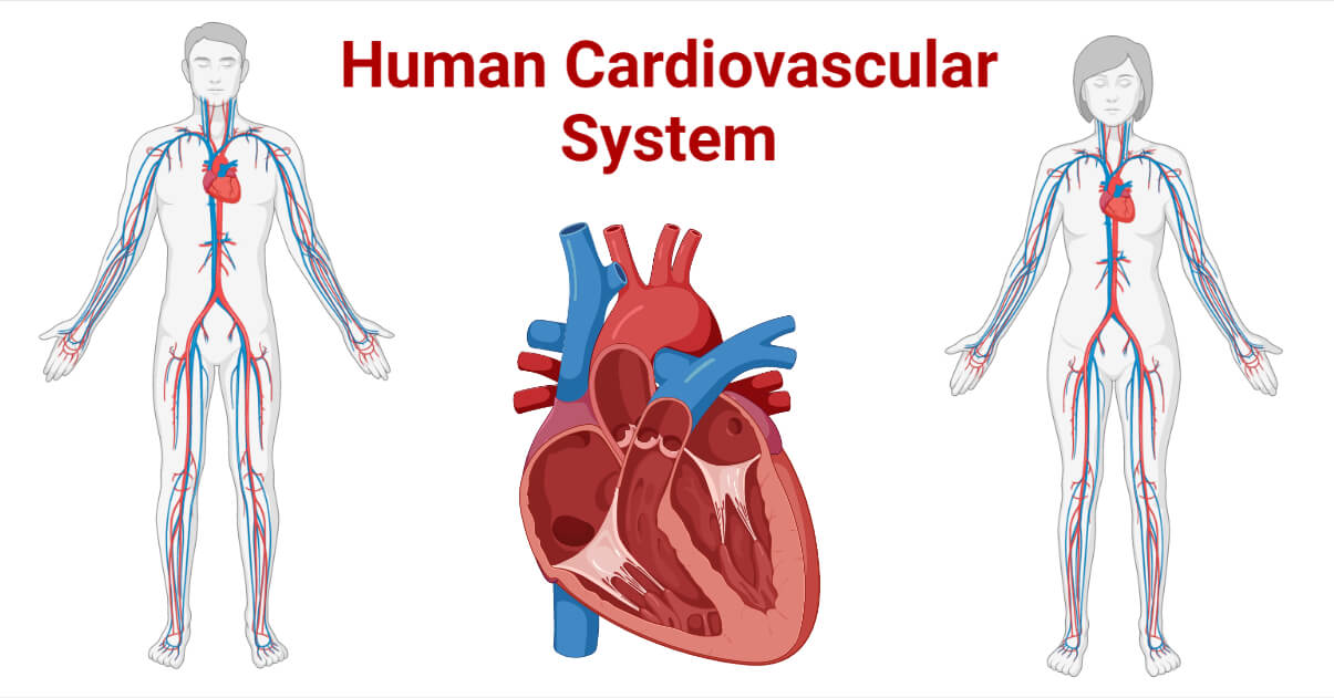
The heart pumps the blood inside the closed blood vessels, and the blood circulates throughout the body, providing nutrients, oxygen, hormones, and other necessary elements to every needy cell of the body and taking back the waste products from the cells to specific organs for excretion. In short, we can say that the cardiovascular system is the transportation pathway, and the blood is the means of transportation inside animals’ bodies. Here, we mainly discuss the human cardiovascular system.
Interesting Science Videos
Organs of the Cardiovascular System
The cardiovascular system comprises the heart and the blood vessels.
A. Heart
The heart is a muscular organ that propels the blood inside the blood vessel for circulation. It is a muscular pump for pumping blood to and from all body parts.
Location of the Heart
Heart lies inside the thoracic cavity behind the sternum, in between the lungs, and just above the diaphragm. It is slightly tilted towards the left part of the thoracic chamber with about the 2/3rd part left of the sternum. It lies in a fluid-filled cavity called the pericardial cavity.
Structure of the Heart
The human heart is slightly larger than or approximately the size of a clenched fist in an adult. It is a nearly cone-shaped structure with a broad base facing upward and right to the sternum and a pointed apex facing downward and left to the sternum. On average the human heart is 13×9×6 cm in dimensions and weighs about 300 grams (0.67 pounds).
Pericardium
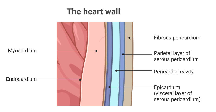
The heart is externally enclosed in a double-layered sac called the pericardium. The pericardium is divided into two layers; first the fibrous pericardium and the internal serous pericardium. The fibrous pericardium is made up of strong connective tissue and provides protection and anchorage with the surrounding structure of the thoracic cavity. The serous pericardium is made of a smooth serous membrane that provides lubrication to the heart to prevent friction during the heartbeat. The serous pericardium is again a double-layered membrane divided into an outer parietal serous pericardium and an inner visceral serous pericardium (also called the epicardium).
The Layers of the heart muscles/wall
The human heart has three distinct layers, namely epicardium, myocardium, and endocardium.
Epicardium is the outermost layer of the heart made up of mesothelium, fats, and connective tissues. It is the visceral layer of the serous pericardium that surrounds the heart and roots of the major coronary vessels. It serves multiple functions like providing physical protection to the heart, signaling for embryonic heart formation and maturation, helping with the heart injury response and regeneration, and secretion of factors for cardiomyocyte proliferation.
The myocardium is the middle thickest muscular layer made up of cardiomyocytes (cardiac muscles). It is responsible for the contraction and relaxation actions of the heart. Additionally, it also serves as a platform for the conduction of cardiac electrical impulses.
The endocardium is the innermost layer of the heart wall covering the internal chambers of the heart and is made up of endothelial cells and elastic fibers. This thin layer is further composed of three sub-layers; connective subendocardial tissue layer, fibro-elastic tissue layer, and endothelium. It serves multiple functions like covering and protecting the valves, acting as a blood-heart barrier and regulating the ionic concentration of heart cells, and preventing the blood from sticking to the heart walls and forming blood clots. The Purkinje fibers are present in the subendocardial layer and help in conducting the cardiac action potential.
Chambers of the heart
Humans have a four-chambered heart i.e. the heart is partitioned into four chambers by the myocardium. The four chambers are grouped as atria and ventricles.
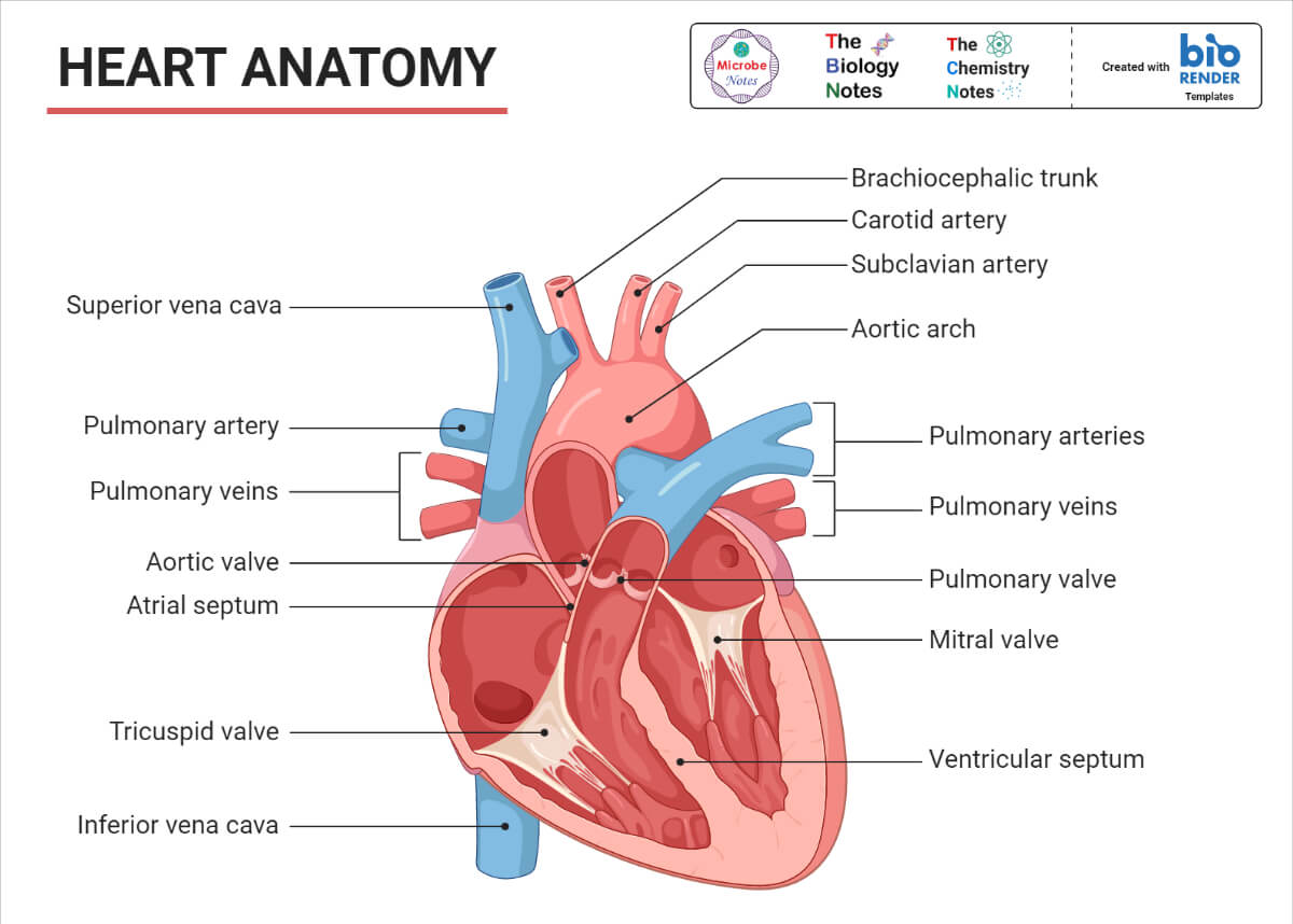
Atria: Atria are the two uppermost blood-receiving chambers of the heart that receives the blood from the veins. These are thin-walled chambers separated by a thin myocardial wall known as the interatrial septum. There are two atria, the right atrium, and the left atrium. The right atrium receives the deoxygenated blood from the superior and inferior vena cava and passes it to the right ventricle. The left atrium receives the oxygenated blood from the pulmonary vein and passes it to the left ventricle for supply to the body.
Ventricles: Ventricles are the two lowermost blood-pumping chambers of the heart that receives blood from the atria and pump outside the heart. These are comparatively larger chambers with thick muscular walls. There is a thick myocardial wall dividing the ventricles into the right and the left ventricle called the interventricular septum. The right ventricle receives the deoxygenated blood from the right atrium and pumps it to the lungs for purification through pulmonary arteries. The left ventricle receives the oxygenated blood from the left atrium and pumps it all over the body through the aorta.
Valves in the heart
Valves are the muscular flaps that maintain the one-way flow of the blood inside the heart i.e. prevent the backward flow of the blood. The human heart has four valves classified into two categories, first the Atrioventricular valves (containing the bicuspid and tricuspid valves), and second the Semilunar valves (containing the pulmonary and aortic valves).
The Atrioventricular (AV) valves are those valves that are located in between the atrium and ventricles and prevent the backward flow of blood from the ventricle to the atrium. The AV valve present between the right atrium and the right ventricle is called the tricuspid valve. There is another AV valve in between the left atrium and the left ventricle known as the bicuspid valve or the mitral valve. As the name suggests, the tricuspid valve has three cusps or leaflets while the bicuspid valve has only two cusps or leaflets. Both of these valves serve the primary function of preventing the return of blood back to the atria from the ventricles.
The Semilunar valves are another set of valves that are located at the base of two large arteries; the aorta and the pulmonary artery. The semilunar valve between the pulmonary artery and the right ventricle is called the pulmonary valve. The semilunar valve present between the aorta and the left ventricle is called the aortic valve. Both of the semilunar valves have three cusps or leaflets and check for the return of blood back to the heart chambers.
Blood Supply in the Heart
The blood inside the heart chambers can’t nourish the heart cells, so like any other organ; the heart also depends on the arteries and veins for blood supply to its cells. The artery that supplies the oxygenated blood along with required nourishments is known as the coronary artery. There are two main coronary arteries, the left, and the right main coronary arteries, arising from just above the aortic valve. The right coronary artery divides and supplies the right atrium, the right ventricle, and the lower part of the left ventricle. The left coronary artery divides and supplies the left atrium, the upper and front parts of the left ventricle, and the septum.
Several cardiac veins drain the deoxygenated blood from the heart. The great cardiac vein, the posterior cardiac vein, the middle cardiac vein, and the small cardiac vein are the major cardiac veins present in a human heart. All these veins drain into an enlarged vessel known as the coronary sinus.
Nerve Supply in the Heart
The heart is influenced by the vagus and the sympathetic nerves that form a network of nerves in the heart called the cardiac plexus. These nerves do not control the heartbeat but can influence the rate and force of heart contraction and relaxation.
Functions of the Heart
The heart is a pumping device and as such its one and only primary function is to pump blood in and out through the blood vessels. In doing so, it helps in transporting essential nutrients, oxygen, hormones, antibodies, antigens, and several other chemical components to every cell and bringing back the excreta from every cell to excretory organs for excretion.
Besides, it produces and maintains blood pressure and manages the blood supply.
| Did you KNOW? Heart pumps about 6000 to 7500 liters of blood a day. Heart beats about 100,000 times a day or 3 billion times in a life of a person. Heart of a newborn beats faster than an adult; about 90 to 190 beats per min. |
B. Blood Vessels
Blood vessels are the closed tubes through which blood circulates. The blood vessels form the network of tubes that carry blood from the heart to all parts of the body and back to the heart from all parts of the body.
Types of Blood Vessels
There are three main types of blood vessels; the arteries, the veins, and the capillaries.
Artery/Arteries
Arteries are the blood vessels that carry the blood away from the heart to various parts of the body. All arteries carry the oxygenated blood except the pulmonary artery which carries the deoxygenated blood from the right ventricle to the lungs for purification. These arteries that transport the oxygenated blood to the body tissues are called systemic arteries.
Arteries are thick muscular-walled blood vessels that are strong to withstand the blood flow at higher pressure. The blood inside arteries flows at a very high pressure of about 120 mm of Hg, hence there is no need for any check valve to prevent the possible backflow of the blood.
Arteries run deep in within the body. On appearance, they look like thick-walled tubes in red color. The wall of an artery is made of three distinct layers;
Tunica Externa: the outermost layer of the arteries, also known as the tunica adventitia. These are made up of fibrous connective tissues and have nerves and capillaries to supply them. This layer support and protect the arteries.
Tunica Media: the middle layer of the arteries. This layer is the thickest and is made up of vascular smooth muscles, elastic fibers, connective tissues, and polysaccharides. This layer helps in maintaining blood pressure.
Tunica Intima: the innermost layer of the arteries. It is the thinnest layer made of a single layer of simple squamous epithelium cells surrounded by a thin layer of connective tissue membrane with elastic fibers.
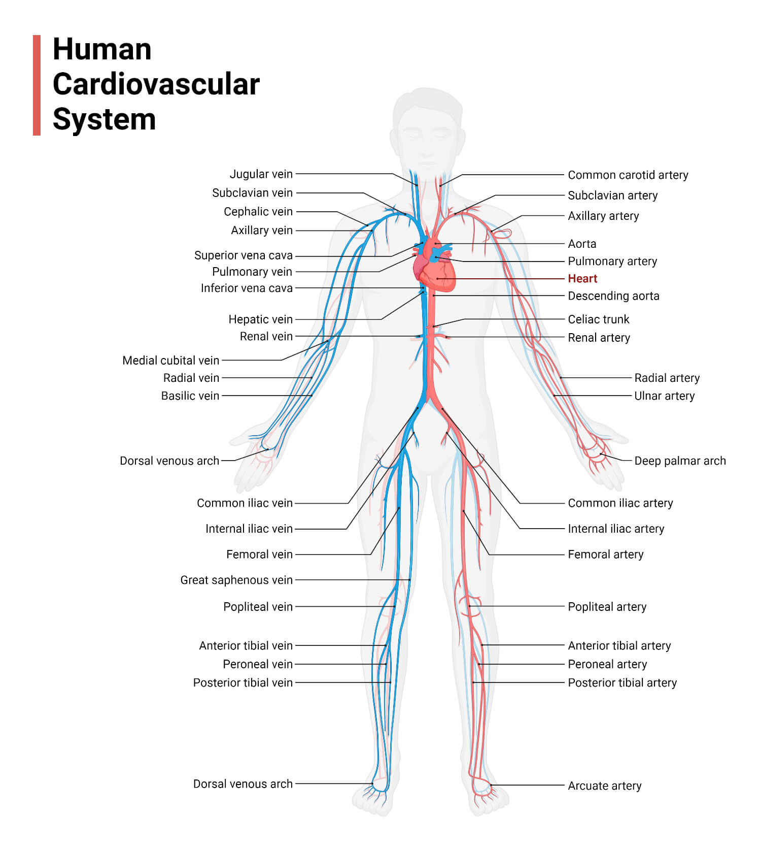
The systemic arteries arise from the aorta and further branch into smaller vessels called the arterioles. These arterioles further branch and become finer until they feed the tissues and cells through capillaries.
The aorta is the largest artery arising directly from the left ventricle and extending downward towards the abdomen. The aorta is anatomically divided into four sections and from each section, a major artery arises and divides to form all arteries of the human body. Some of the major arteries are summarized in the table below.
| The Sections of the Aorta | The Sections of the Aorta | The Sections of the Aorta | The Sections of the Aorta | |
| Ascending Aorta | Aortic Arch | Thoracic Aorta | Abdominal Aorta | |
| Major Arteries Arising from the Sections of the Aorta | The left and the right coronary arteries | – Brachiocephalic trunk – Left common carotid artery – Left subclavian artery – Axillary artery – Branchial artery – Radial and ulnar artery | Intercostal arteries (10 pairs) | – Celiac trunk – Superior mesenteric artery – Renal arteries – Gonadal arteries – Lumbar arteries – Inferior mesenteric artery – Common iliac arteries |
Vein/Veins
Veins are the blood vessels that collect the deoxygenated blood from the various parts (tissues) of the body and transport it back to the heart. All veins carry deoxygenated blood except the pulmonary vein and umbilical vein. The remaining veins that carry deoxygenated blood are collectively called systemic veins. Veins are often closer to the skin and appear blue in color; however, there are deep-seated veins found deep inside the tissues and internal organs.
Comparatively, veins are with thinner walls. The blood flowing through the vein barely crosses the pressure of 10 mm of Hg, so there is no need for a thick wall but contains valves to prevent the backflow of the deoxygenated blood.
Like arteries, the venous walls are made of three layers, namely, the tunica externa, the tunica media, and the tunica intima. The tunica externa is the thickest layer in the veins and the tunica media of the veins have very few smooth muscles.
The veins develop from the smaller and finer branches connected to the capillaries called the venules. The venules collectively join and pour into a larger vessel called the veins which further meet, converge, and drain to the vena cava. There are two major vena cavas, the superior vena cava, and the inferior vena cava. The major veins that converge and drain into the two vena cavas are summarized in the table below.
| The Major Vena Cava | The Major Vena Cava | |
| The Superior Vena Cava | The Inferior Vena Cava | |
| Major Veins Draining into each vena cava | Radial and ulnar veins Cephalic vein Basilic vein Median cubital vein Subclavian vein Vertebral vein Internal jugular vein Brachiocephalic veins Azygos vein | Tibial veins Great saphenous veins Common iliac vein Gonadal vein Renal veins Hepatic portal vein Hepatic vein |
Capillary/Capillaries
Capillaries are tiny and very fine blood vessels of just about 5 to 10 μm in diameter that connect the venules with arterioles. It seems that arterioles are further branched into finer vessels called capillaries which then join and converge to bigger vessels called venules.
These are the sites where there is an exchange of nutrients, gases, water, and other components between cells or tissues and the blood.
The capillary wall is composed of tunica intima only and is very thin so that nutrient and gaseous exchange can take place.
Capillaries are of three types:
Continuous capillaries with uninterrupted lining allow for the exchange of smaller molecules like gaseous molecules, water, and ions.
Fenestrated capillaries with the pores (fenestrae) of 60 to 80 nm covered with the diaphragm of fibrils. These pores allow for the exchange of smaller protein molecules, hormones, and others.
Sinusoidal capillaries with open pores of 30 to 40 μm that allow even RBCs, WBCs, and serum proteins to escape.
Functions of Blood Vessels
- The blood vessels are the pathway for the circulation of blood under the pumping action of the heart. Hence, they are the transporter of nutrients, water, minerals, hormones, etc.
- Capillaries are the site for gaseous exchange and nutrients and other elements exchange.
- They protect from blood loss during circulation and/or injury.
- Indirectly, they are involved in maintaining the body temperature.
| Did you KNOW? Connect all blood vessels end to end and you will get a tube of more than 100,000 Km (or >62,000 miles) that is enough to round the earth 2.5 times. |
Functions of the Cardiovascular System
There are multiple functions of the cardiovascular system, some of which are listed below.
- The most important function is to pump blood throughout the body and collect the blood back for re-circulation.
- Transporting the oxygenated blood and supplying essential elements to every cell and tissue of the body and collecting back the deoxygenated blood along with wastes.
- Maintaining the blood pressure.
- It functions in transporting the blood to the lungs for exchanging the carbon dioxide with oxygen and supplying the oxygen to all the body cells for metabolism (respiration).
- Providing a surface for the exchange of gases, nutrients, wastes, and other elements.
- The valves present in the heart and the veins prevent the backflow of blood.
- The vascular system is responsible for properly transporting the blood and all required nutrients and elements without any blood loss.
- The capillaries of the vascular system form the area for gaseous exchange.
- Receiving the deoxygenated blood and transporting it to the lungs for oxygenation.
- Pumping blood to the kidney for filtration and excretion of wastes.
Diseases of the Cardiovascular System
The cardiovascular system is affected by several disorders and infections, called cardiovascular diseases (CVDs). CVDs are the leading cause of death globally; with about 17.9 million deaths associated with CVDs in 2019. It is reported that almost half of the adults in the USA are affected by any form of CVDs. The diseases of the cardiovascular system can be broadly categorized into infectious cardiovascular diseases and non-infectious cardiovascular diseases.
Infectious Cardiovascular Diseases
These are cardiovascular diseases that are caused by infective microorganisms. The major infectious diseases of the cardiovascular system are:
- Bacterial myocarditis is often associated with bacterial pathogens like Streptococcus spp., Staphylococcus spp., Neisseria spp., Mycobacterium spp., Salmonella spp., and Shigella spp.
- Rheumatic heart disease caused by the Group A Streptococci
- Viral pericarditis and viral myocarditis are often caused by viruses like Adenovirus, Arbovirus, Cytomegalovirus, Influenza virus, Dengue virus, Epstein-Barr virus, Hepatitis-C, Mumps virus, etc.
- Fungal myocarditis is often associated with fungi like Aspergillus spp., Candida spp., Blastomyces spp., Cryptococcus spp., Cocciodioidomyces spp., etc.
Non-infectious Cardiovascular Diseases
Non-infectious CVDs are the leading cause of death globally. Some of the common non-infectious CVDs are:
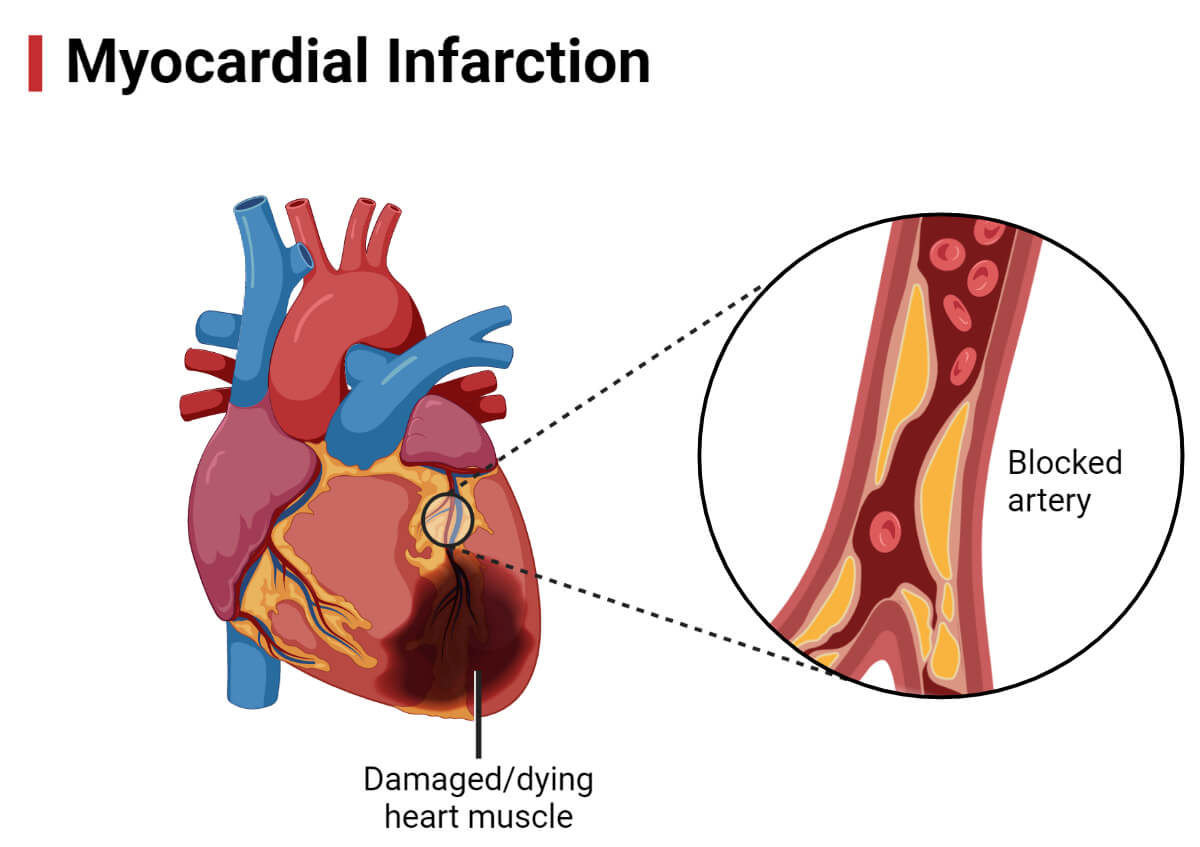
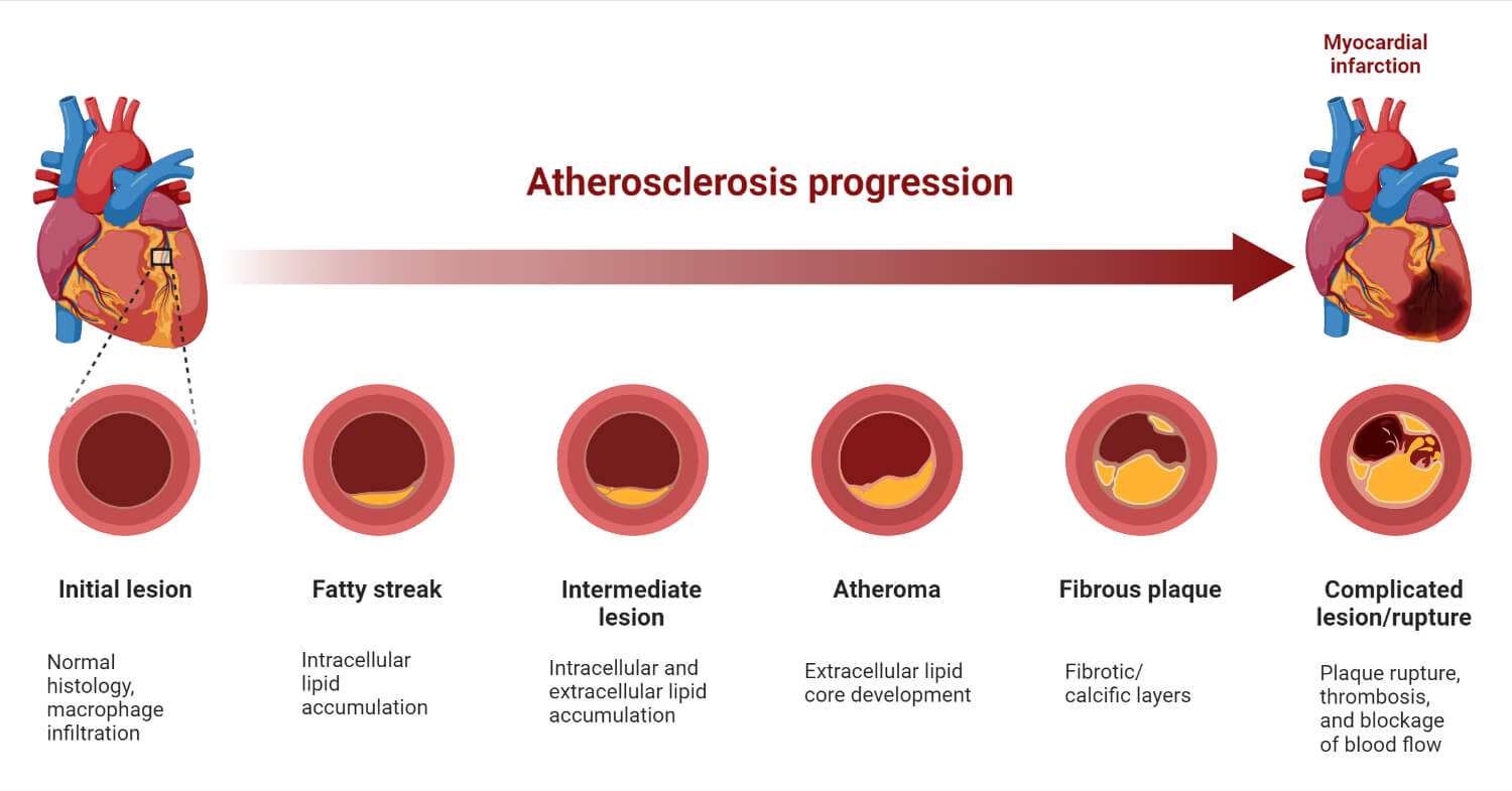
- Heart Attack: The condition when blood flow to a part of the heart is blocked due to blood clots or fatty deposits.
- Ischemic Stroke: The condition when blood flow in the brain is blocked due to a blood clot or obstacle in the blood vessel supplying the brain.
- Heart Failure (Congestive Heart Failure): A condition when the heart fails to pump blood in adequate amounts so that the need for blood and supply of oxygen and other elements are not met.
- Arrhythmia: Irregular/abnormal beating of the heart. Bradycardia is when the heart beats less than 60 times per minute and tachycardia is when the heart beats more than 100 times per minute.
- Valve Disease: Condition when the valve becomes too tight or too loose and fails to check the backflow of the blood.
- Coronary Artery Disease: Condition when the heart’s blood vessels fail to transport blood smoothly due to blockage or inflammation.
- Peripheral Artery Disease: Condition when the systemic blood vessels fail to transport blood smoothly due to blockage or inflammation or constriction.
- Congenital Heart Disease: Malformation of the heart and blood vessels from birth affecting the proper functioning of the cardiovascular system.
- Aortic Disease: The conditions where the aorta experiences some abnormalities like dilation and bulging of the wall (aneurysm).
- Pericardial Diseases: Disorders in the pericardium such as pericarditis (swelling of the pericardium), pericardial effusion and cardiac tamponade (abnormal accumulation of fluid in pericardial sac), and constrictive pericarditis (stiffening and thickening of the pericardium).
- Deep Vein Thrombosis: Condition when blood clot forms in deep veins affecting blood circulation.
- Pulmonary Embolism: Condition due to blockage of an artery supplying to the lungs mostly due to the formation of a blood clot.
- Cerebrovascular Disorder: Conditions or disorders that affect the vesicles supplying the brain.
- Atherosclerosis: Development of abnormalities called lesions in the arterial walls and stiffening of the arterial walls.
- Vasculitis: Inflammation of the blood vessels or thickening of the walls of the blood vessels.
- Angina Pectoris: Pain in the chest due to a low supply of blood in the heart muscles.
- Hypertension: Condition of elevated blood pressure in the arteries (usually 130/80 mm of Hg or 140/90 mm of Hg and above)
- Hypotension: Condition of low blood pressure, usually at or below 90/60 mm of Hg.
References
- Pericardium. (2022, November 28). In Wikipedia. https://en.wikipedia.org/wiki/Pericardium
- Myocardium: definition, structure and function | Kenhub
- Purkinje Fibers : Anatomy, Location & Function – Anatomy Info
- Endocardium: What is it? Types of Layers, Functions and Associated Conditions – Scope Heal
- Endocardium – an overview | ScienceDirect Topics
- Endocardium. (2022, November 16). In Wikipedia. https://en.wikipedia.org/wiki/Endocardium
- Parts Of The Human Heart – Science Trends
- Human heart: Anatomy, function & facts | Live Science
- Human Heart – Anatomy, Functions and Facts about Heart (byjus.com)
- Human cardiovascular system | Description, Anatomy, & Function | Britannica
- 7 Things About Heart and Blood Vessels (softpedia.com)
- What are Blood Vessels? Types, Layers and Functions – An Overview (byjus.com)
- Blood vessel | Definition, Anatomy, Function, & Types | Britannica
- The Types of Blood Vessels in Your Body (thoughtco.com)
- Classification & Structure of Blood Vessels | SEER Training (cancer.gov)
- Sheppard, M. N. (2013). Infectious diseases of the cardiovascular system. Diagnostic Histopathology, 19(3), 99-105. https://doi.org/10.1016/j.mpdhp.2013.01.016
- Schultz, J. C., Hilliard, A. A., & Rihal, C. S. (2009). Diagnosis and Treatment of Viral Myocarditis. Mayo Clinic Proceedings, 84(11), 1001-1009. https://doi.org/10.1016/S0025-6196(11)60670-8
- Khandaker, M. H., Espinosa, R. E., Nishimura, R. A., Sinak, L. J., Hayes, S. N., Melduni, R. M., & Oh, J. K. (2010). Pericardial Disease: Diagnosis and Management. Mayo Clinic Proceedings, 85(6), 572-593. https://doi.org/10.4065/mcp.2010.0046
- Waheed SM, Kudaravalli P, Hotwagner DT. Deep Vein Thrombosis. [Updated 2022 Nov 23]. In: StatPearls [Internet]. Treasure Island (FL): StatPearls Publishing; 2022 Jan-. Available from: https://www.ncbi.nlm.nih.gov/books/NBK507708/
- What is Cardiovascular Disease? | American Heart Association
- Cardiovascular diseases (CVDs) (who.int)
- Cardiovascular Disease: Types, Causes & Symptoms (clevelandclinic.org)
- 15 circulatory system diseases: Symptoms and risk factors (medicalnewstoday.com)
- Blood Vessels: Types, Anatomy, Function & Conditions (clevelandclinic.org)