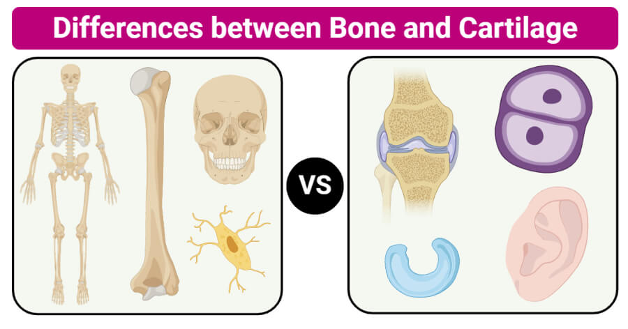
Bone Definition
A bone is a connective tissue where the living cells, tissues, and other components are enclosed within hard non-living intercellular material.
- The two important constituents of bone are collagen and calcium phosphate that distinguish it from other similar structures like the enamel and chitin.
- Bone tissues together make up the human skeleton system and skeletons of other vertebrates.
- These structures exist in different shapes and sizes with different forms of complexities fit for different functions.
- Bone tissues in the hard structure form a honey-comb like a matrix internally composed of two different cells; osteoblasts and osteoclasts.
- Osteoblasts are responsible for making new cells that line and protect the outer surface of the bones, whereas the osteoclasts mineralize the cells to transport minerals into the blood.
- Bones are mineralized tissues consisting of other types of tissues within them like the bone marrow, periosteum, endosteum, and blood vessels.
- Based on the arranged and composition of the tissue, bones are formed of two different tissues; cortical bone and cancellous bone.
- Cortical bone covers the cortex of all bones, which is more rigid and dense. It accounts for about 80% of all the bone mass in humans.
- The cancellous bone, in turn, forms the internal tissue of bones and is spongy and less dense. It is found more at the ends of long bones as it is more flexible than cortical bone.
- Cancellous bone is higher vascular and often contains bone marrow and hematopoietic stem cells that form different blood cells.
- Bones in humans are of five types; long bones, short bones, sesamoid bones, flat bones, and irregular bones.
- Long bones are present on limbs while short bones are present on the wrists and ankles. Flat bones form the skull, and the sesamoid bones are present mostly in joints.
- Bones provide shape and structure to the entire body. They also work together with muscles to help in movement.
- Besides, bone marrow is the site for blood cell formation.
Cartilage Definition
Cartilage is a strong, flexible, fibrous tissue that forms rubber-like padding at the ends of long bones that help in the movement of bones.
- Cartilages are part of connective tissues that also form a structural part of various organs like the ear and nose.
- Cartilages are not as hard, rigid, or dense as bones because they have more collagenous tissue in their matrix. The extracellular matrix is composed of collagen fibers, elastins, and proteoglycans.
- The specialized cells in cartilage are termed chondrocytes which divide to give rise to new cells.
- Cartilages lack blood vessels or nervous supply and the nutrition and oxygen are provided to chondrocytes via diffusion.
- The division and growth of chondrocytes take place at a very slow rate, which is why they don’t increase much in size or mass.
- Based on the number of different components in the cartilage, cartilages are of three types; hyaline cartilage, fibrocartilage, and elastic cartilage.
- Hyaline cartilages are translucent and shiny and are mostly found in ribs, joints, nose larynx and trachea.
- Fibrocartilages are mostly found in joints and have more collagen compared to other cartilages. These are mostly found in the intervertebral discs, the pubic symphysis, and other joints.
- Elastic cartilage is more flexible than other cartilages and has more amount of elastin protein. It is primarily found in the pinna of the ear.
- Cartilages consist of a glycoprotein called lubricin, which helps in bio-lubrication and protects the cartilage against wear and tear.
- It is difficult to repair cartilages as they have limited repair properties. The chondrocytes are localized within lacunae and thus cannot move to the damaged areas. Thus, cartilage damage takes longer to heal.
- The primary function of cartilage is to provide a smooth surface over which other tissues and move and glide easily. Besides, they also provide a site of attachment to the muscles.
Key Differences (Bone vs Cartilage)
Basis for Comparison |
Bones |
Cartilages |
| Definition | A bone is a connective tissue where the living cells, tissues, and other components are enclosed within hard non-living intercellular material. | Cartilage is a strong, flexible, fibrous tissue that forms rubber-like padding at the ends of long bones that help in the movement of bones. |
| Nature | Bones are rigid, non-flexible, and robust. | Cartilages are flexible, soft, and elastic. |
| Types | There are five types of bones:
a) Long bones b) Flat bones c) Short bones d) Irregular bones e) Sesamoid bones |
There are three types of cartilages:
a) Fibrocartilage b) Elastic cartilage c) Hyaline cartilage |
| Blood supply | Bones have rich blood supply with deposition of calcium salts. | Cartilage lacks blood supply except in some cartilage with no deposition of calcium salts. |
| Direction of growth | The growth of bones is bidirectional, i.e. it grows in both directions. | The growth of cartilage is unidirectional, i.e. it grows on only one side. |
| Bone marrow | Bone marrow is present. Bone marrow is a kind of tissue from which all blood cells made. | Bone marrow is absent. |
| Canals | Both the Haversian canal system and the Volkmann canal are present in bones. | Cartilage lacks the Haversian canal system and the Volkmann canal as well. |
| Cells | Bone cells are known as osteocytes. | Cartilage cells are known as chondrocytes. |
| Rigidity | Bones are hard as a result of the deposition of phosphates and carbonates of calcium in the matrix. | Cartilage is soft except for calcified cartilage. |
| Composition | The matrix which forms the bones consist of a protein called ossein and can be both organic or inorganic. | The matrix which forms cartilage consists of a protein called chondrin and is organic. |
| Matrix | In bones, matrix occurs in lamellae and are vascular. | In cartilages, the matrix is said to be as homogeneous mass without lamellae. |
| Lacunae | Lacunae of bones have canaliculi where each lacunae has only one cell. | Lacunae of cartilage do not have canaliculi, and each lacunae has 2-3 cells. |
| Prevalence | Bones replaces cartilage in fetal and childhood period. | Cartilage is mostly prevalent in embryo stages where skeletons are initially built up of cartilages. |
| Appearance | Bone tissues first appear in the 6th to 8th week of the embryo, and ossification of cartilage takes place. | Cartilage stops growing in late teens as chondrocytes stop dividing and regenerate poorly in adults. |
| Functions | Bone provides a framework/shape to the body and protects from mechanical damage. Bone is responsible for RBC and WBC production. | They maintain the shape and flexibility of fleshy appendages and reduces friction at joints. Cartilage also acts as shock absorbers between weight-bearing bones. |
Examples of Bones
Humerus
- The humerus is the longest and largest bone of the upper limb in humans, consisting of a proximal end connected to the shoulder and a distal end connected to the elbow.
- At the proximal end, the head of the humerus joins with the glenoid cavity of the shoulder plate or scapula. At the distal end, the trochlea articulates with the distal end of radius and ulna.
- Humerus, like all other long bones, has an outer cortical bone that is dense and rigid. The outer bone is covered with connective tissue, called the peritoneum.
- Inside the cortical bones is the cancellous bone that is spongy and more flexible. Inside this is the medullary cavity that contains bone marrow which is involved in hemopoiesis.
- Yellow bone marrow is present in the adult, while red bone marrow is present in children.
- The humerus bone is important in muscle movement as several muscles are connected to this bone, helping in contraction and relaxation.
Skull
- The skull is one of the most crucial parts of the skeleton in all vertebrates that provides a protective cage to the brain and some sense organs.
- It is a bony structure, consisting of two parts; cranium and facial bones. The cranium covers the brain and different nerves, whereas the facial bones protect the sense organs and provide facial structure.
- The bones in the skull are connected via fibrous joints that are immovable and are completely fused by adulthood.
- The bones in the skull are mostly flat bones that are similar in composition to the long bones. Most red blood cells in adults are formed in the flat cells.
Examples of Cartilage
Auricular cartilage of the ear
- The cartilage in ears is the auricular cartilage that forms the outermost part of the ears (outer ear).
- It provides shape to the ear while allowing the flexibility and movement.
- Auricular cartilage is an example of elastic cartilage that has a higher concentration of elastin protein.
- Like in all cartilages, this cartilage is also not provided with blood supply or nerve supply.
- It is permanent cartilage that remains the same throughout life while supporting the ear during the development of ear bones.
- Many people tend to pierce the cartilage in the ear for fashion purposes. Even though there are safer ways to pierce the ear, some piecing might lead to infections.
- Infections in the cartilage might lead to significant tissue damage and perichondritis in some cases.
Costal cartilage
- Costal cartilages are the hyaline cartilages of the ribs that serve to elongate the rib bones forward and enable the elasticity of the ribcage.
- These cartilages are only present towards the anterior end of the ribs, providing medial extension.
- As the length of the bones in the ribcage goes on decreasing, the length of these cartilage increases.
- Because these cartilages have higher amounts of elastin protein, they are more flexible and elastic than other cartilages found in the body.
References and Sources
- Tortora GJ and Derrickson B (2017). Principles of Physiology and Anatomy. Fifteenth Edition. John Wiley & Sons, Inc.
- Waugh A and Grant A. (2004) Anatomy and Physiology. Ninth Edition. Churchill Livingstone.
- 3% – https://biodifferences.com/difference-between-bones-and-cartilage.html
- 2% – https://vivadifferences.com/bone-vs-cartilage/
- 1% – https://www.sciencedirect.com/topics/medicine-and-dentistry/cartilage
- 1% – https://www.kenhub.com/en/library/anatomy/the-humerus
- 1% – https://www.difference.wiki/bone-vs-cartilage/
- 1% – https://us.humankinetics.com/blogs/excerpt/muscle-attachments-to-bone
- 1% – https://en.wikipedia.org/wiki/Costal_cartilage
- 1% – https://byjus.com/biology/difference-between-bone-and-cartilage/
- <1% – https://www.youtube.com/watch?v=vDjW00S29l0
- <1% – https://www.sing365.com/wiki/Woven_bone
- <1% – https://www.ncbi.nlm.nih.gov/books/NBK26810/
- <1% – https://www.instructables.com/id/How-to-Properly-Pierce-the-Ear/
- <1% – https://www.healthline.com/health/function-of-bone-marrow
- <1% – https://www.dummies.com/education/science/biology/the-shapes-of-cells/
- <1% – https://www.britannica.com/science/skull
- <1% – https://www.britannica.com/science/bone-anatomy
- <1% – https://quizlet.com/160755554/chapter-7-drag-and-drop-flash-cards/
- <1% – https://quizlet.com/119792893/bones-flash-cards/
- <1% – https://flexikon.doccheck.com/en/Humerus
- <1% – https://en.wikipedia.org/wiki/Long_bone
- <1% – https://en.wikipedia.org/wiki/Cartilages
- <1% – https://en.wikipedia.org/wiki/Cancellous_bone
- <1% – https://bodytomy.com/list-of-all-flat-bones-in-human-body
- <1% – https://answers.yahoo.com/question/index?qid=20110701163845AAyb573
