Blood is a liquid connective tissue made up of blood cells and plasma that circulate inside the blood vessels under the pumping action of the heart.
In the case of human blood, we can say it is red-colored body fluid circulating inside the blood vessels in order to transport the gases (oxygen and carbon dioxide), nutrients, hormones, and other essential chemical components to every body cell and metabolic wastes from every cell to the excretory organs. Simply, blood is a means of transportation present inside our bodies.
In medical terms, blood is referred to as haemo (hemo) or haemato (hemato).
About 7% (8 to 10%) of the body weight of an average human is the weight of blood. In general, an average human (about 70 kg) contains about 5.5 liters of blood. Blood volume per weight is higher in children and comparatively lower in adults. Also, the total volume of blood is low in females than in a male of the same age, weight, and health status.
The blood is slightly basic having a pH of about 7.35 to 7.45. Human blood temperature is slightly higher than the normal body temperature of 370C but rarely exceed 380C in normal condition.
Interesting Science Videos
Components of Blood
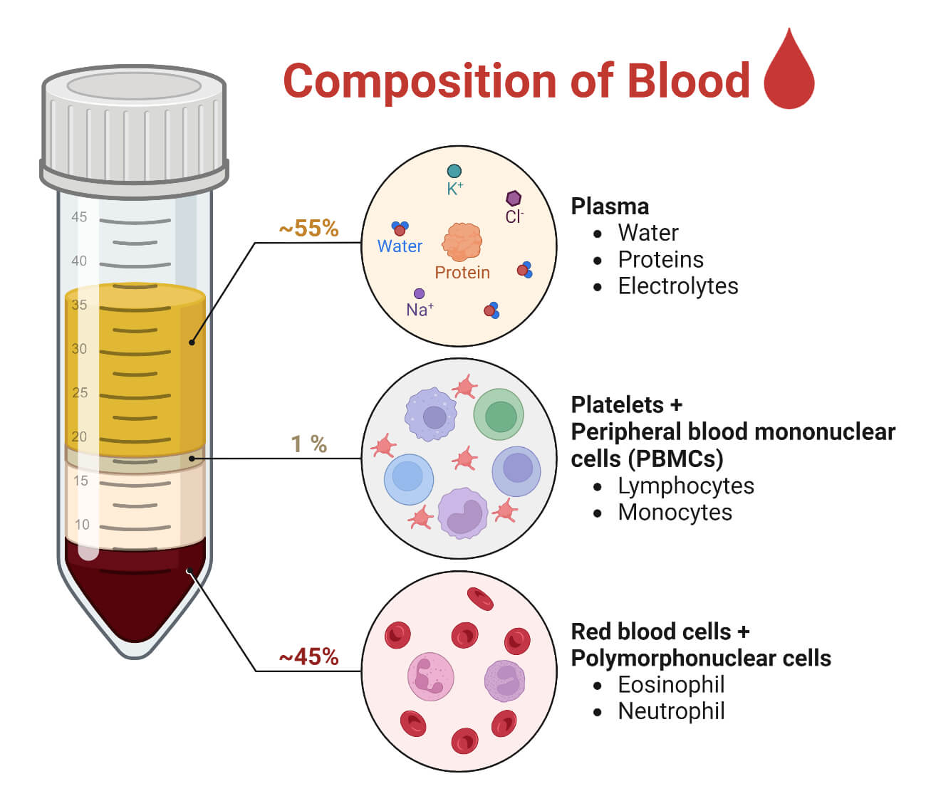
Blood is made up of blood cells (also called hematocytes or hematopoietic cells) and plasma.
Blood Cells (Hematocytes)
These are the cellular components of the blood covering about 45 – 46% of total blood. There are four types of blood cells present in humans, namely, (i) Red Blood Cells (RBCs), (ii) White Blood Cells (WBCs), and (iii) Platelets.
Red Blood Cells
- Red blood cells also called erythrocytes or RBCs are red-colored biconcave cells lacking a nucleus. They are the most abundant cells covering about 99% of the total blood cells. Structurally they are disk or biconcave in structure with about 7 to 7.5 μm diameter and about 2.5 μm thickness at the edge. They have an average life span of 120 days.
- They are comparatively more numerous in males (about 42 to 54% of total blood volume or approx. a total of 4.7 to 6.1 million RBCs per cubic microliter of blood) than in females (about 37 to 47% of total blood volume or approx. a total of 4.2 to 5.4 million RBCs per cubic microliter of blood).
- The most important components of the RBCs are hemoglobin and antigenic glycoproteins (blood group antigens). Hemoglobin is an iron-rich protein that helps to carry oxygen and carbon dioxide gases. These proteins are packed inside the RBC’s cytoplasm and cover about 95% of the dry weight of each RBC.
- The plasma membrane of RBCs contains blood group antigens that are used to classify the blood into various blood groups. The major antigens of concern are A antigen, B antigen, O antigen, and Rhesus (Rh) antigen.
- The major function of RBCs: Transportation of gases (oxygen and carbon dioxide)
White Blood Cells
- White blood cells also called leukocytes or WBCs or polymorphonuclear cells are colorless nucleated defensive blood cells. They are the largest blood cells but are the least in number; on average only about 4000 to 11000 WBCs per cubic microliter of blood. WBCs are polymorphic cells i.e. different types of WBCs have different structures.
- WBCs are immune cells and their major function is to provide defense or immunity to the body.
- The WBCs are of various types, and in a broader sense, they can be categorized into two groups: Granulocytes and Agranulocytes.
- Granulocytes: Also called polymorphonuclear leukocytes are a group of WBCs characterized by multi-lobed nuclei and cytoplasmic granules. There are three types of granulocytes, namely, eosinophils, basophils, and neutrophils.
- Eosinophils are circular granulocytes with bilobed nuclei (roughly of equal size) which take up the acidic red dye and appear red or pink. They are present in low numbers covering only 1 – 6% of total WBCs in blood. Their main role is to defend against parasitic infections and induce inflammation.
- Basophils are circular granulocytes with bilobed nuclei (of distinctly different sizes) which stain blue or bluish-purple due to absorption of the basic methylene blue dye. They are the least abundant WBCs covering less than 1% of the total WBCs. Their main role is as a mediator of inflammation and allergic reactions and the destruction of allergens.
- Neutrophils are small and the most abundant WBCs with multilobed nuclei (up to 6 lobes) which take up both the acidic and basic dye and stain as purple colored. They are most abundant covering about 40 – 75% of total WBCs and are easy to notice due to their multilobed nuclei. Their primary function is to provide defense against bacteria.
- Agranulocytes: These are a group of WBCs characterized by a large single nucleus and cytoplasm lacking granules. There are two types of Agranulocytes; monocytes and lymphocytes.
- Monocytes are the largest types of WBCs having a single round and very large nucleus which stains purple or bluish. They cover about 2 – 10% of total WBCs. Monocytes are further of two types; (i) motile monocytes in circulation known as phagocytes, and (ii) mostly non-motile monocytes in tissue known as macrophages. They are very important for defense against microbial infections, regulation of inflammation, and activation of T-lymphocytes.
- Lymphocytes are the second most abundant type of WBCs characterized by a large kidney-shaped nucleus. They occupy about 20 – 50% of total WBCs. They are further of two types; (i) T-lymphocytes, and (ii) B-lymphocytes. Lymphocytes keep the memory of infection and antigens and produce specific antibodies to fight against the infection.
- The major functions of WBCs are: Providing immunity to defend the body against infections and Regulate the inflammatory response.
Platelets
- Platelets also called thrombocytes are tiny anucleated blood cells regulating hemostasis. They are very small disk-shaped blood cells measuring just about 2 to 4 μm in diameter; however, they are present in the large number of 200000 to 500000 thrombocytes per cubic microliter of blood. They have an average lifespan of 8 to 11 days.
- The main function of thrombocytes is to cause hemostasis (blood clotting).
Blood Plasma
Plasma is the fluid component of blood comprising about 54 to 55% of total blood volume. It is a straw-yellow colored fluid primarily made of water and plasma proteins and a trace amount of other elements like nutrients, hormones, gases, ions, etc. The plasma without the blood clotting proteins is called serum.
The components of human blood plasma are:
- Water accounting about 90 to 92% of the plasma. It maintains the fluidity of the blood, dissolves the components for transport, and also regulates temperature.
- Plasm proteins comprise about 7 to 8% of the total plasma. There are diverse types of plasma proteins like albumins, globulins (antibodies), clotting proteins (like fibrinogen, prothrombin, etc.), lipoproteins, and other conjugated proteins.
- Electrolytes or ions in trace amounts like Na+, K+, Ca++, PO4+++, Mg++, Fe+, etc.
- Nutrients like glucose, amino acids, fatty acids, and vitamins.
- Hormones secreted by different endocrine glands.
- Gases like oxygen and carbon dioxide are in dissolved form and bound form.
- Waste products like metabolic wastes, urea, creatinine, etc.
Formation of Blood Cells
The process of formation of blood cells is called hematopoiesis or hemopoiesis (haemopoiesis) or hematogenesis. On average, 200 billion red blood cells, 10 billion white blood cells, and 400 billion platelets are formed each day in a normal adult body. All of the blood cells are formed from a multipotent hematopoietic stem cell (HSC). HSCs are self-replicating cells having the potential to differentiate and mature into different blood cells and lymphocytes. During the fetal stage hematopoiesis occurs in the yolk sac and aorta-gonad-mesonephros, then in the thymus, spleen, and liver in a developing embryo of 2 to 5 months, and finally, they are formed at bone marrow and lymph nodes in all developed embryo of 5 or more month age, infants and adults. The process of the formation of blood cells inside the bone marrow is called medullary hematopoiesis and the process of the formation of blood cells outside the bone marrow is called extramedullary hematopoiesis.
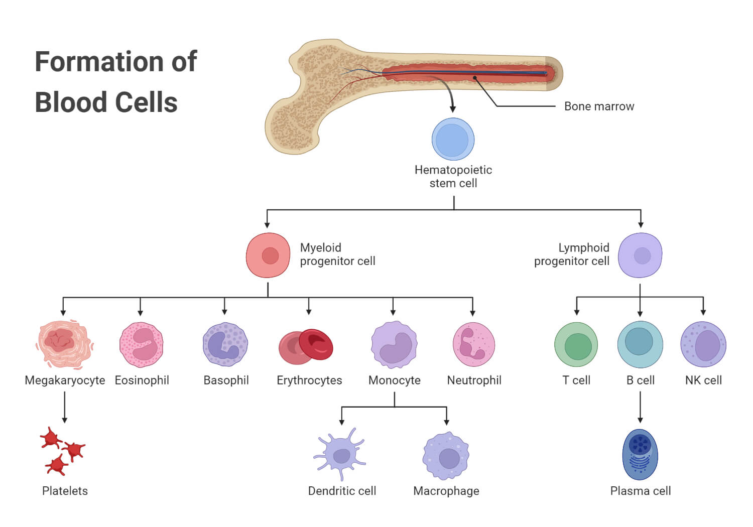
- In the bone marrow, there is an island of hematopoietic tissue where HSC are present. The HSC is developed into a precursor cell or a progenitor cell; based on the influence of the fate-determining factors the HSC differentiates into either the common myeloid progenitor or the common lymphoid progenitor.
- The common myeloid progenitor can differentiate into either RBC or WBC, or platelets, and the common lymphoid progenitor can differentiate into lymphocytes. The duration required for the formation of a blood cell is equal to the life span of that particular blood cell, i.e. for RBC it is 120 days, for WBCs few hours to days, and for platelets, it is 5 – 10 days, so that the blood cells can be replenished continuously.
- Erythropoiesis: It refers to the formation of the RBCs. The common myeloid progenitor develops into the proerythroblast which then simultaneously develops and differentiates into basophilic erythroblast, polychromatic erythroblast, orthochromatic erythroblast, polychromatic erythrocyte, and then finally metamorphose into the erythrocyte (RBC).
- Leukopoiesis: It refers to the formation of leukocytes (WBCs). The common myeloid progenitor can differentiate into the myeloblast which then can differentiate into different granular and agranular leukocytes. The myeloblast develops into a basophil promyelocyte which continuously develops into the basophil myelocyte, basophil metamyelocyte, basophil band, and finally to a basophil. Similarly, the myeloblast can differentiate into neutrophil promyelocyte and eosinophil promyelocyte which undergo similar stages of development like the basophil promyelocyte and finally form neutrophil and eosinophil respectively. Besides, the myeloblast can also develop into a monoblast which then matures into either a monocyte, macrophage, or myeloid dendritic cell.
- Thrombopoiesis: The common myeloid progenitor can also metamorphose into a megakaryoblast which ultimately develops to form a thrombocyte through the promegakaryocyte and megakaryocyte stages sequentially.
Functions of Blood
- The transportation of gases (oxygen and carbon dioxide) using RBCs or hemoglobin and in the dissolved state or combined stage.
- Transportation of nutrients, hormones, ions, and other chemical constituents to the cells for different metabolic processes.
- Collection of the metabolic wastes from all the cells and tissues and transporting them to excretory organs for filtration and excretion.
- Circulating WBCs, antibodies and other components provide immunological functions.
- Platelets and other blood clotting factors circulating induce and maintains hemostasis and break down clots.
- Circulating blood also controls the body temperature.
- Maintains homeostasis and osmolarity of cells and tissues.
What is Blood Circulation?
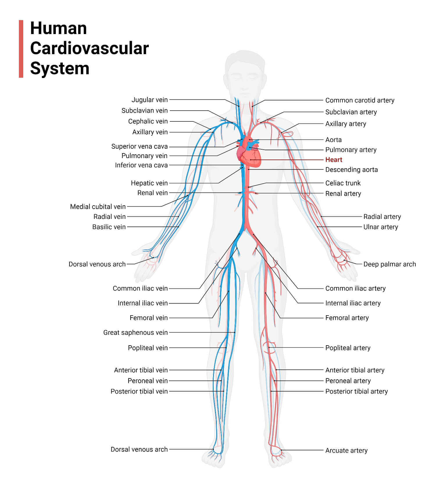
Blood circulation is the movement of blood in the blood vessels due to the mechanical pumping action of the heart. The heart pumps out the oxygenated blood to all the parts of the body via systemic arteries and collects all the deoxygenated blood from various part of the body via systemic veins and pass it to the lungs for reoxygenation. This whole process is called the blood circulation process.
The collection of organs (body system) that are responsible for the circulation of blood is called the blood circulation system. It contains the cardiovascular system and blood. Human blood circulation is a closed type double blood circulation system. The closed type circulation indicates that blood is circulated in closed vessels and returned for recirculation. The double circulation system suggests that there is two separate circulatory pathway; the first includes pulmonary circulation i.e. circulation between the heart and the lungs, and the second includes systemic circulation i.e. circulation between the heart and other organs and tissues.
Types of Blood Circulation
Broadly blood circulation in humans can be divided into two types; pulmonary circulation and systemic circulation.
Pulmonary circulation
It is the process of circulation of blood between the heart and lungs for blood purification (reoxygenation) purposes. The heart collects all the impure blood in the right atrium and sends it to the right ventricle from which it is pumped to the lungs through the pulmonary artery. The pulmonary arteries branch into smaller capillaries in the alveoli of the lungs where the gaseous exchange takes place. The blood from the pulmonary arteries loose carbon dioxide and picks up oxygen from the gas in the alveoli. The capillaries then unite to form pulmonary veins which then carry the oxygenated blood back to the left ventricle of the heart for circulation in the systemic circulation system.
Systemic circulation
It is the process of circulation between the heart and all body tissues through the systemic arteries and veins. The heart receives the oxygenated blood from the pulmonary circulation in the left atrium via the pulmonary veins. The left atrium passed the blood to the left ventricle from which it gets distributed all over the body via the aorta and systemic arteries. The arteries gradually branch into capillaries and transport blood in every tissue and cell. In the systemic capillaries, there occurs the exchange of gases between the blood the body cells, and the exchange of nutrients and metabolic wastes, and other materials. The capillaries then collect the metabolic wastes and carbon dioxide and combine them to form venules. The venules pour in the blood in the veins that finally drain into the vena cava. The vena cava finally returns all the blood to the right atrium to send the blood to purify in the pulmonary circulation. Coronary circulation is a special part of systemic circulation where the blood is supplied to the heart muscles via the coronary arteries and the coronary veins.
Blood Disorders and Diseases of Blood
There are several diseases and illnesses affecting blood and blood circulation. We can broadly categorize those disorders into infectious disorders and non-infectious disorders.
Infectious Blood Disorders
These are the blood disorders caused by infectious microorganisms and/or their products. It is simply called a blood infection and the pathogens involved are blood pathogens. It includes:
- Sepsis: It is the condition when there is the presence of bacteria, fungi, viruses, or parasites in the blood leading to the manifestation of clinical symptoms and the body’s immune response against such pathogens.
- Bacteremia: It means the presence of bacteria in the blood or circulation. Some common bacteria causing bacteremia are Staphylococcus spp., Streptococcus spp., Acinetobacter baumannii, E. coli, Klebsiella spp., Salmonella spp., Pseudomonas aeruginosa, etc.
- Viremia: It is the presence of viruses in the blood. Some common viruses that can be detected in the blood are HIV, Hepatitis B, Hepatitis C, Dengue virus, Measles virus, Chickenpox virus, Ebola virus, Lassa virus, etc.
- Fungemia: It can be defined as the presence of fungi (spores, or vegetative forms including yeasts) in the blood. Some common fungi that can infect the blood are Histoplasma spp., Candida spp., Aspergillus spp., Cryptococcus spp., Saccharomyces spp., etc.
- Parasitemia: It can be defined as the presence of parasites in the blood. Some common parasites that can be detected in the blood are Plasmodium spp., Trypanosoma spp., Babesia spp., Haemoproteus spp., etc.
Non-infectious Blood Disorders
These are blood disorders caused by non-microbial causes. It may be due to defects in the circulation or defects in blood components or other body physiology. Some of the common blood disorders are:
- Anemia: It can be defined as a blood disorder where there is a reduction in the oxygen-carrying capacity of blood (RBCs) and/or a reduction in the quantity and functioning of the hemoglobin. It is assumed that about 1/3rd of the world’s population is affected by this disorder. It can be caused by the disturbance in the formation of RBCs and hemoglobin, the destruction of formed RBCs and hemoglobins, by over bleeding, by over dilution of the blood (hypervolemia), or even intestinal inflammation.
- There are over 400 types of anemia. Clinically they are classified as normocytic anemia (anemia with normal-sized RBCs), megaloblastic anemia (anemia with increased-sized RBCs), and microcytic anemia (anemia with reduced-sized RBCs).
- Scientifically anemia is broadly classified as aplastic anemia (anemia due to damage in the bone marrow), megaloblastic anemia (increased RBCs due to deficiency of vitamin B12 and/or folic acid), hereditary anemia (due to genetic disorder) including most well-known type ‘the sickle cell anemia’, hypochromic anemia (deficiency of hemoglobin), hemorrhagic anemia (due to over bleeding), and anemia due to reduction in the number of RBCs.
- Erythrocytosis: It is a blood disorder when there is an increase in the number of RBCs than usual. It is also known as polycythemia. It may be either the primary erythrocytosis caused by the problem in bone marrow or the secondary erythrocytosis caused by a disorder in other systems except for bone marrow.
- Leukopenia: It is the condition when there is a decrease in the WBCs count in the blood than in normal. Normally, if the WBC count is less than 4.0 × 10⁹ per liter of the peripheral blood then, the condition is defined as leukopenia. If there is a decrease in neutrophils then it is called neutropenia. Similarly, decreased amount of lymphocytes in the blood is termed lymphocytopenia.
- Leukocytosis: It is the condition when there is an increase in the WBCs count in blood than in a normal person. Normally, WBC count above 11.0 × 10⁹/L of peripheral blood is termed leukocytosis. Neutrophilia, Lymphocytosis, and Eosinophilia are the commonly encountered leukocytosis types which mean there is an increase in the number of neutrophils, lymphocytes, and eosinophils respectively.
- Thrombocytopenia: It is the condition when there is a decrease in the number of thrombocytes (platelets) than in normal. Normally, a platelet count of less than 150,000 per μl of blood is stated as thrombocytopenia.
- Thrombocytosis: It is the condition when there is an increase in the number of platelets than in normal.
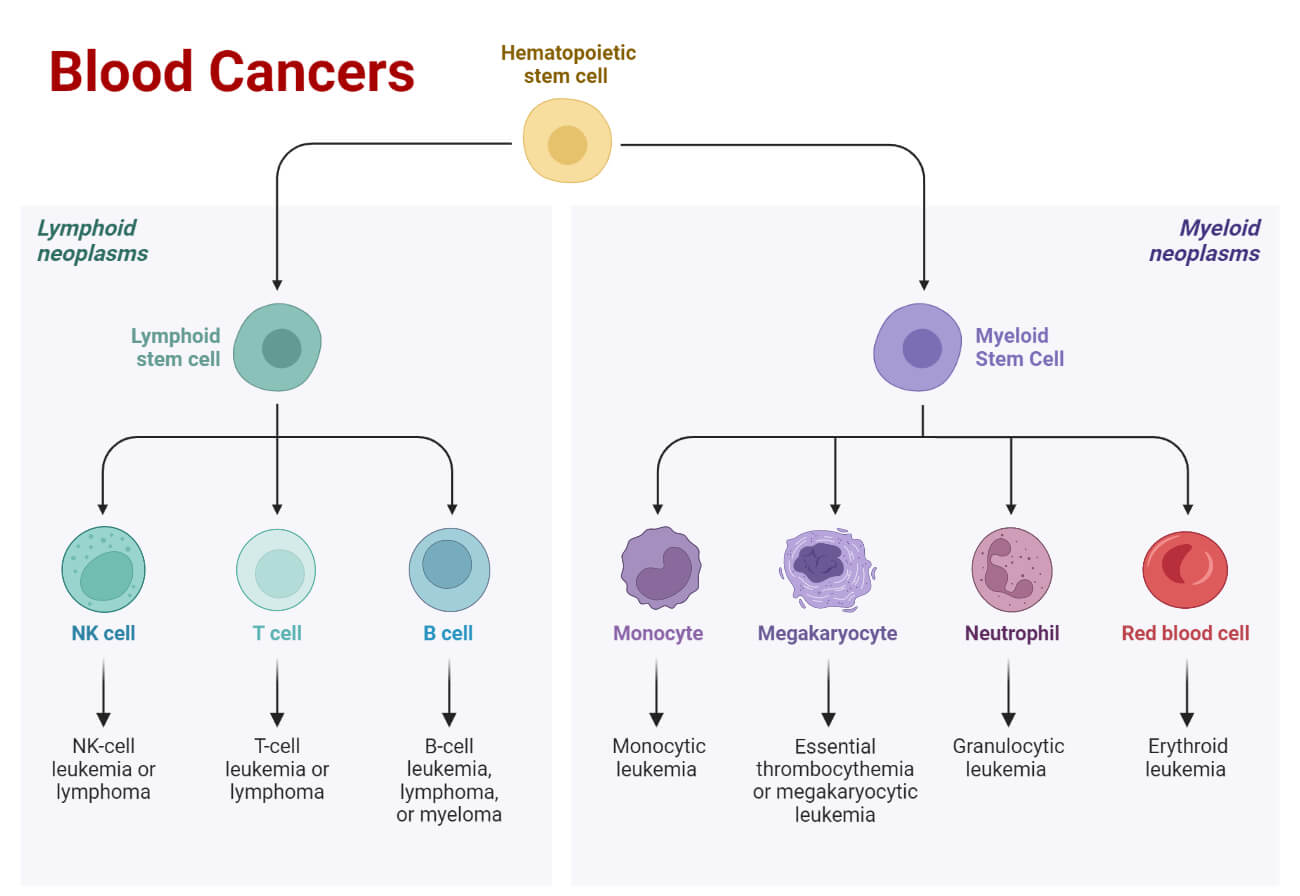
- Blood Cancer: It is the condition when there is an uncontrolled growth of blood cells and blood cell precursors affecting the bone marrow and blood cells. Mainly three types of blood cancer are common; leukemia, myeloma, and lymphoma.
- Leukemia is the most common type of blood cancer where there is an increase in the production of the abnormal WBCs interfering with the bone marrow’s functions including the production of RBCs and platelets. It is extensively associated with the bone marrow and produces a large number of leukemia cells (or blast cells) i.e. undeveloped blood cells.
- Myeloma also referred to as multiple myeloma is a type of blood cancer where there is excessive growth of the plasma cells crowding the bone marrow and affecting the production of RBCs, platelets, and other WBCs.
- Lymphoma is another common type of blood cancer that begins in the lymphoid system and forms tumors from the lymphocytes.
- Hemorrhage: It means the leakage of blood outside of the blood vessels.
- Hematoma: It means the collection of blood in the tissue or organs.
- Hemochromatosis: Excessive amount of iron in the blood is called hemochromatosis.
- Disseminated Intravascular Coagulation: DIC is a condition when there is a continuous hemorrhage and hemostasis in small blood vessels.
- Hemophilia: It is a genetic disorder caused by the deficiency of blood clotting proteins in the blood leading to uncontrolled bleeding and delayed hemostasis.
References
- Ross & Wilson Anatomy & Physiology in Health and Illness. 13th ed. Churchill Livingstone Elsevier. ISBN 978-0-7020-7276-5
- Mank V, Brown K. Leukocytosis. [Updated 2022 Sep 12]. In: StatPearls [Internet]. Treasure Island (FL): StatPearls Publishing; 2022 Jan-. Available from: https://www.ncbi.nlm.nih.gov/books/NBK560882/
- Blood Definition & Meaning – Merriam-Webster
- How much blood is in the human body? | Live Science
- Proportion of white blood cell types in blood – Human Homo sapiens – BNID 102748 (harvard.edu)
- Chapman J, Zhang Y. Histology, Hematopoiesis. [Updated 2022 May 8]. In: StatPearls [Internet]. Treasure Island (FL): StatPearls Publishing; 2022 Jan-. Available from: https://www.ncbi.nlm.nih.gov/books/NBK534246/
- Blood cells and its types with functions (microbiologyinfo.com)
- Understanding the Hematopoiesis Process (verywellhealth.com)
- Jagannathan-Bogdan, M., & Zon, L. I. (2013). Hematopoiesis. Development (Cambridge, England), 140(12), 2463-2467. https://doi.org/10.1242/dev.083147
- Hematopoiesis: Definition, Types & Process (clevelandclinic.org)
- Hematopoiesis – an overview | ScienceDirect Topics
- Composition of Blood and its Functions (byjus.com)
- What is Blood? | OneBlood
- blood – Platelets (thrombocytes) | Britannica
- Blood | Definition, Composition, & Functions | Britannica
- What are the 3 Types of Blood Circulation? – Vascular Health (vascularhealthllc.com)
- Sepsis: Symptoms, Causes, Treatment, Risks, and More (healthline.com)
- Which parasites can be detected in blood? – Studybuff
- Bacteremia: Causes, Symptoms, and Treatments (healthline.com)
- CDC – Bloodborne Infectious Diseases – Stop Sticks : Bloodborne Pathogens – NORA
- Know About Blood Infections – Types, Signs, Symptoms & Treatment | MrMed
- Bloodborne Pathogens: A Complete Guide (hipaaexams.com)
- Bloodborne Pathogens – General Guidance | Occupational Safety and Health Administration (osha.gov)
- https://www.webmd.com/a-to-z-guides/what-is-erythrocytosis
- 8 Types of Anemia | Based on the Blood cell Morphology & Causes (studyread.com)
- Anemia – Symptoms and causes – Mayo Clinic
- Leukopenia: Types, Symptoms, Causes, Treatment & More (healthline.com)
- Platelet Disorders – Thrombocytopenia | NHLBI, NIH
- Thrombocytopenia (low platelet count) – Diagnosis and treatment – Mayo Clinic
- Thrombocytosis: Symptoms, Causes & Treatment (clevelandclinic.org)
- Lymphoma | CDC
- Myeloma | CDC
- Lymphoma – Symptoms and causes – Mayo Clinic
- Blood Cancer: Overview, Symptoms & Types (clevelandclinic.org)
- Platelet Disorders – Thrombocythemia and Thrombocytosis | NHLBI, NIH
- Human Anatomy: Blood – Cells, Plasma, Circulation, and More (webmd.com)