Interesting Science Videos
Habitat of Ascaris lumbricoides
- Ascaris lumbricoides are the largest most familiar endoparasites of the man freely living in the lumen.
- It has also been reported from animals like pigs, cattle, sheep, and squirrels.
- It inhabits the small intestine of more frequently children than adults.
- It is cosmopolitan in distribution.
- Sometimes, it migrates from the intestine to the stomach and comes out through the host’s nostrils.
External Morphology of Ascaris lumbricoides
1. Shape and size
- It is elongated, cylindrical, and gradually tapering at both ends, the anterior end being slender than the posterior.
- It is the largest size nematodes.
- It shows sexual dimorphism. i.e., sexes are separate.
- The female measures 20 to 40 cm in length and 4-6 mm in diameter with a straight tail.
- The male is smaller measures 15-31 cm in length and 2-4 mm in diameter with a ventrally curved tail.
- The cuticle covering the body surface bears a minute transverse striation, giving a worm’s pseudosegmented appearance.
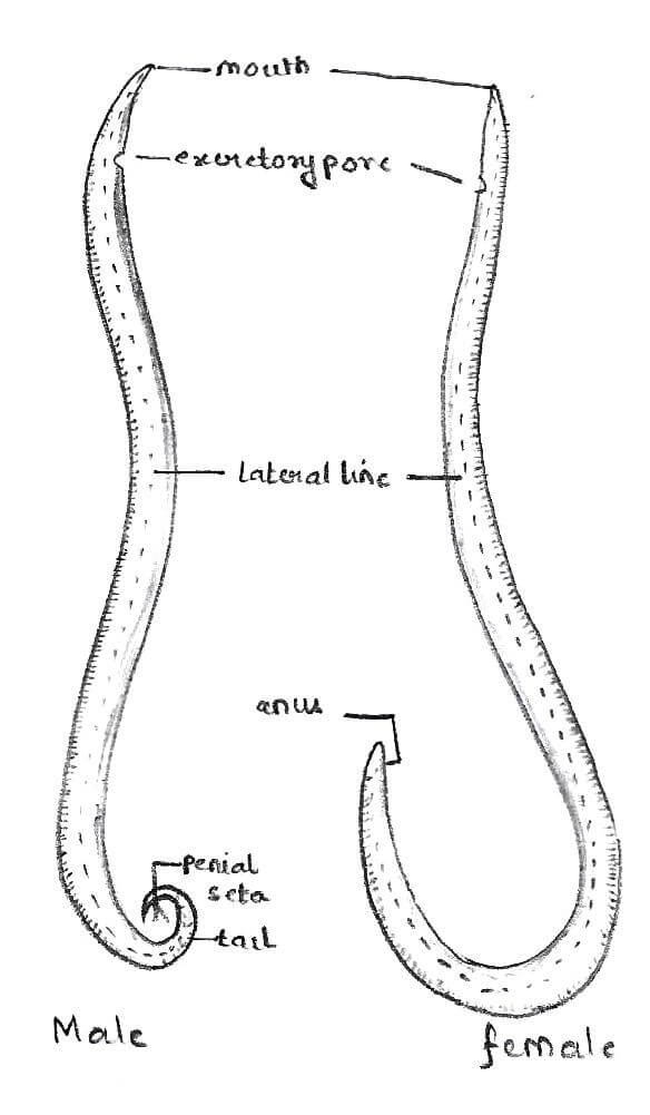
Figure: Male and Female Ascaris lumbricoides. Image Source: Harveen Kaur.
2. Coloration
- Generally, the nematodes have no color.
- The freshly excreted worm is yellowish pink in color, which gradually changes to white.
3. Longitudinal streaks or lines
- The cylindrical body has 4 longitudinal lines -1 narrow mid-dorsal, 1 mid-ventral, and 2 thick ones are lateral.
- The lines are visible externally.
- The dorsal and ventral lines appear pure white, while lateral lines appear brown.
- These lines are thickened all over the body and are simply impressions of the syncytial epidermis.
- The excretory pore is situated mid-ventrally, a little behind the lips.
- The genital pore and anus open separately in the female; the genital pore is situated mid-ventrally at about 1/3rd from the anterior end.
4. Anterior End
- Both the sexes have the same anterior end structures.
- Ascaris have no eyes.
- It is rounded, bearing a small terminal triradiate mouth aperture guarded by 3 broad lips or labia.
- One lip is mid-dorsal and broadly elliptical, and 2 are sub-ventral or latero-ventral and oval.
- Of these, one is on the upper side, and the other two on the two sides of the mouth cavity.
- Dorsal and anterior extension of labial parenchyma bears minute denticles.
- The outer surface of each lip bears minute sensory papillae/ labial.
- Labial papillae are hair-like structures used for tactile reception and are very sensitive to vibrations and touch.
- The dorsal lip has 2 sensory papillae, one on each side.
- Each latero-ventral lip has 1 double sensory papilla in its ventral position.
- These four papillae form an outer labial circle, but nematodes have 6 papillae in the outer labial circle.
- The papillae of the inner labial circles are absent in Ascaris, but other nematodes have 6 papillae in its inner labial circle.
- The latero-ventral lip has lateral papilla, each in lateral and cuticular excavation called amphid.
- Amphids are reduced in parasitic nematodes.
- Just behind the latero-ventral, there is a pair of cervical papillae, one on each side close to the nerve ring.
- Papillae are tangoreceptors (sensory) while amphids are olfactory chemoreceptors.
- The lips bear a fine tooth.
5. Posterior End
- The posterior end of the body shows clear sexual dimorphism.
- In females, the posterior extremity is conical and straight—the midventral transverse apertures, or anus, guarded by one pair of postanal papillae.
- Only the digestive tubes open to the outside through the anus.
- In males, the tail end is curved ventrally in the form of a hook with a conical tip.
- In male anus is replaced by cloaca. It is the common aperture for the rectum and genital tubes.
- Two copulatory setae protrude from the aperture. They are called peneal setae or spicules.
- The setae serve to transfer-sperm into the female vagina during copulation.
- The male tail is characterized by numerous genital papillae ventrally, about 50 pairs of pre-anal papillae, and 5 pairs of postanal papillae.
- Genital papillae of male help in copulation.
- 2 anterior pairs of postanal papillae are double, while the rest are single.
6. Excretory pore
- Excretory pore lies mid-ventrally and near the anterior end.
7. Female gonopore
- At about one-third of the entire length from the anterior end, the body is narrower in female s, and this region is known as the vulvar waist.
- The female genital aperture called vulva or gonopore is situated on the ventral surface of the vulvar waist.
- But the genital pore on the male opens into the cloaca.
Body wall of Ascaris lumbricoides
Ascaris’s body wall is made up of 3 layers, i.e., outer cuticle, middle sub cuticle or epidermis or hypodermis, and inner longitudinal muscle lining of the body cavity.
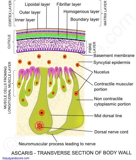
Figure: Body wall of Ascaris. Image Source: Study and Score.
a. Cuticle
- The body is covered with a thick, tough, elastic transparent, and glossy layer cuticle.
- The cuticle is secreted by the underlying epidermis and continuous with the pharynx and rectum’s cuticular lining.
- It is non-cellular and made of albuminous protein, resistant to host digestive juices but permeable to salt and water.
- In a young worm, the cuticle is shed off to permit growth.
- It is divisible into several layers with numerous vertical channels.
- The transverse wrinkles of the cuticle give the characteristic segmental appearance of the body.
- The cuticle also lines the buccal cavity, esophagus, rectum, cloaca, vagina, and excretory pore.
- Under a light microscope, the cuticle can be identified into 4 layers, but in a recent study, the electron microscope has also revealed the fifth lipid layer.
- The lipid layer is about 1000 Aº thick and in the form of a thin osmophilic membrane.
- Cortical or cortex layer which is made of keratin and resistant to the action of host digestive enzymes. It includes an outer cortical layer consisting of discontinuous strips or rings around the body and an inner cortical layer.
- The matrix layer is spongy in consistency and contains Sulphur-rich protein matricin. This layer is elastic and contains several fine fibers. It consists of an outer fibrillar layer, a homogenous layer that shows several radial striations, and a boundary layer resembles the outer cortical layer.
- The fiber layer is the inner layer, but not the last layer of the cuticle ad consists of collagen fibers crossing each other and arranged in 3 strata.
- The basement membrane is a thin layer forming the inner limit of the cuticle.
b. Epidermis or hypodermis
- The hypodermis or epidermis is a thin syncytial layer under the basal lamina.
- It contains many nuclei but no cell walls, the nuclei lie in the longitudinal epidermal chords only.
- This layer projects along the mid-dorsal, mid-ventral, and lateral lines and form epidermal chords.
- Lateral chords are more conspicuous and seen on the surface as yellow lines.
- Excretory canals and lateral nerves run along with the lateral chords.
- while dorsal and ventral chords contain dorsal and ventral nerve, respectively.
- The epidermis of free-living nematodes contains unicellular epidermal glands.
- In the epidermis fat and glycogen are reserved abundantly.
c. Longitudinal muscle/ muscle layer
- A single layer of muscle cells remains attached to the hypodermis. In this layer, circular muscles are altogether absent.
- Longitudinal muscles form a single layer of spindle-shaped muscle cells beneath the epidermis and lining the body cavity.
- The muscle layer is divided into 4 longitudinal columns and strips 2 dorsolateral and 2 ventrolateral.
- Each column contains about 150 muscle cells.
- For the identification of species in nematodes number and disposition of muscle cells play an important role.
- Each muscle cell contains two parts, a muscular part, which is contractile, lies towards the epidermis, and a granular non-contractile protoplasmic part, a club-shaped, bladder-like mass of protoplasm with nucleus and network of supporting fibrils form a fibrous process or muscle tail.
- The muscular portion contains longitudinal contractile fibers arranged at intervals and also the fibers for attachment to the cuticle.
- The protoplasmic part remains inwards and connected with the neuromuscular process.
- Muscle cells of all the cells of 2 dorsolateral columns are ultimately connected with the dorsal nerve cord.
- While those of 2 ventrolateral columns are connected with the ventral nerve cord.
- Dobell (1965) has emphasized that muscle tails are cellular extension, which forms synapsis on the motor nerves of the dorsal and ventral nerve cord.
Reference and Sources
- Kotpal RL. 2017. Modern Text Book of Zoology- Invertebrates. 11th Edition. Rastogi Publications.
- Jordan EL and Verma PS. 2018. Invertebrate Zoology. 14th Edition. S Chand Publishing.
- 6% – https://www.biologydiscussion.com/invertebrate-zoology/phylum-aschelminthe/ascaris-lumbricoides-habitat-sense-organs-and-life-history/29004
- 2% – https://www.notesonzoology.com/phylum-aschelminthes/ascaris-external-features-and-body-wall-phylum-aschelminthes/5904
- 1% – https://www.studyandscore.com/studymaterial-detail/ascaris-general-characters-body-wall-and-body-cavity
- 1% – https://www.biologydiscussion.com/parasitology/parasitic-worm/morphology-of-roundworm-with-diagram/62249
- <1% – https://www.wormatlas.org/hermaphrodite/hypodermis/Hypframeset.html
- <1% – https://www.kikaayacollegeschool.com/biology-o-level-notes-s1-s4
- <1% – https://histology.medicine.umich.edu/resources/small-large-intestine
- <1% – https://en.wikipedia.org/wiki/Epidermis_(botany)
- <1% – https://en.wikipedia.org/wiki/Basement_membrane
- <1% – https://edurev.in/course/quiz/attempt/-1_Test-Ascaris-Lower-Animal/6d6f162b-7c05-43b1-b808-3dfd2df4968b
It’s going to be very interesting to see what happens when the masses discover that their trusted doctors have intentionally ignored probable parasite infections in their patients because, without increasingly heavy parasite loads in the human body, very few, if any, serious diseases would develop.
Great
Ascariasis lumbricoides commonly known as the large intestinal roundworm, roundworm of man causing ascariasis or roundworm infection is the world’s most common intestinal helminth infection, affecting approximately 1 billion people. It is the second most common infection in the United States, after pinworm infection.
Children who put their contaminated hands in their mouths are the most vulnerable to Ascaris lumbricoides infection. Contamination sources range from children’s toys to the soil itself. https://labweeks.com/ascaris-lumbricoides/