Interesting Science Videos
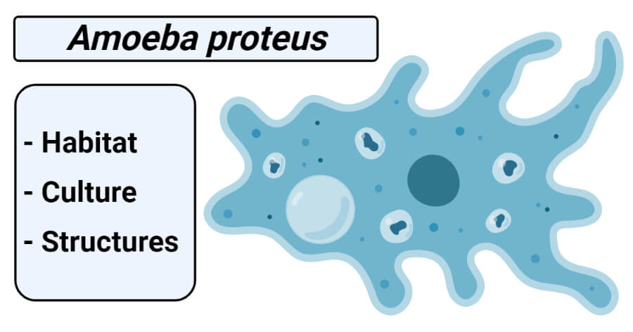
Classification of Amoeba proteus
Phylum: Protozoa
Subphylum: sacromastigophora
Superclass: Sarcodina
Class: Rhizopodea
Subclass: Lobosia
Order: Amoebida
Genus: Amoeba
Species: proteus
Habit and Habitat of Amoeba proteus
- Amoeba proteus is widely distributed and commonly found on the bottom mud or on the underside of aquatic vegetation in freshwater, ponds, ditches, lakes, springs, slow-running streams.
- It is often found in relatively clean ponds with highly oxygenated freshwater.
- It is also found in large “food webbed ecosystems” that contain many algae and plants.
- Due to its adversity to light, it usually sheltered under the sides of lotus ponds or dwell near the bottom of the pond.
- With the help of false feet or pseudopodia, it moves and feeds.
- It has a great power of regeneration.
Culture of Amoeba proteus
- It may be obtained for class study by scraping decaying vegetation from the bottom of a pond.
- Then scrapping, is settle down in the wide-mouth container, Amoeba of different kinds may be found in the sediment and sorted with the help of fine pipette under a binocular microscope.
- A temporary culture of Amoeba can be prepared in the laboratory by the hay-infusion method. In this method decaying weeds or other organic substances such as hay, twigs, dry leaves, seeds, etc. are taken and boiled for 15 minutes with a sufficient amount of freshwater. After boiling, water is filtered, and the filtrate is allowed to cool. To this filtrate, new Amoeba-rich water drops are added and allowed to multiply for 2 to 3 days.
- A culture of Amoeba can be maintained by keeping put some pond water, mud, and leaves in 100 ml of water containing a few grains of wheat. Amoebae will appear after a few days; this shows the presence of cysts in the pond water.
- To make a pure culture, boil four or five grains of wheat in 100 ml of distilled water for 10 minutes and cool for a few days; to this add some Amoebae from the first culture and cover with a glass plate; in ten days many Amoebae will be formed in the pure culture.
Structure of Amoeba proteus
1. Shape and size
- It is a unicellular organism and measures about 250 to 600 µm in maximum diameter and so transparent that is invisible to naked eyes.
- To the naked eyes, the larger A. proteus is just visible as a whitish blob.
- Under the microscope, it appears as an irregular, colorless, jelly-like tiny mass of hyaline protoplasm.
- Amoeba has no fixed shape and the outline of the body continues changing due to the formation of small finger-like outgrowths called pseudopodia. When it withdraws all its pseudopodia, it becomes spherical in shape.
- It has no cell wall; it has a thin delicate outer enclose membrane called the plasmalemma.
- Just beneath this is a non-granular layer, the ectoplasm which encloses the granular endoplasm. However, there is no line of demarcation between the ectoplasm and endoplasm.
- It is considered to have a definite polarity though it is shapeless i.e. it has definite anterior and posterior ends.
- It put out pseudopodia ate anterior end while the posterior end is marked by a wrinkled region called uroid.
2. Pseudopodia
- The pseudopodia are the most defined structures of A. proteus which makes the organism so fascinating.
- Pseudopodia (Gr., pseudos = false; podos = foot) are irregular blunt extensions which are constantly being given out or withdrawn by the body.
- These are of variable size and are capable of protruded or retracted, often with considerable speed.
- They are broad to cylindrical with blunt rounded tips and are composed of both ectoplasm and endoplasm. Such pseudopodia are called lobopodia.
- These are formed as a result of liquefaction and flowing forward of the cytoplasm.
- These “false feet” are used for movement and to engulf prey– making it an essential part of its structure.
- Needless to say, without these structures, the A. proteus would’ve had to adapt using other means to move and gain nutrients or be wiped off the surface of the planet.
3. Plasmalemma
- Amoeba has no pellicle or cell wall.
- The body is covered by a very thin, delicate, invisible, elastic external cell membrane called plasmalemma.
- The thickness of plasmalemma may be from 1/4 µ (0.00025 mm) to 2 microns.
- This membrane is permeable i.e. water and some small soluble molecules can pass freely through it in both directions.
- It is composed of a double layer of lipid and protein molecules.
- The outer layer of plasmalemma is supposed to contain mucoprotein.
- Plasma lemma when broken it can regenerate itself.
- It also retains the protoplasm within the cell.
- An unusual feature of plasmalemma is it contains numerous fine, ridge-like projections on its outer surface that are supposed to be adhesive and binding the organism to its substratum.
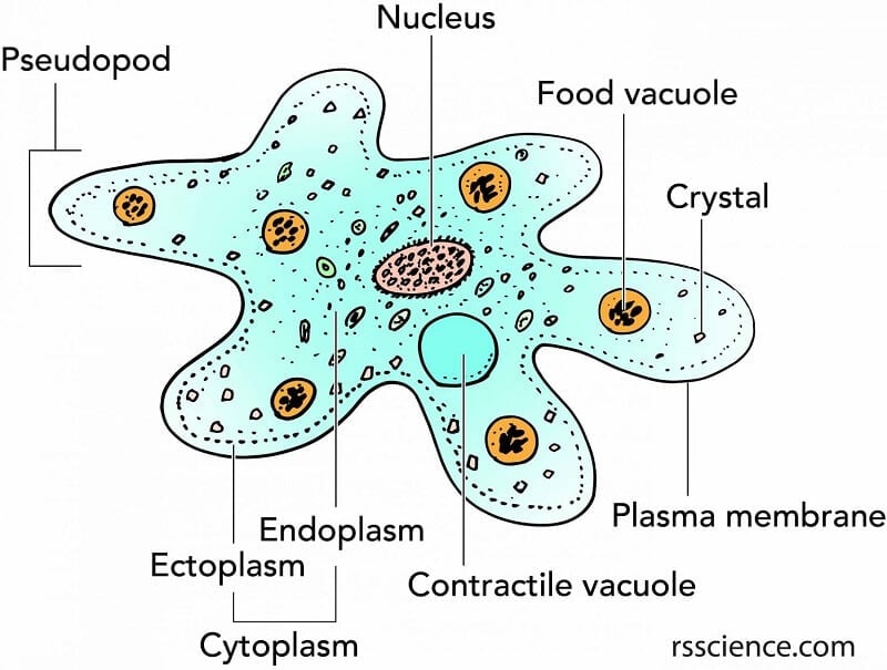
Figure: Structure of Amoeba proteus. Image Source: Rs’ Science.
4. Cytoplasm
- It is a dense mass inside the plasmalemma containing several organelles.
- It consists of 2 parts i.e. an outer ectoplasm and an inner endoplasm.
a. Ectoplasm
- Ectoplasm is an outer clean, thin, and transparent layer lie immediately beneath the plasmalemma.
- It is somewhat rigid and contractile under tension.
- It is thickened into a hyaline cap at the advancing end at the tip of pseudopodia.
- It is considered to be a supporting layer due to the presence of longitudinal rigid.
- Functions: It protects the organelles inside the body. It helps to maintain the shape of the body. It assists the Amoeba in producing pseudopodia.
b. Endoplasm
- Endoplasm is completely surrounded by ectoplasm; the endoplasm forms the bulk of the animal.
- It is fluid-like, granular, and semi-transparent.
- According to Mast, the endoplasm is made up of peripheral viscid or gel state, plasma gel, and a central flowing or sol state plasmasol.
- The plasmagel is granular and more solid but its granules show no movement.
- The plasmasol shows streaming movements and it is a highly granular fluid having various inclusion.
- The plasmagel can be transferred to and plasmasol and vice versa.
- Endoplasm contains the organelles of the cell.
- It supports various physiological functions. Frequent change in the density of the plasmagel and plasmasol helps in producing pseudopodia.
- Besides granules, endoplasm contains a number of important inclusions such as a nucleus, contractile vacuole, food vacuoles, mitochondria, Golgi apparatus, fat globules, and plate-like or bi-pyramidal crystals.
5. Endoplasmic organelles
It contains a number of organelles or structures suspended in it. They are the nucleus, contractile vacuole, food vacuoles, and water globule.
a. Nucleus
- Amoeba proteus contains a single conspicuous nucleus.
- It appears largely flattened, discoidal, and slightly biconcave in young specimens, but it is often folded and convolutes an older specimen.
- Its position is not fixed lies anywhere in the endoplasm.
- It may be difficult to see the nucleus in the body of living Amoeba.
- It can be easily seen in the phase-contrast microscope after fixing and staining Amoeba with a drop of methyl green acetic acid.
- It is granular and refractive to light.
- It is covered by the nuclear membrane made of protein and lipid.
- A honeycomb-like lattice is found below the inner nuclear membrane which play important role in maintaining the flattened form of the nucleus.
- The nucleoplasm is less in number.
- Functions: The nucleus of A. proteus is a membrane-bound organelle that houses most of the cell’s genetic information and controls the actions of the amoeba. It takes an essential part in the reproduction of the cell.
b. Contractile vacuole
- Some bubble-like vacuoles are seen in the endoplasm of Amoeba these are called a contractile vacuole.
- It is singe, clear rounded, transparent, and fast-growing.
- It is filled with watery fluid.
- Its position is not fixed but circulates in the endoplasm
- It is continuously contracted or expanded on the living body of Amoeba, so, named as contractile vacuoles.
- It is surrounded by a crowd of mitochondria, close to which tiny feeder vacuoles of water appear, which then combine to form a larger vacuole.
- Function: It helps in the osmoregulatory and excretory activities of the animals.
c. Food vacuoles
- It is spherical spaces, small and large, contains water and food in various phases of digestion.
- They are scattered in the endoplasm.
- Food vacuoles are formed when Amoeba engulfs food with a drop of water.
- These are non- contractile and of different sizes.
- Digestion takes place inside the food vacuole.
- They diaper with the egestion of non-digestible food from the body.
- Functions: Food vacuoles store the food material. They help in the digestion of food. They have also roles in the excretion of fecal materials.
d. Water globules
- It is the smallest transparent, spherical vacuole filled with water in the body of A. proteus.
- It can be one or more in numbers.
- It is noncontractile.
- Functions: It contains water and keeps the balance of the water constant of the body.
If an A. proteus is examined under an electron microscope the endoplasm shows various inclusions characteristics.
e. Other organelles
- Golgi bodies: It looks like small tubules and vesicles. Function: It helps in the secretion and excretion of food.
- Mitochondria: It is present around the contractile vacuoles and more or less oval and have tubular cristae. Function: They take part in respiration and in energy generation.
- Crystals: Plate-like or bipyramidal crystals are found. It is assumed that these crystals are simply the fecal substances produced during metabolism.
- The contractile vacuole is seen surrounded by several mitochondria and vesicles.
- Lysosome are membrane-bound spherical bodies, scattered in the endoplasm.
- The ribosome is found in endoplasmic vesicles as well as scattered in the endoplasm.
- The endoplasmic reticulum occurs as vesicles form a network of tubules.
Reference
- Kotpal RL. 2017. Modern Text Book of Zoology- Invertebrates. 11th Edition. Rastogi Publications.
- Jordan EL and Verma PS. 2018. Invertebrate Zoology. 14th Edition. S Chand Publishing.
- 12% – https://www.biologydiscussion.com/invertebrate-zoology/protozoa/amoeba-proteus-habitat-structure-and-metabolism/28183
- 4% – https://davidwangblog.wordpress.com/structure-and-functions/
- 2% – https://www.brainkart.com/article/Body-structure-of-Amoeba-and-its-functions_16485/
- 2% – https://www.biologydiscussion.com/parasites/the-structure-and-life-cycle-of-amoeba-with-diagram/2724
- 1% – https://www.thoughtco.com/endoplasmic-reticulum-373365
- <1% – https://www.qsstudy.com/biology/reproduction-of-amoeba
- <1% – https://www.ncbi.nlm.nih.gov/pmc/articles/PMC5065387/
- <1% – https://www.britannica.com/science/lysosome
- <1% – https://quizlet.com/94834338/ch-5-biology-study-flash-cards/
- <1% – https://byjus.com/biology/amoeba/
I want learn more about biology